J Korean Ophthalmol Soc 2019;60(4):369-373 ISSN 0378-6471 (Print)⋅ISSN 2092-9374 (Online)
https://doi.org/10.3341/jkos.2019.60.4.369
Case Report
성인의 눈물샘에서 발생한 안와 림프관 기형 1예
A Case of Adult Orbital Lymphatic Malformation in the Lacrimal Gland
송원석1⋅조성진2⋅최연주1
Won Seok Song, MD1, Sung Jin Cho, MD2, Youn Joo Choi, MD1
한림대학교 강동성심병원 안과학교실1, 한림대학교 강동성심병원 병리학교실2 Department of Ophthalmology, Hallym University Kangdong Sacred Heart Hospital1, Seoul, Korea
Department of Patholology, Hallym University Kangdong Sacred Heart Hospital2, Seoul, Korea
Purpose: When there is a mass in the superior temporal orbit area, a lacrimal gland tumor should be suspected. We report a rare case of orbital lymphatic malformation that was histologically diagnosed in a patient with typical clinical features of the lacrimal gland.
Case summary: A 55-year-old female with no underlying disease and no ophthalmic history visited our clinic with a right upper eyelid edema associated with an enlarged painless eyelid mass 1 month prior to her visit. The patient stated that she discovered the mass 1 year previously.The palpebral lobe of the lacrimal gland protruded slightly with congestion of the surrounding conjunctiva. Enhanced computed tomography showed a 3 cm well-defined heterogeneous mass in the right lacrimal gland area and several well-defined round calcifications within the mass. Orbital tissue or bone involvement was not observed. The pleo- morphic adenoma of the lacrimal gland was the most clinically suspicious, so complete resection of the mass was performed using lateral orbitotomy. Histopathologically, lymphangioma (lymphatic malformation) originating from the lacrimal gland was diagnosed.
Conclusions: Orbital lymphatic malformation can occur in the lacrimal gland. The present case showed that differential diagnosis can reveal the presence of an adult lacrimal gland tumor.
J Korean Ophthalmol Soc 2019;60(4):369-373
Keywords: Lacrimal gland tumor, Orbital lymphangioma, Orbital lymphatic malformation
■Received: 2018. 10. 4. ■ Revised: 2018. 11. 9.
■Accepted: 2019. 3. 23.
■Address reprint requests to Youn Joo Choi, MD Department of Ophthalmology, Kangdong Sacred Heart Hospital, Hallym University College of Medicine, #150 Seongan-ro, Gangdong-gu, Seoul 05355, Korea Tel: 82-2-2224-2274, Fax: 82-2-470-2088 E-mail: [email protected]
*Conflicts of Interest: The authors have no conflicts to disclose.
ⓒ2019 The Korean Ophthalmological Society
This is an Open Access article distributed under the terms of the Creative Commons Attribution Non-Commercial License (http://creativecommons.org/licenses/by-nc/3.0/) which permits unrestricted non-commercial use, distribution, and reproduction in any medium, provided the original work is properly cited.
앞쪽 안와의 외상측에 발생하는 종괴로서 가장 먼저 생 각해야 할 질환은 눈물샘종양이다. 눈물샘종양은 크게 비 상피병변과 상피성병변으로 나눌 수 있으며, 증상의 발현 기간, 통증 및 주변 연부조직의 양상, 영상의학적 소견을 통해서 질환을 감별하고, 경계가 명확한 종괴의 경우 완전
절제하여 조직학적 확진이 이루어진다. 본 증례에서는 임 상적으로 전형적인 눈물샘의 다형샘종 양상을 보인 환자에 서 조직학적으로 안와 림프관 기형(orbital lymphatic mal- formation)으로 진단된 드문 증례를 보고하고자 한다.
증례보고
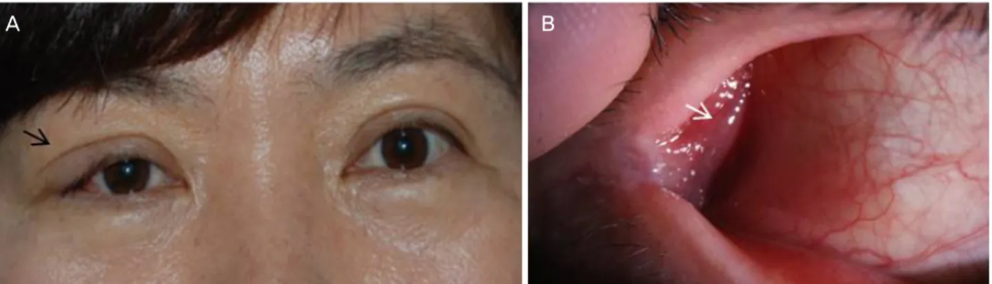
1년 전부터 발견된 통증 없는 우상안검 종괴가 있었던 55세 여자 환자가 1개월 전부터 크기가 커지면서 눈꺼풀부 종이 발생하여 내원하였다(Fig. 1A). 기저 질환 및 특별한 안과적 과거력은 없었다. 우상안검 외측에서 중간 정도 단 단함의 종괴가 촉지되었으며 눈꺼풀을 들어보았을 때 눈물 샘의 눈꺼풀엽이 약간 돌출되고 주변 결막의 충혈 소견이
A B
Figure 1. Clinical photograph. (A) Moderate firm, palpable mass with eyelid swelling (black arrow) is seen at right upper eyelid. (B) Prominent palpebral lobe of lacrimal gland with mild conjunctival injection (white arrow) is seen.
A B
Figure 2. Preoperative enhanced computed tomography images of coronal view (A) and axial view (B). A 3 cm well-defined hetero- geneous mass at right lacrimal gland area, and several well-marginated round calcifications within mass.
있었다(Fig. 1B). 이후 촬영한 조영증강 안와 전산화단층촬 영에서 우측 눈물샘의 3 cm 크기의 경계가 명확한 비균질 성 종괴가 관찰되었다. 군데군데 석회화 소견이 보였으며, 주변 안와조직이나 골 침범 소견은 보이지 않았다(Fig. 2).
1년여간에 걸쳐 서서히 발생한 무통성 종괴라는 임상양 상 및 앞쪽 안와 상외측에 경계가 잘 지어지는 안와종양이 라는 영상 소견을 토대로 눈물샘종양 중 다형샘종을 임상 적으로 가장 의심하였고, 이에 외측 안와절개술을 통한 종 양 전절제 생검을 시행하였다. 수술 시 예상과는 다르게 종 괴는 단단한 덩어리가 아닌 매우 얇은 피막으로 둘러싸인 말랑한 혈관 덩어리였으며, 정상 조직으로 여겨지는 눈물 샘 조직 사이에 위치하고 있었다. 이에 출혈이 발생하지 않 게 조심하여 종괴를 주변 조직과 분리하였다(Fig. 3A). 스
폰지 같이 말랑한 종괴는 크기가 약 2.0 × 1.5 cm로 측정되 었으며, 조직검사 소견상 얇은 내피세포로 둘러싸인 낭성 조직이 불규칙하게 확장되어 있었고, 점액성 퇴화(myxoid degeneration) 소견을 보여 오랜 기간동안 발생한 병변임을 시사하였다. 정상적인 눈물샘 조직이 확장된 낭종병변에서 근접한 부위뿐 아니라 낭종 사이사이에서도 관찰되었고, 활동성 염증 소견이나 혈관의 이형성은 관찰되지 않았다 (Fig. 3B-D). 확장된 낭 내부에는 혈구세포가 보이지 않았 으며, 이러한 소견을 종합적으로 눈물샘에서 기원한 림프 관종, 즉 림프관 기형으로 최종 진단되었다. 수술 후 촬영 한 전산화단층활영에서 안와종양이 완전히 제거되었음을 확인하였으며, 이후 1년 반 동안 재발 소견 없이 경과 관찰 중이다(Fig. 4).
A B
C D
Figure 3. Gross findings and pathologic findings of the excised mass. (A) Gross findings of the excised mass measuring approximately 2.0 × 1.5 cm in diameter. (B-D) Pathologic findings. Microscopic examination of the removed tumor showed multiple dilated cystic spaces (red arrows) lined with thin-walled endothelium (yellow star) with myxoid degeneration. Note that normal lacrimal glandular cells (black arrows) are observed in the mass (B: Hematoxylin and eosin stain [H&E stain], ×10, C: H&E stain, ×40, D: H&E stain, ×100).
A B
Figure 4. Post-operative enhanced computed tomography images of coronal view (A) and axial view (B). Orbital tumor was com- pletely removed after the operation.
고 찰
경계가 비교적 명확하고 주변 골 조직에 침윤이 없는 무 통성의 눈물샘종양의 경우, 다형생종이 임상적으로 가장 흔한 추정 진단이 된다.1,2 따라서 본 증례의 경우에도 이러 한 임상적 추정 진단을 갖고 수술을 시행하였다. 그러나 수 술 중 소견은 전형적인 다형샘종의 양상과는 확연히 달랐 으며, 조직검사 결과 눈물샘에서 발생한 림프관 기형으로 확진되었다.
눈물샘에서 발생한 림프관 기형(림프관종)은 문헌 보고 상 급격한 안구돌출과 안구운동장애 양상으로 나타난 6세 소아 환자 1예만이 지금까지 보고되었는데, 외상이나 상부 기관지 감염력 없이 발생하였다. 또한 안와 전산화단층영 상 소견상 눈물샘을 침범한 경계가 명확하고 조영증강이 균일하지 않은 안와종양 소견으로 보여, 처음에는 염증성 거짓안와종양의 가능성으로 경구 스테로이드 치료를 시도 하였다가 호전 소견이 없어 절개생검을 시행하여 확진한 경우였다.3 그러나 증례보고에서 제시한 병리사진은 눈물 샘의 조직은 같이 보이지 않고 림프관종 자체의 병리 소견 만 제시되어 있어서, 림프관 기형과 눈물샘의 조직학적 관 계를 명확히 보여주지 못한 한계가 있었다.3 본 증례는 Fig.
3에서 제시된 바와 같이 절제된 덩어리 내부에 정상 눈물 샘 조직과 함께 그 사이마다 내피세포로 둘러싸인 확장된 낭종성 공간이 관찰되고, 낭종 내부는 혈구가 포함되지 않 고 점액성 퇴화 소견을 보여, 눈물샘 내부에서 기원한 림프 관 기형이 오랜 기간에 걸쳐 변성된 것임을 확인할 수 있었 다.
림프계와의 정상적인 교류의 장애로 인해 발생하는 림프 관계의 선천 기형인 이 질환의 명명에 있어서는 아직까지 도 혼동의 여지가 있다. 임상에서는 전통적으로 “림프관종”
이라는 용어를 써왔으며 임상의에게는 이 용어가 아직까지 익숙한 용어일 수 있으나, 최근에는 “림프관 혹은 림프정맥 관계의 기형(lymphatic or lymphaticovenous malformation)”
으로 명명하는 것이 병인에 근거한 보다 정확한 명칭이라 는 견해로 바뀌었다. Orbital society는 안와의 혈관 기형을 혈관 내부의 flow의 종류에 따라 type 1 (no flow), type 2 (venous flow), type 3 (arterial flow)로 분류한 바 있다.4 International Society for the Study of Vascular Anomalies 의 분류에 따르면, 림프정맥관계의 기형은 slow flow이며, 형태학적인 분류로는 부피 2 cm3 이상의 낭종 포함 여부에 따라 대낭성(macrocystic), 소낭성(microcystic), 혼합성(mixed) 으로 분류할 수 있고, 발생 위치에 따라 표재성(superficial, 피하 또는 눈알결막), 심부성(deep), 혼합성(combined), 복 합성(complex, 두경부, 뇌 등 안와를 넘는 병변을 포함하는
경우)으로 나눌 수 있다.5 또한 조직병리학적 소견과 혈류 역동학적 평가에 근거하여 순수 림프관 기형(pure lymphatic malformation)과 복합 림프관 기형(combined lymphaticove- nous malformation)으로 구분하였고, 복합 림프관 기형은 다시 정맥우위(venous dominant)와 림프관계 우위(lymphatic dominant)로 분류하였다.5 안와의 경우 피부, 결막, 눈물샘 그리고 시신경초를 제외한 부위는 정상적으로 림프관이 발 달되어 있지 않기 때문에 안와에서 발생하는 대부분의 혈 관기형은 안와의 정맥에서 기원하게 되며, 따라서 안와의 순수 림프관 기형은 비교적 드물며 이 경우 피부나 결막에 기원한 경우가 대부분이므로 대개 앞쪽 안와의 불규칙한 덩어리로 보이는 경우가 많았다.4-6 본 증례의 경우는 눈물 샘에서 기원한 순수 림프관 기형이라고 할 수 있으며, 눈물 샘에서 기원한 만큼 비교적 경계가 잘 지어진 눈물샘의 덩 어리 양상으로 발현되었다고 할 수 있다.
안와 림프관 기형은 전형적으로는 10-20대 이내에 발견 되지만 70대 이상의 노년기에 임상적으로 발현된 증례들도 보고된 만큼, 성인에서 발생한 안와종양의 감별 진단에 있 어서 림프관 기형을 배제해서는 안된다.7 Bailey et al7은 문 헌 검색상 30세 이후에 발견된 성인의 안와 림프관 기형은 비교적 비슷한 임상양상을 보여줌을 강조한 바 있다. 대개 정상적인 시력을 보이며, 매우 서서히 발생하는 안구돌출 이 가장 흔한 임상양상이었고, 안구운동장애와 통증이 각 각 그 다음을 차지하였으며, 출혈로 인한 급격한 안구돌출 은 매우 드물었다. 발생한 위치는 표재성 및 심부성이 다양 하게 보고되었지만 수술적 치료로서 부분절제술이 많이 시 행되었고, 재발을 보인 경우는 드물었다. 본 증례의 경우 환자 안구돌출과 같은 소견은 보이지 않으며, 흔히 보고되 지 않는 눈물샘의 림프관종으로 덩이의 위치도 전형적이지 않아 림프관종을 의심하기 어려웠다. 하지만 눈물샘에 발 생한 순수 림프관 기형인 만큼 수술적으로 접근이 어렵지 않았고, 술 중 출혈도 심하지 않아 종양을 완전 절제할 수 있었고 술 후 1년간 재발 소견은 보이지 않고 있다. 결론적 으로 안와 림프관 기형은 눈물샘에서도 기원할 수 있으며, 따라서 성인의 눈물샘종양의 드문 감별 질환의 하나로 고 려할 수 있음을 본 증례를 통해 제시하고자 한다.
REFERENCES
1) von Holstein SL, Rasmussen PK, Heegaard S. Tumors of the lac- rimal gland. Semin Diagn Pathol 2016;33:156-63.
2) Andreasen S, Esmaeli B, Holstein SL, et al. An update on tumors of the lacrimal gland. Asia Pac J Ophthalmol (Phila) 2017;6:159-72.
3) Bagheri A, Amoohashemi N, Salour H, Yazdani S. Lacrimal gland lymphangioma: report of a case and review of literature. Orbit
= 국문초록 =
성인의 눈물샘에서 발생한 안와 림프관 기형 1예
목적: 안와 외상측의 종괴가 발견될 때 우선적으로 눈물샘종양을 의심할 수 있다. 본 증례에서는 임상적으로 전형적인 눈물샘의 다형 샘종 양상을 보인 환자에서 조직학적으로 안와 림프관 기형(orbital lymphatic malformation)으로 진단된 드문 증례를 보고하고자 한 다.
증례요약: 기저 질환 및 특별한 안과적 과거력이 없는 55세 여자 환자가 1년 전부터 발견된 통증 없는 우상안검 덩이가 1개월 전부터 크기가 커지면서 눈꺼풀부종이 발생하여 내원하였다. 우상안검 외측에서 덩이가 촉지되었으며 눈물샘의 안검엽이 약간 돌출되고 주 변 결막의 충혈 소견이 있었다. 조영증강 안와 전산화단층촬영에서 우측 눈물샘의 3 cm 크기의 경계가 명확한 석회화 소견이 동반된 비균질성 덩이가 관찰되었고 주변 안와조직이나 골 침범 소견은 보이지 않았다. 이에 다형샘종 의심하에 외측 안와절개술을 통한 종양 전절제 생검을 시행하였다. 조직검사 소견상 눈물샘에서 기원한 림프관종, 즉 림프관 기형으로 진단되었다.
결론: 안와 림프관 기형은 눈물샘에서도 발생할 수 있으며 따라서 성인의 눈물샘종양의 감별 질환의 하나로 고려할 수 있음을 본 증례를 통해 제시하고자 한다.
<대한안과학회지 2019;60(4):369-373>
송원석 / Won Seok Song
한림대학교 강동성심병원 안과학교실 Departments of Ophthalmology, Hallym University Kangdong Sacred Heart Hospital 2012;31:197-9.
4) Harris GJ, Sakol PJ, Bonavolontà G, De Conciliis C. An analysis of thirty cases of orbital lymphangioma. Pathophysiologic consid- erations and management recommendations. Ophthalmology 1990;97:1583-92.
5) Rootman J, Heran MK, Graeb DA. Vascular malformations of the orbit: classification and the role of imaging in diagnosis and treat-
ment strategies*. Ophthalmic Plast Reconstr Surg 2014;30:91-104.
6) Nassiri N, Rootman J, Rootman DB, Goldberg RA. Orbital lym- phaticovenous malformations: current and future treatments. Surv Ophthalmol 2015;60:383-405.
7) Bailey ST, Wojno TH, Shields CL, Grossniklaus HE. Late-onset presentation of orbital lymphangioma. Ophthalmic Plast Reconstr Surg 2007;23:100-3.