CASE REPORT
내혈관 및 내시경적 방법으로 치료한 췌장염을 동반한 췌십이지장동맥류 파열
박찬, 박동은
원광대학교 의과대학 외과학교실
Ruptured Pancreaticoduodenal Artery Aneurysm with Pancreatitis Treated Using Endovascular and Endoscopic Methods
Chan Park and Dong Eun Park
Department of Surgery, Wonkwang University School of Medicine, Iksan, Korea
Pancreaticoduodenal artery aneurysm (PDAA) is a rare form of abdominal visceral aneurysm that accounts for approximately 2% of all cases. Most cases of PDAA are associated with celiac artery stenosis (CAS). Regardless of the size, there is a risk of rupture. Therefore, treatment should be performed immediately after discovery, even though the need to treat the accompanying CAS, if present, is controversial. The authors report a case of ruptured PDAA and accompanying pancreatitis treated using endovascular and endoscopic methods without treatment of CAS. A 50-year-old man was admitted to the emergency department of Wonkwang University Hospital with epigastric pain and hypovolemic shock. CT revealed a ruptured PDAA and a large volume hemoperitoneum. Emergency angiog- raphy was performed, and angioembolization of the PDAA was performed successfully. Follow-up CT revealed infection and pan- creatitis, which were treated by surgical drainage and pancreatic duct stenting with ERCP. Because the degree of stenosis was not severe, it was decided to follow-up the accompanying CAS. After discharge, the patient was followed up without complications. (Korean J Gastroenterol 2021;77:194-198)
Key Words: Median arcuate ligament syndrome; Hemoperitoneum; Aneurysm, ruptured
Received February 19, 2021. Revised March 10, 2021. Accepted March 12, 2021.
CC This is an open access article distributed under the terms of the Creative Commons Attribution Non-Commercial License (http://creativecommons.org/licenses/
by-nc/4.0) which permits unrestricted non-commercial use, distribution, and reproduction in any medium, provided the original work is properly cited.
Copyright © 2021. Korean Society of Gastroenterology.
교신저자: 박동은, 54538, 익산시 무왕로 895, 원광대학교병원 외과
Correspondence to: Dong Eun Park, Department of Surgery, Wonkwang University Hospital, 895 Muwang-ro, Iksan 54538, Korea. Tel: +82-63-859-1490, Fax:
+82-63-855-2386, E-mail: [email protected], ORCID: https://orcid.org/0000-0002-7839-6338 Financial support: This paper was supported by Wonkwang University in 2020.
Conflict of interest: None.
INTRODUCTION
Pancreaticoduodenal artery aneurysm (PDAA) is a rare type of vascular disease that accounts for 2% of all abdominal visceral aneurysms. The condition is very dangerous, with a high mortality rate if discovered in a ruptured state. Although the risk of rupture is not related to size, it is crucial to treat it immediately upon discovery. Because the pathophysiology of PDAA has been confirmed to be associated with celiac ar-
tery stenosis (CAS), there is some controversy regarding the treatment of accompanying CAS if it occurs. This paper re- ports a case of a ruptured PDAA and accompanying pan- creatitis, treated with endovascular and endoscopic methods, without a treatment for CAS.
CASE REPORT
A-50-year-old man with no significant/relevant medical his-
A C
B
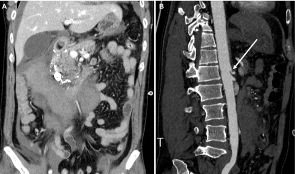
Fig. 1. Initial computed tomography (CT) performed in the emergency department. (A) Axial CT demonstrating contrast extravasation of the inferior pancreaticoduodenal artery (PDA) (arrow), which appears to be due to an aneurysmal sac, and large acute hematoma in the right anterior pararenal space and perihepatic space are shown. (B) CT revealing a mild hypodense lesion at the pancreas head, suggesting acute pancreatitis (arrow). (C) Maximum intensity projection image revealing aneurysmal dilatation of the inferior PDA (arrow).
A
B
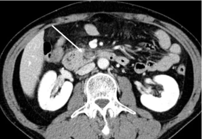
Fig. 2. Arteriography after initial resuscitation. (A) Celiac artery stenosis (CAS) (arrowhead) and hepatic arterial blood flow from the pancreaticoduodenal artery (PDA) are shown (arrow). (B) Large pseudoaneurysm and extravasation in the superior anterior PDA were detected (arrow).
tory was admitted to Wonkwang University Hospital with sud- den-onset epigastric abdominal pain, which began after per- forming push-ups, but no trauma. Upon admission, a physical examination revealed a mildly distended abdomen with epi- gastric tenderness. The patient’s blood pressure was 50/40 mmHg, with a heart rate of 107 beats/min. Rapid fluid re- suscitation restored the patient’s circulation status. After the blood pressure was stabilized, contrast-enhanced CT was performed. Contrast extravasation of the inferior pan- creaticoduodenal artery (PDA) was observed, which appeared to be due to the aneurysmal sac (Fig. 1A). A large-volume hemoperitoneum in the right anterior pararenal space and perihepatic space was evident (Fig. 1A, B). A mild hypodense lesion at the head of the pancreas, suggesting acute pan- creatitis, was also observed (Fig. 1C). Laboratory investigations performed 2 hours after admission revealed a hemoglobin level of 9.1 g/dL. Emergency angiography was performed im- mediately to determine the source of bleeding and control it. A 6 Fr sheath was placed into the right common femoral artery, and a right heart catheter and microcatheter were then
A B
Fig. 3. Vascular-aorta computed tomography angiography after 2 days of transarterial embolization. (A) Coronal image revealing slightly decreased size of hematoma in the anterior pararenal space. (B) Celiac artery stenosis is also apparent (arrow).
A
B
Fig. 4. Magnetic resonance imaging and endoscopic retrograde cholangiopancreatography (ERCP) performed to determine the etiology of the elevated serum amylase and lipase and treat pancreatitis (A) increased signal intensity in the T1-weighted image at the head of the pancreas (arrow). (B) ERCP with plastic stenting in the pancreatic duct was performed.
used to perform the celiac, gastroduodenal, and pan- creaticoduodenal arteriographies. The angiographic findings re- vealed severe CAS and compensation of hepatic arterial blood flow from the PDA (Fig. 2A). In addition, extravasation of the contrast material in the superior anterior PDA was detected (Fig. 2B). After selecting superior anterior PDA, stent-graft in- sertion was attempted, but it was difficult to proceed due to the vascular tortuosity. Collateral circulation appeared to be sufficient on superior mesenteric arteriography. For that reason, angioembolization was performed using a mixture of N-butyl cyanoacrylate and lipiodol (1:4). No procedure-related complications were encountered. The patient was hospitalized in the intensive care unit and followed-up carefully because of the large volume of hemoperitoneum and decreased hemo- globin levels. Octreotide acetate administration and fasting with total parenteral nutrition were administered to prevent the exacerbation of acute pancreatitis. Two days later, abdomi- nal CT angiography was repeated, which revealed a slight decrease in the hemoperitoneum in the anterior pararenal space (Fig. 3A). CAS was also observed (Fig. 3B). A fever of 38.4℃ was recorded on day 18 of hospitalization.
Percutaneous drainage of the anterior pararenal hematoma was performed under the suspicion of infection in the hematoma. Nevertheless, the fever and leukocytosis persisted, and a pre-rectal abscess was found on CT performed under
Fig. 5. Computed tomography performed 6 months after discharge.
Marked decreased size of the previously noted mass-like lesion with mild haziness in the retromesenteric space, suggesting an improving state of organizing hematoma with adjacent fibrotic changes (arrow).
suspicion of other infection foci. On day 37 of hospitalization, laparoscopic irrigation and drainage were performed.
Subsequently, the hematoma improved gradually, but the se- rum levels of amylase and lipase increased gradually to 195 IU/L and 450 IU/L, respectively. ERCP was performed under the suspicion of acute necrotizing pancreatitis on MRCP (Fig.
4A). There was no significant leakage or dilatation in the main pancreatic duct. On the other hand, an examination of amylase and lipase in the drainage tube placed after surgery could not exclude continuous pancreatic juice leakage. Therefore, plastic stenting was performed because of the concerns about acute pancreatitis exacerbation and the potential worsening of the intraperitoneal infection (Fig. 4B). The patient’s labo- ratory findings stabilized, and he was discharged on day 62 of hospitalization without any related symptoms. A CT scan performed 6 months later revealed marked improvement in the organizing hematoma with adjacent fibrotic changes (Fig. 5).
DISCUSSION
Abdominal visceral aneurysms are rare. Approximately 60%, 20%, 5.5%, and 2% of abdominal visceral aneurysms occur in the splenic artery, hepatic artery, superior mesenteric artery, and PDA, respectively.1 These aneurysms are typically asymptomatic but present with gastrointestinal bleeding or hemorrhagic shock when ruptured.2 Among these, true PDAAs
are rare; 63% are associated with celiac trunk lesions, such as median arcuate ligament syndrome, but the underlying mechanism is unclear.3 The aneurysmal dilatation of the rep- resentative arteries is believed to be caused by the increase in collateral flow due to stenosis or occlusion of the major aortic branches.4 Chronic increased blood flow in the small peripancreatic arteries results in local arterial hypertension that weakens and dilates the arterial wall, leading to a true aneurysm. A recent study using an electric circuit model con- firmed that in patients with concurrent CAS and PDAAs, either of these could come first and predispose the other.5
Although true PDAAs are rare, approximately 50% present with rupture, resulting in a 26% mortality rate.3 PDAAs are different from other abdominal visceral aneurysms because of the low correlation between the diameter of the aneurysm and the possibility of rupture.6 Typically, aneurysms rupture into the retroperitoneal space and cause acute abdominal pain. Hematoma that accumulates in the retroperitoneal space and wraps around the head of the pancreas, or organiz- ing infection, can cause compression symptoms and provoke pancreatitis. In the present case, the findings in the follow-up CT scan suggest acute pancreatitis through this mechanism.
No treatment guidelines have been established for the man- agement of PDAA. Most investigators agree that the size of the aneurysmal sac is not a risk factor for rupture. Nevertheless, it should be treated immediately after discovery. The treatment modalities are largely divided into surgical and endovascular.
Surgical treatments include resection, ligation, and bypass.
Although surgery is considered to be the initial definitive treat- ment for PDAA, it is associated with higher procedure-related morbidity and mortality than endovascular treatment.7,8 With the rapid advances and development of embolization materials and super-selective techniques, surgical treatment is being performed in limited circumstances, such as hemorrhagic shock or the failure of endovascular treatment.
Median arcuate ligament (MAL) syndrome (MALS) is a dis- order caused by compression of the celiac artery root, result- ing in decreased blood flow. Although CAS has a significant causal relationship with the formation of PDAA, it remains controversial whether MAL release should be performed.
Some authors argue that transarterial embolization (TAE) with- out revascularization can lead to a recurrence of PDAA or ischemic dysfunction of a related organ, such as the liver, spleen, or duodenum, as a result of the absence of the major
collateral vessels.9,10 On the other hand, several authors have reported that revascularization after TAE may not be neces- sary because there was no recurrence of the aneurysm after TAE during the follow-up period.11 This is supported by the need for MAL release in the treatment of CAS, the difficulty of endovascular treatment at the celiac artery orifice, and the higher probability of conversion to open surgery than lapa- roscopy when surgery is performed.11 According to a study based on three-dimensional CT, MALS can be divided into three categories according to the stenosis rate and length:
type A, <50% and ≤ 3 mm; type B, 50-80% and 3-8mm; type C, 80-100% and ≥8 mm.12 In the present case, the stenosis was considered to be type B. Furthermore, persistent intra- peritoneal hematoma and infection occurred after the procedure. When considering surgical treatment, extensive surgery such as celiac revascularization should be considered if the MAL release is insufficient. Therefore, considering the general condition of the patient, it was decided to follow up closely without performing surgery, and there was no sig- nificant recurrence according to CT performed later.
In conclusion, PDAAs caused by CAS should be treated re- gardless of the size and symptoms because of the possibility of rupture. Endovascular treatment is now considered to be a more appropriate first-line measure because surgical treat- ment results in higher mortality and morbidity. There is no consensus regarding the necessity of active treatment of ac- companying MALS; however, deciding whether to proceed will depend on the degree of CAS. Careful evaluation and multi- disciplinary discussion are required for optimal management of these aneurysms.
REFERENCES
1. Stanley JC, Wakefield TW, Graham LM, Whitehouse WM Jr, Zelenock GB, Lindenauer SM. Clinical importance and manage- ment of splanchnic artery aneurysms. J Vasc Surg 1986;3:
836-840.
2. Corey MR, Ergul EA, Cambria RP, et al. The natural history of splanchnic artery aneurysms and outcomes after operative intervention. J Vasc Surg 2016;63:949-957.
3. de Perrot M, Berney T, Deléaval J, Bühler L, Mentha G, Morel P.
Management of true aneurysms of the pancreaticoduodenal arteries. Ann Surg 1999;229:416-420.
4. Kalva SP, Athanasoulis CA, Greenfield AJ, et al. Inferior pan- creaticoduodenal artery aneurysms in association with celiac axis stenosis or occlusion. Eur J Vasc Endovasc Surg 2007;
33:670-675.
5. Yoon HJ, Choi JS, Shin WY, Lee KY, Ahn SI. Causal relationship be- tween celiac stenosis and pancreaticoduodenal artery aneur- ysm: interpretation by simulation using an electric circuit.
Biomed Res Int 2020;2020:2738726.
6. Sakatani A, Doi Y, Kitayama T, et al. Pancreaticoduodenal artery aneurysm associated with coeliac artery occlusion from an aortic intramural hematoma. World J Gastroenterol 2016;22:
4259-4263.
7. Coll DP, Ierardi R, Kerstein MD, Yost S, Wilson A, Matsumoto T.
Aneurysms of the pancreaticoduodenal arteries: a change in management. Ann Vasc Surg 1998;12:286-291.
8. Ricci G, Riu P, Attinà GM, et al. Endovascular treatment of rup- tured pancreaticoduodenal artery aneurysm: the importance of collateral vessels. A case report. Int J Surg Case Rep 2017;
41:205-208.
9. Murata S, Tajima H, Fukunaga T, et al. Management of pan- creaticoduodenal artery aneurysms: results of superselective transcatheter embolization. AJR Am J Roentgenol 2006;187:
W290-W298.
10. Takeuchi Y, Morikage N, Samura M, et al. Treatment options for celiac stenosis and pancreaticoduodenal artery aneurysms. Ann Vasc Surg 2017;41:281.e21-281.e23.
11. Sgroi MD, Kabutey NK, Krishnam M, Fujitani RM.
Pancreaticoduodenal artery aneurysms secondary to median arcuate ligament syndrome may not need celiac artery re- vascularization or ligament release. Ann Vasc Surg 2015;29:
122.e1-122.e1227.
12. Sugae T, Fujii T, Kodera Y, et al. Classification of the celiac axis stenosis owing to median arcuate ligament compression, based on severity of the stenosis with subsequent proposals for man- agement during pancreatoduodenectomy. Surgery 2012;151:
543-549.