MOK, a Pharmacopuncture Medicine, Reduces Inflammatory Response through Inhibiting the Proinflammatory Cytokine Production in LPS-stimulated Mouse Peritoneal Macrophages ※
Ji Hye Hwang
1, Min Sub Hwang
2, Yong-ki Park
1,3,*1
Korean Medicine R&D Center, Dongguk University
2
Dept. of Acupuncture & Moxibustion Medicine, College of Korean Medicine, Dongguk University
3
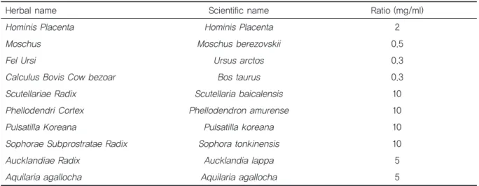
Department of Herbology, College of Korean Medicine, Dongguk University
[Abstract]
Objectives : In this study, we investigated the anti-inflammatory and anti-oxidative effects of MOK, a pharmacopuncture medicine, in lipopolysaccharide (LPS)-stimulated mouse peritoneal macrophages.
Methods : Peritoneal macrophages were isolated from ICR mice. Primary macrophages were treated with MOK extract (1.25, 2.5, 5, 10, and 20 mg/ml) for 30 min and then stimulated with LPS (1 μg/ml) for the indicated times. Cytotoxicity was measured using MTT and LDH assays.
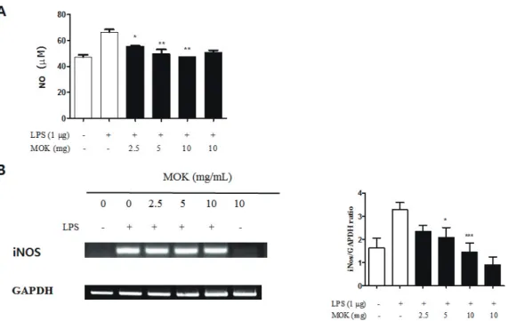
Nitric oxide (NO) production in culture supernatants was measured using the Griess assay.
The mRNA expression of iNOS, COX-2, proinflammatory cytokines (TNF-α, IL-1β, and IL-6) and antioxidant enzymes (HO-1 and MnSOD) was measured by RT-PCR.
Results : Treatment with MOK extract (2.5, 5, and 10 mg/ml) significantly decreased LPS-induced NO production in peritoneal macrophages through inhibition of iNOS expression. The expres- sion of COX-2, TNF-α, IL-1β, and IL-6 mRNA was also decreased in LPS-stimulated macrophages upon treatment with MOK extract. MOK treatment also increased the expression of HO-1 and MnSOD mRNA in macrophages.
Conclusion : These results indicate that MOK exerts anti-inflammatory and antioxidant effects by regulating the transcription levels of inflammatory mediators and antioxidant proteins in activated macrophages.
※ This research was supported by the Korean Health Technology R&D Project through the Korean Health Industry Development Institute (KHIDI) funded by the Ministry of Health& Welfare, Republic of Korea (grant number : HI16C0622).
✱ Corresponding author : Department of Herbology, College of Korean Medicine, Dongguk University, Dongdaero 123, Gyeongju 38066, Republic of Korea
Tel : +82-54-770-2661 E-mail : [email protected] Key words :
Antiinflammation;
Antioxidation;
LPS;
MOK;
Peritoneal macrophages;
Pharmacopuncture
Received : 2017. 01. 11.
Revised : 2017. 02. 02.
Accepted : 2017. 02. 06.
On-line : 2017. 02. 20.
This is an Open-Access article distributed under the terms of the Creative Commons Attribution Non-Commercial License (http://creativecommons.org/licenses/by- nc/3.0) which permits unrestricted non-commercial use, distribution, and reproduction in any medium, provided the original work is properly cited.