Introduction
Majority of the patients with chronic stroke expe- rience neurological deficits in their limbs, and the functional recovery of the impaired limbs is often poor. The neurological deficits that occur in stroke patients result in many activity limitations, including paralysis, muscle weakness, limited range of motion, contracture, and misalignment of the neurological system (Shumway-Cook and Woollacott, 2007).
Sequentially, functional limb problems appear more significant in their gait pattern. Therefore, gait re- storation is one of the main goals of stroke re- habilitation (Duncan et al, 2007).
In general, a variety of techniques, including ther- apeutic massage, myofascial release, range of motion (ROM) exercises as well as stretching and strength- ening exercises, have been used for functional im- provement of the impaired limb in stroke patients.
Although previous studies have failed to establish Corresponding author: Seung-chul Chon [email protected]
This work was supported by the Korea Foundation for the Advancement of Science & Creativity (KOFAC), and funded by the Korean Government (MOE).
The Effects of Sciatic Nerve Mobilization on Hamstring Flexibility, Lower Limb Strength and Gait Performance in
Patients With Chronic Stroke
Yun-hyeok Shin1, MSc, PT, Seung-chul Chon2, PhD, PT
1Dept. of Rehabilitation Medicine, Chungnam National University Hospital
2Dept. of Physical Therapy, College of Medical Science, Konyang University
Abstract
1)The purpose of this study was to evaluate the effects of mobilization of the sciatic nerve on hamstring flexibility, lower limb strength, and gait performance in patients with chronic stroke. This study was a randomized clinical trial with a crossover design. Sixteen subjects were recruited for this study. The subjects were randomly divided into two intervention groups and underwent either of the following two interventions: sciatic nerve mobilization or static stretching of the hamstring. We assessed hamstring flexibility, lower limb strength, and gait performance using a digital inclinometer, a hand-held dynamometer, and the 10-meter walk test, respectively. Subjects had a 24-hour rest period between each session in order to minimize carryover effects. Measurements for each test were assessed prior to and immediately after the intervention sessions. Using a two-way analysis of variance test with repeated measures, data from the two trials were analyzed by comparing the differences between both techniques.
The level of statistical significance was set at .05. Sciatic nerve mobilization resulted in significantly better knee extensor strength (p=.023, from 15.32±5.98 to 18.16±6.95 ㎏) and knee flexor strength (p=.011, from 7.80±4.80 to 8.15±4.24 ㎏) in the experimental group than in the control group. However, no significant effects of static stretching of the hamstring were observed on hamstring flexibility from the ankle plantar flexion (p=.966) and ankle neutral positions (p=.210) and on gait performance (p=.396). This study indicated that the sciatic nerve mobilization technique may be more effective in muscle activation of the knee extensor muscle and knee flexor muscle than hamstring static stretching technique in patients with chronic stroke.
Key Words: Gait performance; Hamstring flexibility; Lower limb strength; Nerve mobilization;
Static stretching; Stroke.
clinical evidence for the superiority of any particular technique, technique selection depends on the clini- cian’s decision-making process and the therapeutic aim (Jette et al, 2005). In particular, nerve mobi- lization technique, also called neurodynamics, is a treatment modality that is used in relation to pathol- ogies of the nervous system. The lack of evidence makes adoption of effective therapeutic approaches for patients with functional impairments of the mus- culoskeletal system an issue for most clinicians.
Nerve mobilization involves the stretching and re- laxing of the nerves in order to maintain normal muscle tone and ensure a desirable ROM (Butler and Jones, 1991). The main theoretical objective of this technique is the restoration of the balance between the movement of neural tissues and adjacent me- chanical borders, allowing reduced intrinsic pressures on the neural tissue, thus stimulating optimum phys- iologic function (Gifford et al, 1998). Recently, there has been a shift away from a mechanical rationale in order to comprise physiological concepts such as structure and function of the nervous system. Nerve mobilization is now a more recognized terminology that refers to the cooperative physiological, bio- mechanical and morphological functions of the nerv- ous system (Shacklock, 2008).
The hamstring muscle is an essential element in adjusting knee joint extension, which is related to gait performance. Shortening of the hamstring muscle may reduce the ROM of the knee joint and affect the physiological movement of the lumbopelvic system, causing gait problems with asymmetric weight bear- ing and pelvic inclination (Cleland et al, 2006). For example, a reduction in the flexibility of the ham- string muscles while using the hip extensors predom- inantly leads to the overuse of the hamstring as well as insufficient activation of the gluteus maximus and abdominal muscles. This results in a variety of com- pensatory mechanisms that lead to instability of the trunk muscles in stroke patients (Sahrmann, 2002).
Despite previous studies on the effects of upper limb nerve mobilization on stroke patients’ upper
limb muscle flexibility, strength, and other functions (Shacklock, 2008), there are few studies on the ef- fects of nerve mobilization of the lower limb muscles on the flexibility, strength, and walking ability of patients with stroke hemiparesis (Butler and Jones, 1991). In addition, although one study investigated mainly the influence of nerve mobilization on periph- eral nervous system disorders, studies on the effec- tive treatments for improving lower limb function among central nervous system disorders, are lacking.
This study investigated the effects of sciatic nerve mobilization on the lower limbs of stroke patients by measuring flexibility, strength, and gait function.
Methods
Subjects
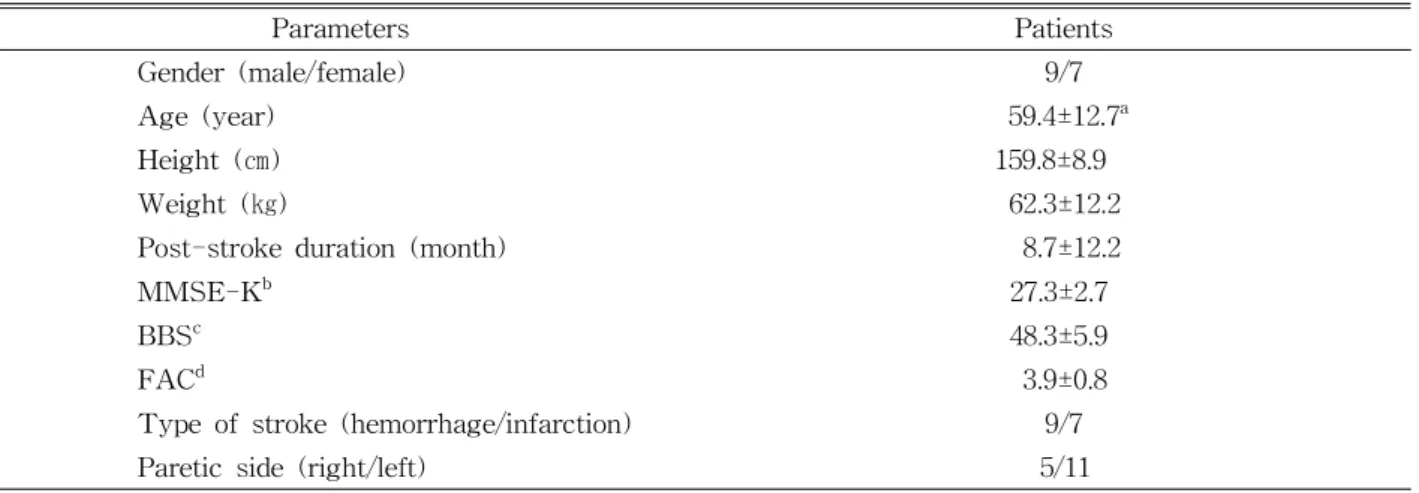
The sixteen patients enrolled in this study were selected from among the stroke inpatients and out- patients at the university-affiliated hospital. Before participation, the protocol and procedures of the study were explained to all subjects prior to getting their informed consent. The inclusion criteria were as fol- lows: (1) diagnosis of cerebral infarction or cerebral hemorrhage, (2) classified as ≥grade 3 (fair) as per the manual muscle test of the affected lower limb muscle, (3) no pain in the lower limbs for at least 6 months (Tully et al, 2005), (4) adequate cognitive function as demonstrated by a score of at least 24 points on the Korean version of the Mini-Mental State Examination, (5) performing gait over 10 meters independently, with or without an assistive device, (6) Berg Balance Scale score less than 41, (7) and a functional ambulation category score over 3. The ex- clusion criteria were as follows: (1) contracture of the lower limb joint, (2) having undergone a surgery of the lower limb, (3) orthopedic injury to the lower limbs, (4) history or present diagnosis of additional musculoskeletal diseases, and (5) hemineglect, visual problems, or other pains. Table 1 presents the demo- graphic and clinical characteristics of the patients.
Procedures
The sixteen subjects were allocated to both the groups (two interventions) alternately, or on alter- nate days for 30 minutes a day, 3 days a week, all 32 sessions for 11 weeks. The procedures included two techniques: (1) sciatic nerve mobilization (experimental group), (2) hamstring static stretching (control group). These techniques were crossed over randomly with just one technique per day for each day to avoid the carryover effect of each technique.
Randomization for the application of the two techni- ques was done by blindly drawing one card from an envelope containing two cards marked 1 and 2.
Intervention instructions were provided as detailed verbal commands, while intervention procedures were checked using a stopwatch. All procedures were performed at a regular frequency and according to a metronome for the affected lower limb. The application time for all compo- nents was about 30 minutes. During both techniques, the patients were instructed to breathe comfortably and not apply any force on the lower limbs.
Sciatic nerve mobilization
The sciatic nerve mobilization technique consisted of four components for relaxation of the sciatic nerve.
First, while in a supine position and with the neck and trunk in a neutral position, the lower limb on the
unaffected side was fixed in order to make them immobile. The lower limb on the affected side was placed in full straight leg raise (SLR) for 20 seconds.
Second, ankle joint dorsiflexion accompanied the per- formance of the SLR with a slight vibration. Third, hip joint adduction and internal rotation were applied for 40 seconds. Fourth, in order to promote tension of the sciatic nerve and enable it to reach the maximum level, cervical flexion was sequentially applied (Figure 1). This process was sequentially repeated for 10 mi- nutes (3 sets) (Butler and Jones, 1991).
Hamstring static stretching
The hamstring static stretching technique con- sisted of four components for relaxation of the ham- string muscle. First, while in a supine position and
Parameters Patients
Gender (male/female) 9/7
Age (year) 59.4±12.7a
Height (㎝) 159.8±8.9
Weight (㎏) 62.3±12.2
Post-stroke duration (month) 8.7±12.2
MMSE-Kb 27.3±2.7
BBSc 48.3±5.9
FACd 3.9±0.8
Type of stroke (hemorrhage/infarction) 9/7
Paretic side (right/left) 5/11
amean±standard deviation,bmini-mental state examination (Korean version), cBerg balance scale, dfunctional ambulation category.
Table 1. Demographic and clinical characteristics of the patients (N=16)
Figure 1. Sciatic nerve mobilization.
with the neck and trunk in a neutral position, the lower limb on the unaffected side was then fixed in order to prevent bending. Second, hip flexion posi- tions were held for 20 seconds without rotating the hip joint. Third, the knee joint was gradually ex- tended up to the subject’s pain threshold while keep- ing the hip joint of the affected side at 90 degrees for 40 seconds. Fourth, the unaffected side of lower limb was fixed with a Velcro strap in order to hold the pelvic neutral position (Bandy et al, 1998). This process were repeated for 10 minutes (3 sets) (Butler and Jones, 1991).
Measurements
Hamstring flexibility, lower limb strength, and gait performance were assessed using a digital in- clinometer, hand-held dynamometer, and the 10-meter walk test, respectively. All measures were performed in the affected lower limb. The measurements were conducted immediately before and after the intervention. The two examiners who performed the measurements were blinded to study inter- ventions in order to rule out potential bias (Decoster et al, 2005).
Hamstring flexibility
An electronic inclinometer (Dualer IQ the smarter inclinometer, JTECH medical, Salt Lake, USA) was used to measure the available ROM. This measure- ment device has high reliability. Intraclass correlation coefficient of the electronic inclinometer was ≥.95 (Kolber and Hanney, 2012). The subject was placed in a neutral supine position with the affected hip and knee joint flexed to 90 degrees by using an ex- perimental frame constructed to maintain the hip flexion position at 90 degrees, at the lateral knee joint. The pelvis and the contralateral knee joint of the lower limb were fixed with Velcro straps during measurement. The subjects then passively extended their knees to the maximum while holding the hip joint at 90 degrees at the ankle plantar flexion or ankle neutral positions, respectively. The subjects
were instructed to give a verbal notification if pain was evoked during the hamstring flexibility meas- urement process. The hamstring flexibility with ankle plantar flexion or ankle neutral positions was per- formed individually for 3 times and the average val- ue was used for the analysis.
Lower limb strength
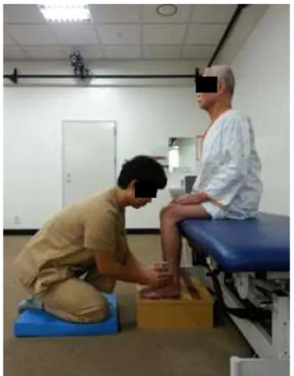
A hand-held dynamometer (Microfet2, Hoggan Health Industries, West Jordan, USA) was used to measure the strength of knee extensor and knee flexor muscles, respectively. The reliability of the hand-held dynamometer was .99 (Kim and Lee, 1996). During both the measurements, the subject was in a neutral sitting position with the affected hip and knee joint flexed to 90 degrees. The strength measure point of the knee extensor and flexor muscles was identified on the anterior and posterior sides of the bimalleolar line, respectively, while in the sitting position (Douma et al, 2014) (Figure 2). The subjects were instructed to maintain the lower limb posture using the maximum eccen- tric contraction for 5 seconds, in order to offer re- sistance to the examiner for 5 seconds to be offer- ing resistance of the examiner’s hand-held dyna- mometer (Burns and Spanier, 2005). If a patient could not maintain the starting sitting position, the data were excluded from analysis. The test was re-
Figure 2. Knee extensor strength measure.
peated 5 times and the average value was calcu- lated after discarding the minimum and maximum values (Hung et al, 2010).
Gait performance
Gait performance was assessed using the 10-meter walk test. We chose this test because it can be easily administered and has been performed in clinical circumstance (Dean et al, 2001). The pa- tients were instructed to walk a total of 14 meters in order to minimize the acceleration and deceler- ation values. The test was repeated 3 times, and the average value was used for analysis. One-minute break times between measurements alleviated fatigue.
The reliability for this test were high (r=.89~1.00) (Dalgas et al, 2012).
Statistical analysis
Descriptive statistics included mean and standard deviations. The normal distribution of the sample was tested using the Kolmogorov-Smirnov test, which showed a normal distribution of the all variables. Two-way analysis of variance with re- peated measures was used to assess the main effects (group and time effects) and their interaction effects on hamstring flexibility, lower limb strength, and gait performance between the sciatic nerve mobilization and hamstring static stretching. The collected data were analyzed using SPSS ver. 18.0 (SPSS Inc., Chicago, IL, USA). The level of statistical sig- nificance was set at a p value of <.05.
Results
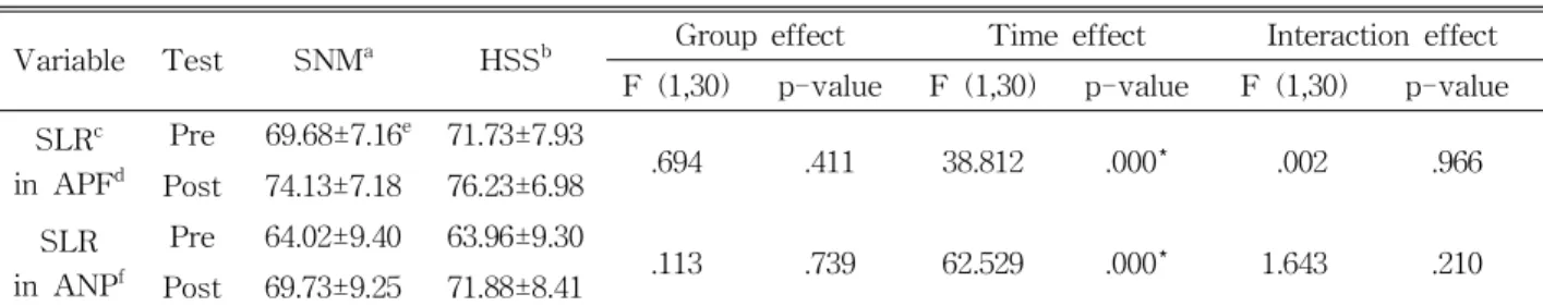
There was no significant group main effect for hamstring flexibility in the ankle plantar flexion (F=.694, p=.411) or ankle neutral positions (F=.113, p=.739), respectively. However, there were sig- nificant time main effects for hamstring flexibility in the ankle plantar flexion (F=38.812, p<.001) and ankle neutral positions (F=62.529, p<.001), respectively. There was no significant group×time interaction effect for hamstring flexibility in both, the ankle plantar flexion (F=.002, p=.966) and ankle neutral positions (F=1.643, p=.210), respectively (Table 2).
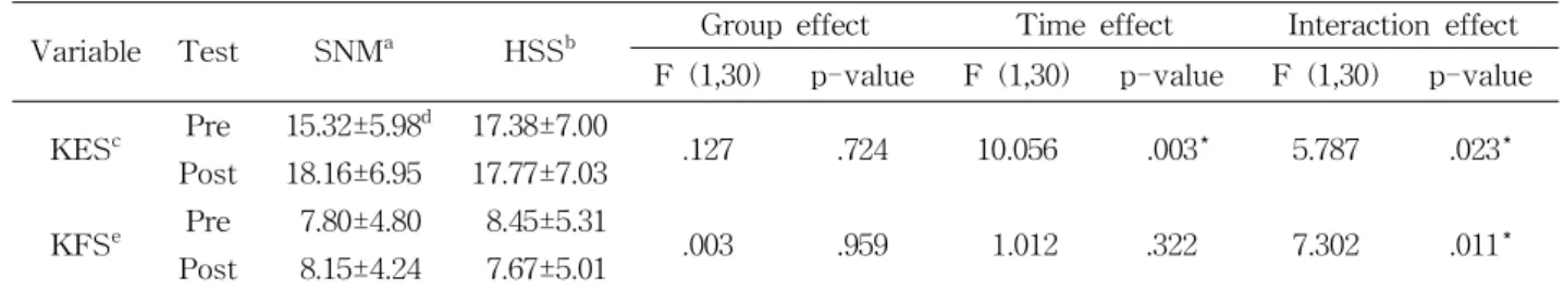
There was no significant group main effect for knee extensor (F=.127, p=.724) or knee flexor strengths (F=.003, p=.959), respectively. There was a significant time main effect for the knee extensor strength (F=10.056, p=.003) but not for the knee flexor strength (F=1.012, p=.322). There were sig- nificant group×time interaction effects for both knee extensor (F=5.787, p=.023) and knee flexor strengths (F=7.302, p=.011), respectively (Table 3).
Although there was no significant group main ef- fect for the 10-meter walk test, there was a sig- nificant time main effect for the 10-meter walk test (F=.001, p=.974). There was a significant time main effect for 10-meter walk test (F=6.407, p=.017). No significant group×time interaction effect was ob- served for 10-meter walk test (F=.743, p=.396) (Table 4).
Variable Test SNMa HSSb Group effect Time effect Interaction effect
F (1,30) p-value F (1,30) p-value F (1,30) p-value SLRc
in APFd
Pre 69.68±7.16e 71.73±7.93
.694 .411 38.812 .000* .002 .966
Post 74.13±7.18 76.23±6.98 SLR
in ANPf
Pre 64.02±9.40 63.96±9.30
.113 .739 62.529 .000* 1.643 .210
Post 69.73±9.25 71.88±8.41
asciatic nerve mobilization,bhamstring static stretching, cstraight leg raise, dankle plantar flexion,emean±standard deviation, fankle neutral position, *p<.05.
Table 2. Comparisons of hamstring flexibility (degree) in ankle plantar flexion and ankle neutral position be- tween sciatic nerve mobilization technique and hamstring static stretching technique (N=16)
Discussion
This randomized crossover study compared the ef- fects of sciatic nerve mobilization and hamstring static stretching techniques on hamstring flexibility, lower limb strength and gait performance in hemiplegic pa- tients with chronic stroke. Most of all, the results of this research revealed a main finding. The sciatic nerve mobilization technique was greater than the hamstring static stretching technique for the improve- ment in the knee extensor and knee flexor strengthening. Thus, sciatic nerve mobilization techni- que reported a positive effect on the muscle activation of the knee joint muscles as well as hamstring flexi- bility in stroke patients.
The effectiveness of the sciatic nerve mobilization technique is also elucidated by quantitative measurements. Before the intervention, the knee measurements were approximately 69 and 64 degrees, respectively, and increased after the intervention to 74 degrees (7% increase) and 69 degrees (8% increase), respectively. This improvement in hamstring muscle flexibility has been reported in previous studies
(Castellote-Caballero et al, 2013). However, the differ- ences were not statistically significant in these cases.
It has been reported that sciatic nerve mobilization improves the long-term adaptability and flexibility of the sciatic nerve (Cleland et al, 2006). However, Fasen et al (2009) reported that the SLR passive measure- ments used in this study are more suitable than the active measure method for assessing paralytic stroke hemiplegia, and embrace the concept of measurement of sciatic nerve mobilization technique. Additionally, the increase in flexibility may be inferred to influence improvement by not only muscle elasticity, but also connective tissue extensibility (Fasen et al, 2009).
Certainly, these results have important clinical implications. After the sciatic nerve mobilization inter- vention, the knee extensor strength and knee flexor strength measurements were approximately increased to 3 ㎏ (from 15 to 18 ㎏) and 1 ㎏ (from 7 to 8 ㎏), respectively. Muscle strength increases during the ap- plication of nerve mobilization; this has been demon- strated in previous studies (Boyd et al, 2009; Hall et al, 1998). Nerve mobilization reduces intra-neural pressure, leading to increased blood flow to the
Variable Test SNMa HSSb Group effect Time effect Interaction effect
F (1,30) p-value F (1,30) p-value F (1,30) p-value KESc Pre 15.32±5.98d 17.38±7.00
.127 .724 10.056 .003* 5.787 .023*
Post 18.16±6.95 17.77±7.03 KFSe Pre 7.80±4.80 8.45±5.31
.003 .959 1.012 .322 7.302 .011*
Post 8.15±4.24 7.67±5.01
asciatic nerve mobilization, bhamstring static stretching, cknee extensor strength,dmean±standard deviation, eknee flexor strength, *p<.05.
Table 3. Comparisons of lower extremity strength (㎏) between sciatic nerve mobilization technique and ham-
string static stretching technique (N=16)
Variable Test SNMa HSSb Group effect Time effect Interaction effect
F (1,30) p-value F (1,30) p-value F (1,30) p-value 10MWTc Pre 26.79±26.21d 25.98±24.97
.001 .974 6.407 .017* .743 .396
Post 22.58±18.79 23.91±20.96
asciatic nerve mobilization,bhamstring static stretching, c10-meter walk test, dmean±standard deviation, *p<.05.
Table 4. Comparisons of gait performance (seconds) between sciatic nerve mobilization technique and ham-
string static stretching technique (N=16)
nerves. Hence, this mechanism improves axoplasmic flow and nerve conduction (Nee and Butler, 2006). In this study, sciatic nerve mobilization techniques sig- nificantly improved knee flexion and extension strengths compared to the conventional technique:
hamstring static stretching. It supports the belief that sciatic nerve mobilization techniques activate neuro- transmission fibers related to motor function and sen- sory disorders, and improvement and motor abilities of the lower limbs (Cleland et al, 2006). Additionally, this technique is neurophysiologically attributable to accel- erated quadriceps muscle-mediated hamstring relaxa- tion, which is a function of the antagonistic muscle (Fox, 2006).
Before the intervention, timed 10-meter walk test was approximately 26 seconds, and decreased to ap- proximately 22 seconds (15% decrease) after the intervention. Improvements in walking in the ex- perimental group may be attributed to the strengthen- ing of the lower limb muscles after the sciatic nerve mobilization technique on the time main effect, al- though there was no significant difference in gait performance. Recent research examining the relation- ship between functional ambulation and lower limb strength in stroke patients reported a similarly strong correlation (Mentiplay et al, 2015). Previous study in- dicates muscle activity increases during the application of nerve mobilization technique (Boyd et al, 2009) and muscle activation increases as a consequence of a spinal reflex response to the nociceptive input to avoid nerve injury (Hall et al, 1998). Additionally, the fact that sciatic nerve mobilization can contribute to the recovery of normal hamstring muscle length is thought to have caused the improvements in knee ex- tensor strength. Thus, the sciatic nerve mobilization technique may account for the recovery of normal hamstring muscle length, improvements in lower limb strengthening, and functional gait ability in stroke patients.
Despite the positive effects of sciatic nerve mobi- lization technique, this research study has several limi- tations that can be avoided in further studies. First, the
small sample size may be a major limiting factor in establishing the generality of the results to all patients with chronic stroke. Secondly, this study suggests that there are immediate effects of a single dose of sciatic nerve mobilization technique and the potential for mus- cle strengthening effect with treatment. We recognize that the findings of this study cannot describe the long-term effects of the sciatic nerve mobilization tech- nique owing to the lack of follow up data. Additionally, it may be difficult to understand the applicability of our findings beyond our sciatic nerve mobilization technique. Finally, because the sciatic nerve mobi- lization technique was applied after a one-day interval, it may be difficult to certify the independence of their effects completely. Robust studies with a larger sample size and a longer followup period are necessary in or- der to confirm our findings.
Conclusion
This study suggests that the sciatic nerve mobi- lization technique may be more beneficial than the hamstring static stretching technique in the manage- ment of hamstring flexibility, lower limb strength and associated gait performance in patients with chronic stroke. Most of all, the improvement in the lower limb strength was greater with the sciatic nerve mobi- lization technique compared to the hamstring static stretching technique. Certainly, our quantitative devices are useful in providing numerical data for confirmation of the improvement in flexibility and strength after the intervention. This study provides useful information for clinicians and researchers who want to explore addi- tional therapeutic options for improving motor function of the lower limb during chronic stroke rehabilitation.
References
Bandy WD, Irion JM, Briggler M. The effect of stat- ic stretch and dynamic range of motion training
on the Flexibility of the hamstring muscles. J Orthop Sports Phys Ther. 1998;27(4):295-300.
Boyd BS, Wanek L, Gray AT, et al.
Mechanosensitivity of the lower extremity nerv- ous system during straight-leg raise neuro- dynamic testing in healthy individuals. J Orthop Sports Phys Ther. 2009;39(11):780-790. http://
dx.doi.org/10.2519/jospt.2009.3002
Burns SP, Spanier DE. Break-technique handheld dynamometry: Relation between angular velocity and strength measurements. Arch Phys Med Rehabil. 2005;86(7):1420-1426.
Butler DS, Jones MA. Mobilisation of the Nervous System. 1st ed. Melbourne, Churchill Livingstone, 1991:68-69.
Castellote-Caballero Y, Valenza MC, Martin-Martin L, et al. Effects of a neurodynamic sliding tech- nique on hamstring flexibility in healthy male soccer players. A pilot study. Phys Ther Sport.
2013;14(3):156-162. http://dx.doi.org/10.1016/j.ptsp.
2012.07.004
Cleland JA, Childs JD, Palmer JA, et al. Slump stretching in the management of non-radicular low back pain: Pilot clinical trial. Man Ther.
2006;11(4):279-286.
Dalgas U, Severinsen K, Overgaard K. Relations be- tween 6 minute walking distance and 10 meter walking speed in patients with multiple sclerosis and stroke. Arch Phys Med Rehabil. 2012;93(7):
1167-1172. http://dx.doi.org/10.1016/j.apmr.2012.02.
026
Dean CM, Richards CL, Malouin F. Walking speed over 10 metres overestimates locomotor capacity after stroke. Clin Rehabili. 2001;15(4):415-421.
Decoster LC, Cleland J, Altieri C, et al. The effects of hamstring stretching on range of motion: A systematic literature review. J Orthop Sports Phys Ther. 2005;35(6):377-387.
Douma RK, Soer R, Krijnen WP, et al. Reference values for isometric muscle force among work- ers for the netherlands: A comparison of refer- ence values. BMC Sports Sci Med Rehabil. 2014;
6(1):10. http://dx.doi.org/10.1186/2052-1847-6-10 Duncan PW, Sullivan KJ, Behrman AL, et al.
Protocol for the locomotor experience applied post-stroke (LEAPS) trial: A randomized con- trolled trial. BMC Neurol. 2007;7:39.
Fasen JM, O’Connor AM, Schwartz SL, et al. A randomized controlled trial of hamstring stretch- ing: Comparison of four techniques. J Strength Cond Res. 2009;23(2):660-667. http://dx.doi.org/
10.1519/JSC.0b013e318198fbd1
Fox M. Effect on hamstring flexibility of hamstring stretching compared to hamstring stretching and sacroiliac joint manipulation. Clinical Chiropractic.
2006;9(1):21-32.
Gifford L. Neurodynamics. In: Pitt-Brooke J, Reid H, Lockwood J, et al eds. Rehabilitation of Movement: Theoretical basis of clinical practice.
1st ed. London, WB saunders, 1998.
Hall T, Zusman M, Elvey R. Adverse mechanical tension in the nervous system? Analysis of straight leg raise. Man Ther. 1998;3(3):140-146.
Hung CJ, Jan MH, Lin YF, et al. Scapular kinematics and impairment features for classifying patients with subacromial impingement syndrome. Man Ther. 2010;15(6):547-551. http://dx.doi.org/10.1016/
j.math.2010.06.003
Jette DU, Latham NK, Smout RJ, et al. Physical therapy interventions for patients with stroke in inpatient rehabilitation facilities. Phys Ther.
2005;85(3):238-248.
Kim JW, Lee KM. Evaluation of isometric shoulder strength in korean adults using a hand-held dynamometer. Ann Rehabil Med. 1996;20(1):
186-193.
Kolber MJ, Hanney WJ. The reliability and con- current validity of shoulder mobility measure- ments using a digital inclinometer and goni- ometer: A technical report. Int J Sports Phys Ther. 2012;7(3):306-313.
Mentiplay BF, Adair B, Bower KJ, et al. Associations between lower limb strength and gait velocity following stroke: A systematic review. Brain Inj.
2015;29(4):409-422. http://dx.doi.org/10.3109/02699052.
2014.995231
Nee RJ, Butler D. Management of peripheral neuro- pathic pain: Integrating neurobiology, neuro- dynamics, and clinical evidence. Phys Ther Sport.
2006;7(1):36-49.
Shacklock M. Neural mobilization: A systematic re- view of randomized controlled trials with an analysis of therapeutic efficacy. J Man Manip Ther. 2008;16(1):23-24.
Sahrmann SA. Does postural assessment contribute to patient care? J Orthop Sports Phys Ther.
2002;32(8):376-379.
Shumway-Cook A, Woollacott MH. Motor Control:
Translating research into clinical practice. 3rd ed. Philadelphia, PA, Lippincott Williams &
Wilkins, 2007:121-122.
Tully EA, Fotoohabadi MR, Galea MP. Sagittal spine and lower limb movement during sit-to-stand in healthy young subjects. Gait Posture. 2005;
22(4):338-345.
This article was received August 4, 2015, was re- viewed August 4, 2015, and was accepted September 21, 2015.