Collagen-induced Arthritis Rat Model에서 염증성 통증에 대한 봉독약침의 진통효과 및 기전연구:
5HT-3 & Muscarinic
Cholinergic Mechanisms에 대한 연구
서병관ᆞ박동석ᆞ백용현 경희대학교 한의과대학 침구학교실
배경 및 목적 : 봉독약침요법(bee venom pharmacopuncture, BVP)은 rheumatoid arthritis(RA)의 치료에 활용되고 있으나, RA로 인한 염증성 통증에 대한 봉독약침의 진통효과와 specific mechanism은 아직까지 명확하게 밝혀지지 않았다. 이에 본 연구에서는 RA animal model로서 collagen-induced arthritis(CIA) rat model에서 봉독약침의 a1-adrenergic, 5HT-3 그리고 muscarinic cholinergic mechanism을 확인하고자 한다.
방법 : CIA를 유도하기 위하여 male Sprague–Dawley rat에 freund’s incomplete adjuvant에 乳化시킨 bovine type II collagen을 주입하고 14일 후 booster injection 시행하였다. 진통효과는 tail flick latency (TFL)로 평가하였다.
결과 : 관절염의 유도 이후 염증성 통증 역치는 시간이 지나면서 낮아지며, 5주 이후로는 통증 역치에 큰 변화가 없이 유지되었다. 첫 번째 immunization으로부터 5주 경과 후 족삼리(ST36)에 봉독약침처치(0.25 mg/
kg)를 시행하여 유의한 진통효과를 관찰하였다. 또한 봉독약침의 진통효과는 ondansetron(5HT-3 receptor antagonist,
1)
Antinociceptive Effect and the Mechanism of Bee Venom Pharmacopuncture on Inflammatory Pain in the Rat Model of Collagen-induced Arthritis:
Mediation by 5HT-3 & Muscarinic Cholinergic Receptors Seo Byung-kwan, Park Dong-suk and Baek Yong-hyeon
Dept. of Acupuncture & Moxibustion, College of Oriental Medicine, Kyung Hee University
*
This work was supported by the Korea Research Foundation Grantfunded by the Korean Government (MOEHRD,Basic Research Promotion Fund) (331-2008-1-E00442).
․Acceptance : 2011. 1. 7. ․Adjustment : 2011. 2. 2. ․Adoption : 2011. 2. 6.
․Corresponding author : Baek Yong-hyeon, Department of Acupuncture, Kyung Hee University Hospital at Gangdong, 149 Sangil-dong, Gangdong-gu, Seoul 134-727, Republic of Korea.
Tel. 82-2-440-6224 E-mail : [email protected]
국문초록
Original Article
0.5㎎/kg, i.p.), atropine(muscarinic cholinergic receptor antagonist, 1㎎/kg, i.p.)의 전처치에 의하여 억제되 었으나, prazosin(a1-adrenergic receptor antagonist, 1㎎/kg, i.p.)의 전처치에 의해서는 억제되지 않았다.
결론 : 봉독약침은 CIA로 인한 염증성 통증에 유의한 진통효과를 나타내며 그 analgesic mechanism은 5HT-3와 muscarinic cholinergic receptor에 의하여 매개되며 a1-adrenergic receptor에 의하여 매개되지는 않았다.
핵심 단어 : 봉독약침(BVP), collagen-induced arthritis(CIA), ondansetron, atropine, prazosin, tail Flick latency(TFL)
Ⅰ. Introduction
Rheumatoid arthritis (RA) is a kind of autoim- mune disease that is characterized by progressive joint destruction, deformity, disability with swelling and pain in multiple joints
1). As a remedy of RA, non-steroidal anti-inflammatory drugs (NSAIDs) are recommended in medical field, but long term management with NSAIDs may result in serious side effects, such as gastrointestinal ulcer and renal morbidity
2). Therefore, another treatment without side effects is needed in the management of inflam- matory pain induced by RA.
Bee Venom Pharmacopuncture (BVP) has been used to relieve pain and to treat inflammatory dis- eases such as rheumatoid arthritis (RA) in humans
3)and experimental animals
4,5). Related with the anti- nociceptive mechanisms of BVP, there were several studies showing that the analgesic effect of BVP was mediated by α2-adrenergic and 5HT-1 receptors in models of neuropathic pain, acetic acid-induced visceral pain, and formalin pain
6-9). And, in the rat model of collagen-induced arthritis (CIA) as an animal model of RA
10), BVP showed antinociceptive effect which is mediated by α2- adrenergic receptor
11). While Bee Venom Pharma- copuncture (BVP) shows the analgesic effect in RA, the antinociceptive mechanisms related with the inflammatory pain by using the bovine type II collagen-induced RA model have not been fully studied.
The primary purpose of this study was to determine whether Bee Venom Pharmacopuncture (BVP) is able to show the antinociceptive effect on inflammatory pain in the rat model of collagen- induced arthritis (CIA). A second goal of this study was to clarify whether the antinociceptive effect of BVP is related to the activation of α1-adrenoceptor, 5HT-3 and muscarinic cholinergic receptors. These receptor types are thought to play important roles in spinal analgesic mechanisms associated with the descending pain modulatory system.
Ⅱ. Materials and Methods
A. Subjects
Young adult male Sprague-Dawley rats (Sam:
acN (SD)BR, 180~200g, n=110) were housed in group cages (4~5per cage) with water and food available ad libitum. The room was light/dark (08 : 00~20 : 00h light, 20 : 00~08 : 00h dark) controlled and kept at 2124℃. All experiments were con- ducted in accordance with the guidelines of the In- ternational Association for the Study of Pain (IASP)
12).
B. The induction of collagen-induced arthritis (CIA)
CIA induction was performed as described by
Trentham et al.
10). 1.0ml of an emulsion containing
500mg of bovine type II collagen (Chondrex Inc.,
Washington, USA) in a 0.3% acetic acid solution (Cosmo Bio., Tokyo, Japan) and 500mg of Freund’s incomplete adjuvant (Chondrex Inc, Washington, USA) were intradermally injected into the base of the tail. Two weeks after the first injection, 0.5ml of the same emulsion was intracutaneously injected into the Lt. plantar surface of the rat. On the basis of the arthritis evaluation method by Trentham et al.
10), the rats with the sum total score over 10 were selected as experimental animals. The degree of arthritis severity was scored on a scale of 0~4 in each limb of the rat, where 0 = no inflammation, 1 = unequivocal inflammation of 1 joint, 2 = unequivocal inflammation of at least 2 joints of the limb or moderate inflammation of 1 joint, 3 = severe infla- mmation of ≥ 1 joint and 4 = maximum inflam- mation of ≥ 1 joint in the limb.
C. Behavior Assessments (Tail Flick Latency; TFL)
For assessing the antinociceptive effect, a tail flick unit (Ugo Basile Model 7360, Comrio, Italy) was used to evaluate the pain threshold by using the change of tail flick latency (TFL). The light of the tail flick unit was turned out as soon as the rat flicked its tail and the time lapse between the onset of irradiation and the flick of the tail was read. The intensity of the light bulb was set so the baseline reaction time was 12±0.3s. For proper application of tail flick test and BVP, the rat was restrained in a plastic holder (5.3×15cm in diameter×length) and the tail was laid on the light bulb. When TFL exceeded 20s during an experimental procedure, the light bulb was switched off to minimize tissue damage of the tail. The degree of analgesia was expressed as a percentile change in TFL and was determined as follows
13,14).
Acquired TFL change (%) =
post. acup. TFL - baseline TFL
× 100 baseline TFL
5 weeks after the first administration of bovine
type II collagen, the behavioral test was performed with the tail flick unit prior to (as baseline test) and 10, 20, 30, 45 and 60min after BVP treatment.
Experimental rats were properly fitted in plastic holders with their tails protruding outside, and were allowed to adapt to the environment for 60min/day for 7 days. This procedure was also for adaptation to the restraint that the animals were submitted to during the BVP treatment
15).
D. Bee venom treatment and agonists/
antagonists pretreatment
Whole Bee Venom (Sigma, St. Louis, MO) dis- solved in saline was administrated subcutaneously and bilaterally, at a dose of 0.25mg/kg
9), into an acupoint (Zusanli, ST
36). The Zusanli acupoint was chosen because it is traditionally used for the relief of inflammatory pain. This acupoint is located at the anterior tibial muscle and about 10mm below the knee joint. Control animals were injected bilaterally into the Zusanli acupoint with an equal volume of dimethylsulfoxide (DMSO). Bee venom treatment was conducted 5 weeks after the first administration of bovine type II collagen.
In order to reveal the related analgesic mech- anism, the selective α1-adrenoceptor agonist pheny- lephrine (ICN Biomedicals, Ohio, USA; 2mg/kg, i.p.) and antagonist prazosin (ICN Biomedicals, Ohio, USA;
1mg/kg, i.p.), 5HT-3 agonist m-chlorophenyl- biguanide (ICN Biomedicals, Ohio, USA; 1mg/kg, i.p.) and antagonist ondansetron (ICN Biomedicals, Ohio, USA; 0.5mg/kg, i.p.), muscarinic cholinergic agonist neostigmine (ICN Biomedicals, Ohio, USA;
100㎍g/kg, i.p.) and antagonist atropine (ICN Bio- medicals, Ohio, USA; 1mg/kg, i.p.) were injected intraperitoneally 20 minutes before BVP treatment.
Drugs were dissolved in DMSO.
E. Experimental groups
Experimental groups were divided into eleven groups:
a. CIA induction group (CIA, n=10);
b. non-treatment arthritic group (None-Tx, n=10);
c. DMSO-treated / Zusanli acupoint arthritic group (Z-DMSO, n=10);
d. BV-treated / Zusanli acupoint arthritic group (Z-BVP, n=10);
e. DMSO pretreatment / BV-treated / Zusanli acupoint arthritic group (BVP+DMSO, n=10);
f. phenylephrine pretreatment / BV-treated / Zusanli acupoint arthritic group (BVP+Phenyl, n=10);
g. prazosin pretreatment / BV-treated / Zusanli acupoint arthritic group (BVP+Prazo, n=10);
h. m-chlorophenyl-biguanide pretreatment / BV- treated / Zusanli acupoint arthritic group (BVP +m-chlo, n=10);
i. ondansetron pretreatment / BV-treated / Zusanli acupoint arthritic group (BVP+ondan, n=10);
j. neostigmine pretreatment / BV-treated / Zusanli acupoint arthritic group (BVP+Neosti, n=10);
k. atropine pretreatment / BV-treated / Zusanli acupoint arthritic group (BVP+Atrop, n=10);
F. Statistics
All data are represented as means ± standard error of mean. The significance of statistical dif- ferences were determined using non-parametric Friedman’s rank test followed by Dunnett’s post- hoc test in a group, non-parametric Mann-Whitney
U-test between two groups and non-parametricKruskal-Wallis ANOVA followed by Dunnett’s post- hoc test among groups. p<0.05 was considered significant.
Ⅲ. Results
A. Induction of Inflammatory Pain by CIA
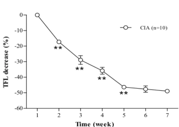
The changes of TFL after induction of CIA are shown in Fig. 1. After induction of CIA, there were statistically significant decreases of TFL until 5 weeks. After 5 weeks, there were no significant decreases in TFL. This means that inflammatory
pain was induced by CIA and that the pain reached its maximum value starting from the 5
thweek. On the basis of this result, BVP treatment into Zusanli (ST
36) was conducted at 5 weeks after the induction of CIA (Fig. 1).
1 2 3 4 5 6 7
-60 -50 -40 -30 -20 -10 0
CIA (n=10)
Time (week)
**
** **
**
TFL decrease (%)
Fig. 1. Changes of tail flick latency (TFL) after induction of collagen-induced arthritis (CIA)
After induction of arthritis, TFL decreased as time passed and reached its minimum value starting from the 3rd week. Each datum is represented as mean±SE of TFL on each tested week. Asterisks indicate significantly different values from the previous week.
* : p<.05. **: p<.01.
Friedman’s rank test followed by Dunnett’s post-hoctest.
B. Antinociceptive Effect of Bee Venom Pharmacopuncture (BVP) Treatment
The antinociceptive effects of Zusanli BVP in CIA are shown in Fig. 2. In the Zusanli BVP treatment group (Z-BVP, n=10), there were marked increases in TFL. Z-BVP showed significant incre- ases in TFL compare to the Zusanli DMSO treatment (Z-DMSO, n=10) and non-treatment group (None- Tx, n=10) at 10, 20, 30, 45 and 60min after the initiation of BVP. There were no significant differ- ences between Z-DMSO and None-Tx. This means that Zusanli DMSO treatment did not show antino- ciceptive effect.
The maximal analgesic effect of BVP was seen
at 30min after the initiation of BVP injection and
this antinociceptive effect is maintained for at least
60minutes. These results indicate that the treatment
of BVP can relieve the inflammatory pain in the
CIA animal model.
1 2 3 4 5 6 -10
0 10 20 30 40 50 60 70
***
***
## **
### ##
##
###
***
***
Time (min)
TFL increase (%)
None - T x (n=10) Z - BVA (n=10) Z - DMSO (n=10)
Fig. 2. Effects of Z-BVP on the antinociceptive effect in CIA
Z-BVP (●, n=10), group of BVP into a Zusanli (ST36) acupuncture point. Z-DMSO (▽, n=10), group of DMSO injection into a Zusanli (ST36) acupuncture point. None-Tx (○, n=10), group without any treatment. The Z-BVP group shows the significant increase of TFL after bee venom treatment (Mann-Whitney U-test).
** : p<.01. *** : p<.001.
Significantly different from Z-DMSO group.
## : p<.01. ### : p<.001.
Significantly different from None-Tx group.
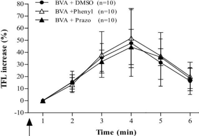
C. Involvement of α1-adrenoceptor on BVP Effect in CIA
The effects of α1-adrenoceptor agonist and anta-
1 2 3 4 5 6
-10 0 10 20 30 40 50 60 70 80
Time (min)
TFL increase (%)
BVA + DMSO (n=10) BVA +Phenyl (n=10) BVA + Prazo (n=10)
Fig. 3. Effect of α1-adrenoceptor agonist and an- tagonist pretreatment (arrow) on BVP-induced anti- nociceptive effect in CIA
BVP+DMSO(●, n=10), group of DMSO pretreatment BVP into a Zusanli (ST36). BVP+Phenyl (△, n=10), group of agonist phenylephrine pretreatment BVP into a Zusanli (ST36). BVP+Prazo (▲, n=10), group of antagonist prazosin pretreatment BVP into a Zusanli (ST36). There are no significant differences between BVP+DMSO and BVP+
Prazo.
gonist on BVP-induced antinociception in CIA are shown in Fig. 3. There were no significant differ- ences between the antagonist prazosin pretreatment
Zusanli BVP treatment group (BVP+Prazo, n=10)and the DMSO pretreatment Zusanli BVP treatment group (BVP+DMSO, n=10). This result shows that the antinociception of BVP treatment was not blocked by α1-adrenoceptor antagonist pretreatment.
In the phenylephrine and prazosin only treatment group, there were no statistically significant differ- ences in TFL and likewise in the saline treatment group (data not shown).
D. Involvement of 5HT-3 receptor on BVP Effect in CIA
The effects of 5HT-3 receptor agonist and anta- gonist on BVP-induced antinociception in CIA are shown in Fig. 4. There were statistically significant differences in TFL between the antagonist ondan- setron pretreatment Zusanli BVP treatment group (BVP+ondan, n=10) and the DMSO pretreatment
1 2 3 4 5 6
-10 0 10 20 30 40 50 60 70 80
* *** ** *
Time (min)
TFL increase (%)
BVA + DMSO (n=10) BVA + m-chlo (n=10) BVA + ondan (n=10)
Fig. 4. Effects of 5HT-3 receptor agonist and antagonist pretreatment (arrow) on BVP-induced antinociceptive effect in CIA
BVP+DMSO (●, n=10), group of DMSO pretreatment BVP into a Zusanli (ST36). BVP+m-chlo (△, n=10), group of agonist m-chlorophenyl-biguanide pretreatment BVP into a Zusanli (ST36). BVP+ondan (▲, n=10), group of ant- agonist ondansetron pretreatment BVP into a Zusanli (ST36). Asterisks indicate significantly different values bet- ween BVP+DMSO and BVP+ondan.
* : p<.05. ** : p<.01. *** : p<.001.
Significantly different from BVP+DMSO group.
Zusanli BVP treatment group (BVP+DMSO, n=10)
at 10, 20, 30 and 45min after the initiation of BVP.
This results show that the 5HT-3 receptor anta- gonist ondansetron pretreatment significantly blocked the BVP antinociceptive effects. There was no syner- gistic effect in the m-cholorophenyl-biguanide pre- treatment Zusanli BVP treatment group (BVP+m- chlo, n=10). In the m-cholorophenyl-biguanide and ondansetron only treatment group, there were no statistically significant differences in TFL and likewise in the saline treatment group (data not shown).
E. Involvement of muscarinic cholinergic receptor on BVP Effect in CIA
The effects of muscarinic cholinergic receptor agonist and antagonist on BVP-induced antinociception in CIA are shown in Fig. 5. There were statistic- ally significant differences in TFL between the an- tagonist atropine pretreatment Zusanli BVP treatment group (BVP+Atrop, n=10) and the DMSO pretreatment
Zusanli BVP treatment group (BVP+DMSO, n=10)at 10, 20, 30 and 45min after the initiation of BVP.
BVA + DMSO (n=10) BVA + Neosti (n=10) BVA + Atrop (n=10)
1 2 3 4 5 6
-10 0 10 20 30 40 50 60 70 80
* *** ** *
Time (min)
TFL increase (%)
Fig. 5. Effects of muscarinic cholinergic receptor agonist and antagonist pretreatment (arrow) on BVP- induced antinociceptive effect in CIA
BVP+DMSO (●, n=10), group of DMSO pretreatment BVP into a Zusanli (ST36). BVP+Neosti (△, n=10), group of agonist neostigmine pretreatment BVP into a Zusanli (ST36). BVP+Atrop (▲, n=10), group of antagonist atropine pretreatment BVP into a Zusanli (ST36). Asterisks indicate significantly different values between BVP+DMSO and BVP+Atro.
* : p<.05. ** : p<.01. *** : p<.001.
Significantly different from BVP+DMSO group.