• 검색 결과가 없습니다.

Heterogeneous Porous WO<sub>3</sub>@SnO<sub>2</sub> Nanofibers as Gas Sensing Layers for Chemiresistive Sensory Devices

N/A
N/A
Protected

Academic year: 2021

Share "Heterogeneous Porous WO<sub>3</sub>@SnO<sub>2</sub> Nanofibers as Gas Sensing Layers for Chemiresistive Sensory Devices"

Copied!
7
0
0

로드 중.... (전체 텍스트 보기)

전체 글

(1)

http://dx.doi.org/10.5369/JSST.2018.27.5.345 pISSN 1225-5475/eISSN 2093-7563

Heterogeneous Porous WO 3 @SnO 2 Nanofibers as Gas Sensing Layers for Chemiresistive Sensory Devices

Peresi Majura Bulemo

1,2

, Jiyoung Lee

1

, and Il-Doo Kim

1,3,+

Abstract

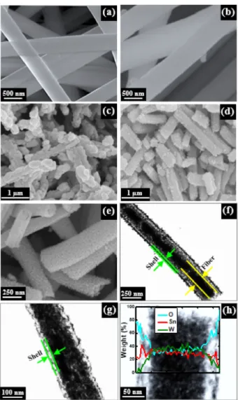
We employed an unprecedented technique to synthesize porous WO

3

@SnO

2

nanofibers exhibiting core-shell and fiber-in-tube con- figurations. Firstly, 2-methylimidazole was uniformly incorporated in as-spun nanofibers containing ammonium metatungstate hydrate and the sacrificial polymer (polyacrylonitrile). Secondly, the 2-methylimidazole on the surfaces of nanofibers was complexed with tin(II) chloride (SnCl

2

) via simple impregnation of the as-spun nanofibers in ethanol containing tin(II) chloride dihydrate (SnCl

2

·2H

2

O). The presence of vacant p-orbitals in tin (Sn) and the nucleophilic nitrogen on the imidazole ring allowed for the reaction between SnCl
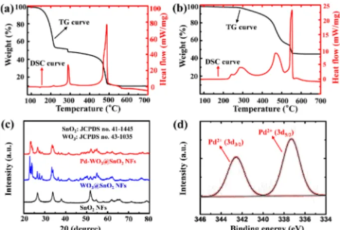
2

and 2-methylimidazole, forming adducts on the surfaces of the as-spun nanofibers. The calcination of these nanofibers resulted in porous WO

3

@SnO

2

nanofibers with a higher surface area (55.3 m

2

·g

−1

) and a better response to 1-5 ppm of acetone than pristine SnO

2

NFs synthesized using a similar method. An improved response to acetone was achieved upon functionalization of the WO

3

@SnO

2

nano- fibers with catalytic palladium nanoparticles. This work demonstrates the potential application of WO

3

@SnO

2

nanofibers as sensing lay- ers for chemiresistive sensory devices for the detection of acetone in exhaled breath.

Keywords: WO

3

@SnO

2

heteronanostructures, adducts, core-shell, fiber-in-tube, gas sensors, exhaled breath

1. INTRODUCTION

One-dimensional (1D) semiconducting metal oxide (SMO)- based heteronanostructures, especially those in core-shell configurations, exhibit versatility and applicability in gas sensing applications owing to their interfacial heterojunction barriers [1- 2]. More importantly, the combination of different SMOs with different work functions allows for the tunability of the gas- sensing properties [3]. Apart from the transduction of the molecular interaction of gases with individual material components, heterojunctions (such as n-n or p-n interfaces) in hybrids offer synergistic functionalities that amplify their sensing

capability. Typically, strain-induced defects originating from lattice mismatch are effective electron- and hole-trapping regions that cause the depletion of charge carriers in the vicinity of the interfaces. Of particular interest here are the electron kinetics at the interfaces of the contact materials. Lattice mismatch between the core-shell components creates a high density of states originating from surface strains (defects), which are dependent on core size and shell thickness [4]. Because of the trapped electrons, the electron barrier extends into the interface-forming elements.

In an effort to pursue the advantages of this interfacial phenomenon, several composite materials have been widely synthesized and used in gas sensing applications. In particular, few reports have been made on using SnO

2

and WO

3

heteronanostructures as sensing layers, and few have shown amplified responses to several gases, such as H

2

S, NH

3

, NO, and NO

2

[5-9]. However, their performance toward low gas concentrations (< 5 ppm) has not been significantly investigated.

Nonetheless, a combination of SnO

2

and WO

3

is an ideal choice for the suppression of moisture poisoning in pristine SnO

2

systems because WO

3

does not show cross-sensitivity to moisture [10-12].

Moreover, the enhancement of porosity and surface area further improves the interaction of SnO

2

-WO

3

nanostructures with the target gases.

In this work, porous 1D WO

3

@SnO

2

fibrous nanostructures exhibiting core-shell and fiber-in-tube configurations were

1

Department of Materials Science and Engineering, Korea Advanced Institute of Science and Technology, 291 Daehak-ro, Yuseong-gu, Daejeon 34141, Republic of Korea

2

Department of Mechanical and Industrial Engineering, University of Dar es Salaam, P. O. Box 35131, Dar es Salaam, Tanzania

3

Advanced Nanosensor Research Center, KI Nanocentury, Korea Advanced Institute of Science and Technology, 291 Daehak-ro, Yuseong-gu, Daejeon 34141, Republic of Korea

+

Corresponding author: [email protected]

(Received: Sep. 8, 2018, Revised: Sep. 16, 2018, Accepted: Sep. 17, 2018)

This is an Open Access article distributed under the terms of the Creative Commons Attribution Non-Commercial License(http://creativecommons.org/

licenses/bync/3.0) which permits unrestricted non-commercial use, distribution,

and reproduction in any medium, provided the original work is properly cited.

(2)

rationally synthesized via an electrospinning-impregnation technique followed by heat treatment in air. For the first time, we have incorporated 2-methylimidazole in the electrospinning solution containing ammonium metatungstate hydrate and polyacrylonitrile (PAN) and used it as an agent for the incorporation of SnCl

2

on as-spun nanofibers (henceforth referred to as A-NFs) using a simple impregnation technique. We will hereafter refer to the impregnated A-NFs as I-NFs. Although the use of 2-methlimidazole and SnCl

2

to synthesize Sn-based infinite coordinated polymer (ICP) structures has not been reported, the two can undergo fast reactions to form adducts. In our case, the surfaces of the I-NFs contain the SnO

2

forming precursor, whereas the WO

3

precursor is encapsulated in the interior. The postcalcined nanostructures (WO

3

@SnO

2

NFs) were subsequently functionalized with palladium (Pd) by employing a metal mirror reaction technique, followed by heat treatment to stabilize and oxidize the Pd nanoparticles (NPs). Finally, the Pd-functionalized 1D WO

3

@SnO

2

nanostructures (henceforth referred to as Pd- WO

3

@SnO

2

NFs) were investigated to determine their gas sensing performance toward various trace concentrations (1-5 ppm) of analyte gases.

2. EXPERIMENTAL

2.1 Synthesis of WO

3

@SnO

2

NFs

Unless otherwise specified, all chemicals were purchased from Sigma-Aldrich and used without further purification. A-NFs were synthesized from a viscous electrospinning solution containing 2.7 g of N,N-dimethylformamide (DMF, anhydrous, 99.8%), 70 mg of ammonium metatungstate hydrate [(NH

4

)

6

H

2

W

12

O

40

·xH

2

O,

≥ 85% WO

3

basis], 0.28 g of polyacrylonitrile (PAN, M

w

~150,000 g·mol

−1

), and 0.45 g of 2-methylimidazole (M

w

= 82.1 g·mol

−1

). Firstly, (NH

4

)

6

H

2

W

12

O

40

·xH

2

O was dissolved in DMF, and the mixture was vigorously stirred at room temperature until a homogeneous solution was obtained. Typically, 60 min were sufficient to attain a clear homogeneous solution when stirring at 500 rpm. Afterwards, the templating polymer (PAN) was added.

The temperature of the mixture was elevated to 60 °C, followed by stirring at 500 rpm for 2 h. Finally, 2-methylimidazole was added, and the solution was further stirred at 500 rpm for another 2 h to obtain a viscous electrospinning solution. The obtained solution was loaded into a 12-mL syringe with a single-nozzle needle (25-gauge) prior to electrospinning. The electrospinning equipment consisted of a power supply, a syringe pump, and a

rotary collecting drum (rotating at 50 rpm, clockwise). The solution-loaded syringe was fixed on the electrospinning machine, with the gap between the needle tip and the stainless-steel collector (on the rotary drum) maintained at 18 cm. Electrospinning was performed by applying a voltage of 8.5 kV at the tip of the needle while the collector was grounded. The feed pump was adjusted to eject the solution at a rate of 0.2 mL·min

−1

. The obtained A-NFs (in the form of a fiber web) were left to dry in air. A brief exposure of the A-NFs to ambient air allowed for additional evaporation of the solvent. Then, the A-NFs were impregnated with a solution containing tin(II) chloride dihydrate (SnCl

2

·2H

2

O).

To achieve this, 0.3 g of tin(II) chloride dihydrate (SnCl

2

·2H

2

O, M

w

= 225.63 g·mol

−1

) was dissolved in 6 mL of ethanol, followed by the immersion of the A-NFs for 20 min. Afterwards, the obtained NFs (i.e., the I-NFs) were filtered and dried overnight at 50 °C, followed by heat treatment in air at 600 °C for 2 h to obtain WO

3

@SnO

2

NFs. The temperature was increased at a steady ramping rate of 10 °C·min

−1

from room temperature.

2.2 Functionalization of WO

3

@SnO

2

NFs with Pd NPs

The postcalcined WO

3

@SnO

2

NFs were decorated with Pd NPs via a metal mirror reaction technique [13]. Two samples of Pd- WO

3

@SnO

2

NFs were prepared by varying the amount of PdCl

2

(M

w

= 177.33 g·mol

−1

, Kojima Chemicals) in ethanol. Precisely, 2.5 and 5.0 mM PdCl

2

were separately stirred (speed = 500 rpm) at 60°C for 20 min, followed by the addition of 0.1 g of WO

3

@SnO

2

NFs to each solution. To attach Pd NPs on the surfaces of the NFs, a small amount of butylamine (equal to that of PdCl

2

) was added and the mixtures were gently stirred for 15 min at the same temperature. The butylamine slowly reduced the Pd

2+

ions in the solution to form Pd NPs. Afterwards, the Pd- WO

3

@SnO

2

NFs were filtered and dried at 50 °C for 12 h in a convection oven. The actual quantities of Pd in the two samples decorated using 2.5 and 5.0 mM PdCl

2

were determined via X-ray fluorescence (XRF) spectroscopy (ZSX Primus II, Rigaku) and were found to be 0.36 wt% and 0.75 wt% with respect to WO

3

@SnO

2

, respectively.

2.3 Material characterization

The morphology and lattice parameters of the synthesized

structures were analyzed via scanning electron microscopy (SEM,

XL-30 SFEG, Philips) and field-emission transmission electron

microscopy (FE-TEM, Tecnai G2 F30 S-Twin, FEI). To study the

(3)

thermal properties of the structures, thermogravimetric (TG) and differential scanning calorimetry (DSC) analyses (Labsys Evo, Setaram) were conducted. X-ray diffraction (XRD, D/Max-2500, Rigaku) employing CuKα radiation (λ = 1.5406 Å) was used to confirm the crystal structure of the NFs. The surface areas and the pore size distribution were investigated using the Brunauer- Emmett-Teller (BET) and the Barrett-Joyner-Halender (BJH) methods (TriStar II 3020, Micromeritics), respectively. The elemental distribution of the synthesized materials was investigated using the scanning transmission electron microscopy (STEM) mode on the FE-TEM. The chemical bonding states were investigated via X-ray photoelectron spectroscopy (XPS, Sigma Probe, Thermo VG Scientific).

2.4 Sensor preparation and sensing tests

The sensors were prepared as follows. First, 2.0 mg of the NFs (SnO

2

NFs, WO

3

@SnO

2

NFs, and Pd-WO

3

@SnO

2

NFs) was dispersed via sonication in 80 µL of ethanol for 1 min. Then, the paste (two drops from a 1-µL pipette) was drop-coated onto the surfaces of alumina substrates (2.5-mm long, 2.5-mm wide, and 0.2-mm thick) to make uniform sensing layers. Each substrate consisted of two interdigitated parallel gold electrodes (25-µm thick and spaced at 70 µm) on the top surface and platinum microheaters on the bottom surface. Four gas sensors based on pristine SnO

2

NFs, WO

3

@SnO

2

NFs, 0.36 wt% Pd-decorated WO

3

@SnO

2

NFs (henceforth referred to as 0.36Pd-WO

3

@SnO

2

NFs), and 0.75 wt% Pd-decorated WO

3

@SnO

2

NFs (henceforth referred to as 0.75Pd-WO

3

@SnO

2

NFs) were prepared. The sensor based on SnO

2

NFs was used for comparative purposes.

Gas-sensing measurements were conducted using five gases, namely hydrogen sulfide (H

2

S), acetone (CH

3

COCH

3

), ammonia (NH

3

), nitrogen monoxide (NO), and nitrogen dioxide (NO

2

), in a highly humid ambient (relative humidity, RH = 90%). Sensing measurements were performed using a homemade sensing equipment consisting of a data acquisition system (34972A, Agilent) for measuring the instantaneous resistance, a temperature control unit (E3647A, Agilent), and a 16-channel sensor test chamber (34902A, Agilent). The complete experimental setup was in accordance with that of a past report [14]. Sensing tests were carried out in the temperature range of 300-450 °C. Prior to the tests, the sensors were stabilized in a humid air ambient to obtain a stable resistance. The analytes were then introduced in turns at different concentrations (1-5 ppm), in periods of 10 min for the analyte gas and 10 min for air. The sensing equipment included solenoid valves and mass flow controllers to adjust the

flow and concentrations of the analyte gases introduced into the sensing chamber. All analytes were diluted with air to attain 20 µmol of balance air per mole.

3. RESULTS AND DISCUSSIONS

3.1 Morphology of the synthesized structures Scheme 1 shows a conceptualized illustration of the proposed synthetic approach for the formation of WO

3

@SnO

2

NFs. Upon electrospinning with the solution containing (NH

4

)

6

H

2

W

12

O

40

·xH

2

O, PAN, and 2-methylimidazole, well-aligned 1D NFs (i.e., A-NFs, represented as cross-sections) were obtained (Fig. 1a). When the A-NFs were impregnated in a solution containing SnCl

2

·2H

2

O, their surfaces turned whiter, indicating that the SnCl

2

in the solution complexed with surface-exposed 2-methylimidazole to form adducts [15]. It should be noted that our preliminary experimental investigations indicated that even solutions of 2-methylimidazole and SnCl

2

·2H

2

O (using ethanol and ethanol/

DMF as solvents, respectively) spontaneously turned turbid when mixed. Typically, because of the vacant p-orbital in Sn, fast reactions are to be expected between dichlorostannylene (SnCl

2

) and 2-methylimidazole. In our case, SnCl

2

is likely to react with the nucleophilic nitrogen on the imidazole rings to achieve a stable octet configuration of Sn [16]. It is worth mentioning that, when forming the adducts, 2-methylimidazole, which is the bidentate ligand, coordinates with SnCl

2

in two positions to adopt a trigonal pyramid configuration, whereas the singlet electron pair in Sn remains inert.

Scheme. 1. Schematic illustration of the synthetic procedure for syn-

thesizing WO

3

@SnO

2

NFs with (a) core-shell and (b)

fiber-in-tube morphologies. AMH and MI stand for 2-

methylimidazole and ammonium metatungstate hydrate,

respectively.

(4)

Fig. 1b shows the impregnated A-NFs (i.e., I-NFs). Because (NH

4

)

6

H

2

W

12

O

40

·xH

2

O has poor solubility in ethanol, it remained intact in the core even after the impregnation of the A-NFs. In addition, the reaction of SnCl

2

and 2-methylimidazole occurs very fast, and we suppose that the formed adducts prevent any escape of (NH

4

)

6

H

2

W

12

O

40

·xH

2

O and PAN from the interior. As observed in Figs. 1a-b, the size of the I-NFs was larger than that of the A- NFs, implying that adducts of 2-methylyimidazole and SnCl

2

formed on the surfaces of the A-NFs, whereas the cores consisted of (NH

4

)

6

H

2

W

12

O

40

·xH

2

O. The direct calcination of A-NFs (without impregnation) resulted only in a poor 1D WO

3

morphology as PAN and 2-methylimidazole, which largely

formed the backbone of the A-NFs, decomposed (Fig. 1c). This can be attributed to the fact that the amount of (NH

4

)

6

H

2

W

12

O

40

·xH

2

O was insufficient to provide a stable 1D WO

3

morphology upon calcination. On the other hand, when as- spun NFs (similar to A-NFs but without the WO

3

precursor) were impregnated in a solution containing SnCl

2

·2H

2

O (for 20 min), followed by drying (for 12 h) and calcination (at 600 °C for 2 h), a discernable 1D morphology of SnO

2

NFs was observed (Fig.

1d). These two cases suggest the possibility of forming porous stable 1D morphologies of WO

3

@SnO

2

NFs upon calcination of I-NFs.

Fig. 1e shows the morphology of the WO

3

@SnO

2

NFs after calcination at 600 °C for 2 h. The WO

3

@SnO

2

NFs exhibited both core-shell and fiber-in-tube morphologies (Figs. 1f-g). A STEM analysis indicated that the core of the NFs (illustrated in Scheme 1a) and the fiber in the tube (illustrated in Scheme 1b) were composed of WO

3

, whereas the shell was largely composed of SnO

2

(Fig. 1h). Because of a high ramping rate (10 °C·nm

−1

) and an absence of PAN in the shell of the I-NFs, the SnO

2

precursor readily oxidized to form a solid shell compared with the WO

3

precursor in the interior. Afterwards, as the PAN in the core decomposed, either the oxidizing WO

3

precursor concomitantly aggregated, forming the fiber in the tube or remained staggered, forming the porous WO

3

core encapsulated with the SnO

2

shell.

The porosity in the shells of the WO

3

@SnO

2

NFs can also be attributed to an outward diffusion of the decomposed PAN. As shown in Figs. 2a-b, the WO

3

@SnO

2

NFs exhibited a mesoporous morphology with a surface area of 55.3 m

2

·g

−1

, which was nearly twice that of pristine SnO

2

NFs (28.0 m

2

·g

−1

), whereas its pore size and pore volume were 10.3 nm and 0.1 cm

3

·g

−1

, respectively. The higher surface area originated from the enhanced porosity upon the decomposition of PAN and organic matter.

To investigate the thermal behavior of the A-NFs and I-NFs, the samples were heated in air from room temperature to 800 °C.

Thermogravimetric (TG) and differential scanning calorimetry (DSC) analyses (Figs. 3a-b) revealed that organic matter completely decomposed as the temperature reached approximately 500 and 550 °C, respectively. This implies that the adducts on I-NFs were slightly less thermally stable compared with mere 2-methyimidazole and PAN in bare A-NFs. Beyond these temperatures, constant weights of crystallized WO

3

(10%) and WO

3

@SnO

2

NFs (45%) were obtained. To investigate the crystalline nature of the WO

3

@SnO

2

NFs, we carried out an XRD analysis of postcalcined WO

3

@SnO

2

NFs and SnO

2

NFs. As shown in Fig. 3c, all the diffraction peaks of the WO

3

@SnO

2

NFs belonged to pure cassiterite SnO

2

(JCPDS no. 41-1445) and Fig. 1. SEM images of (a) as-spun NFs (A-NFs) containing PAN, 2-

methylimidazole, and (NH

4

)

6

H

2

W

12

O

40

·xH

2

O, (b) I-NFs, (c)

WO

3

NFs upon direct calcination of A-NFs prior to impreg-

nation, (d) SnO

2

NFs obtained upon calcination of impreg-

nated NFs without (NH

4

)

6

H

2

W

12

O

40

·xH

2

O, and (e)

WO

3

@SnO

2

NFs upon calcination of I-NFs. TEM images of

WO

3

@SnO

2

NFs showing (f) fiber-in-tube and (g) core-shell

configurations. (h) Linescan profile of WO

3

@SnO

2

NFs.

(5)

monoclinic WO

3

(JCPDS no. 43-1035). When the WO

3

@SnO

2

NFs were decorated with Pd NPs followed by a heat treatment (500 °C for 1 h), the component phases in the WO

3

@SnO

2

NFs were maintained with the exception of the oxidation of Pd to PdO, which was confirmed by the binding energies of the Pd 3d

3/2

and Pd 3d

5/2

peaks at 337.2 and 342.4 eV, respectively (Fig. 3d). Note that no traces of PdO were identified in the XRD patterns, possibly because of the small amount of Pd NPs. A corroboration via TEM analysis revealed that the WO

3

@SnO

2

NFs consisted of dominant lattice fringes assigned to the SnO

2

(110) and WO

3

(202) planes spaced at 0.332 and 0.262 nm, respectively.

3.2 Investigation of gas sensing capability

To investigate the gas sensing performance of SnO

2

NFs, WO

3

@SnO

2

NFs, 0.36Pd-WO

3

@SnO

2

NFs, and 0.75Pd- WO

3

@SnO

2

NFs, the operating temperature of the four prepared

sensors was carefully controlled and maintained in the range of 300-450 °C. Exposure of the sensors to various gases (H

2

S, CH

3

COCH

3

, NH

3

, NO, and NO

2

) in this range of temperature allowed for the determination of the optimal operating temperature. Optimal sensing was observed at 350 °C toward acetone (Fig. 4a). At this temperature, the response (defined as R

a

/ R

g

, where R

a

and R

g

are the resistances of the sensor upon exposure to air and the target gas, respectively) of the 0.75Pd- WO

3

@SnO

2

NFs was 25.4 toward 5 ppm of acetone. The responses of the unfunctionalized SnO

2

NFs and the WO

3

@SnO

2

NFs were 7.3 and 14.4, respectively. We expected an increase in response in the order of SnO

2

NFs < WO

3

@SnO

2

NFs <

0.75Pd-WO

3

@SnO

2

NFs, owing to the possible n-n junction in the WO

3

@SnO

2

NFs and the additional catalytic activity of the Pd NPs in the 0.75Pd-WO

3

@SnO

2

NFs [17]. Typically, at n-n junctions of SnO

2

and WO

3

, charge separation (electrons and holes) occurs, leading to the creation of a barrier potential at the interfaces [5-6]. It is worth mentioning that heterojunctions are usually associated with a high density of states that originate from surface strains due to lattice mismatch, allowing for electron trapping and the depletion of charge carriers in the vicinity of the interfaces. Because of this, the gas sensing performance of the WO

3

@SnO

2

NFs was expected to be higher than that of the SnO

2

NFs. The higher surface area of the WO

3

@SnO

2

NFs (55.3 m

2

·g

−1

) compared with that of pristine SnO

2

NFs (28.0 m

2

·g

−1

) implies that the former possess many more reaction sites than the latter. With exception of H

2

S, for which the 0.75Pd-WO

3

@SnO

2

NFs sensor also exhibited an enhanced response (R

a

/R

g

= 18.3), the responses of the sensors to other gases (NO, NO

2

, and NH

3

), were considerably low (R

a

/R

g

< 2) (Fig. 4b), unlike in previous reports that indicated appreciable responses in WO

3

@SnO

2

NFs- based sensors toward NO, NO

2

, and NH

3

[5-6]. The response to 1 ppm of acetone was 6.4, indicating that the sensor has the capability to detect trace concentrations of acetone in exhaled human breath. Note that concentration of acetone is typically in the range of 300-900 ppb in healthy human breath and approximately 1.8 ppm in the breath of people with diabetes [18].

The sensitivity of the sensor to trace concentration (1 ppm) was coupled with a fast response (48 s) compared with the response times at higher concentrations (Fig. 4c). Typically, a fast sensor response is critical, especially for the real-time analysis of exhaled breath.

The sensing mechanism of the sensors can be explained in terms of the resistance variations upon cyclic exposure of the sensors to air and the target gas. The abundant oxygen in air can be adsorbed on the surface of the WO

3

@SnO

2

NFs according to Fig. 2. (a) Nitrogen (N

2

) adsorption-desorption isotherms and (b) the

corresponding BJH desorption pore size distribution of

WO

3

@SnO

2

NFs.

(6)

Equation 1 when the sensors are exposed to air, accompanied with the withdrawal of electrons from the conduction band of WO

3

and SnO

2

[17, 19]. The withdrawal of electrons forms an electron- depleted region, with a consequent increase in resistance.

As shown in Fig. 4d, the resistance of the materials in background air increased in the order of SnO

2

NFs < WO

3

@SnO

2

NFs < 0.75Pd-WO

3

@SnO

2

NFs. This indicates that more electrons were withdrawn in 0.75Pd-WO

3

@SnO

2

NFs than in the other materials owing to the catalytic effect of Pd. Because of the

heterojunction, the WO

3

@SnO

2

NFs showed higher resistance than pristine SnO

2

NFs. When the sensors were exposed to acetone, the withdrawn electrons were restored into the conduction bands of SnO

2

and WO

3

according to Equation 2 [20].

O

2

+ 2e

→ 2O

(ads.)

(1)

CH

3

COCH

3(gas)

+ 2O

(ads.)

→ CH

3

O

+ C

+

H

3

+ CO

2(gas)

+ 2e

(2)

4. CONCLUSIONS

A facile method was employed to synthesize WO

3

@SnO

2

NFs with different morphologies i.e., core-shell and fiber-in-tube structures. The reactivity of SnCl

2

with 2-methylimidazole, the low solubility of PAN, and the insolubility of ammonium metatungstate in ethanol allowed for the synthesis of I-NFs, which resulted in mesoporous structures with a high surface area (pore size = 10.3 nm and surface area 55.3 m

2

·g

−1

) upon heat treatment in air. The functionalization of the WO

3

@SnO

2

NFs with Pd NPs improved their response (R

a

/R

g

= 6.4) to low concentrations of acetone (1 ppm), and they showed a fast response (48 s). This acetone-sensing property demonstrates that the proposed sensor (0.75Pd-WO

3

@SnO

2

NFs) can be used for the detection of acetone in the breath of people with diabetes. However, further investigation may be required to improve the discrimination of acetone from H

2

S.

ACKNOWLEDGMENTS

The authors acknowledge the Ministry of Science, ICT and Future Planning of the Korea government for the financial support provided through the Wearable Platform Materials Technology Center (funded by the National Research Foundation of Korea, under grant No. 2016R1A5A1009926), the Biomedical Treatment Technology Development Project (grant No. 2015M3A9D7067418), the National Research Foundation of Korea (grant No. NRF- 2015R1A2A1A1A6074901, the BRL Program through grant No.

2014R1A4A1003712), and the Global Frontier Project (CISS- 2011-0031870).

REFERENCES

[1] I. S. Hwang, S. J. Kim, J. K. Choi, J. Choi, H. Ji, G. T. Kim, G. Cao, and J. H. Lee, “Synthesis and Gas Sensing Char- acteristics of Highly Crystalline ZnO-SnO

2

Core-Shell Fig. 3. Thermal decomposition of (a) the A-NFs and (b) the I-NFs.

(c) XRD patterns of the SnO

2

NFs, the WO

3

@SnO

2

NFs, and the Pd-WO

3

@SnO

2

NFs. (d) High-resolution XPS spectra of the Pd 3d region.

Fig. 4. Gas sensing characteristics of the synthesized materials. (a)

Time-dependent response-recovery profiles of the sensors for

various concentrations of acetone at 350 °C. (b) The response

of the 0.75Pd-WO

3

@SnO

2

NFs to 5 ppm of different gases at

350 °C. (c) Variation of the response and recovery times of

the 0.75Pd-WO

3

@SnO

2

NFs for various concentrations (1-5

ppm) of acetone. (d) Baseline resistances of the SnO

2

NFs,

the WO

3

@SnO

2

NFs, and the 0.75Pd-WO

3

@SnO

2

NFs upon

exposure to air at 350 °C.

(7)

Nanowires”, Sens. Actuators, B, Vol. 148, No. 2, pp. 595- 600, 2010.

[2] T. Li, W. Zeng, and Z. Wang, “Quasi-One-Dimensional Metal-Oxide-Based Heterostructural Gas-Sensing Materi- als: A Review”, Sens. Actuators, B, Vol. 221, pp. 1570- 1585, 2015.

[3] S. Park, H. Ko, S. Kim, and C. Lee, “Role of the Interfaces in Multiple Networked One-Dimensional Core-Shell Nano- structured Gas Sensors”, ACS Appl. Mater. Interfaces, Vol.

6, No. 12, pp. 9595-9600, 2014.

[4] G. A. Beane, K. Gong, and D. F. Kelley, “Auger and Carrier Trapping Dynamics in Core/Shell Quantum Dots Having Sharp and Alloyed Interfaces”, ACS Nano, Vol. 10, No. 3, pp. 3755-3765, 2016.

[5] H. Y. Li, Z. X. Cai, J. C. Ding, and X. Guo, “Gigantically Enhanced NO Sensing Properties of WO

3

/SnO

2

Double Layer Sensors with Pd Decoration”, Sens. Actuators, B, Vol.

220, pp. 398-405, 2015.

[6] A. Sharma, M. Tomar, and V. Gupta, “Low Temperature Operating SnO

2

Thin Film Sensor Loaded With WO

3

Micro-discs with Enhanced Response for NO

2

Gas”, Sens.

Actuators, B, Vol. 161, No. 1, pp. 1114-1118, 2012.

[7] N. Van Toan, C. M. Hung, N. Van Duy, N. D. Hoa, D. T.

T. Le, and N. Van Hieu, “Bilayer SnO

2

-WO

3

Nanofilms for Enhanced NH

3

Gas Sensing Performance”, Mater. Sci. Eng., B, Vol. 224, pp. 163-170, 2017.

[8] L. Yin, D. L. Chen, H. W. Zhang, G. Shao, B. B. Fan, R.

Zhang, and G. S. Shao, “In Situ Formation of Au/SnO

2

Nanocrystals on WO

3

Nanoplates as Excellent Gas-Sensing Materials for H

2

S Detection”, Mater. Chem. Phys., Vol. 148, No. 3, pp. 1099-1107, 2014.

[9] Y. Gui, F. Dong, Y. Zhang, Y. Zhang, and J. Tian, “Prepa- ration and Gas Sensitivity of WO

3

Hollow Microspheres and SnO

2

Doped Heterojunction Sensors”, Mater. Sci. Semi- cond. Process., Vol. 16, No. 6, pp. 1531-1537, 2013.

[10] M. Morimitsu, Y. Ozaki, S. Suzuki, and M. Matsunaga,

“Effects of Surface Modification with Platinum and Ruthe- nium on Temperature and Humidity Dependence of SnO2- Based CO Gas Sensors”, Sens. Actuators, B, Vol. 67, No. 1- 2, pp. 184-188, 2000.

[11] J. Y. Shen, L. Zhang, J. Ren, J. C. Wang, H. C. Yao, and Z.

J. Li, “Highly Enhanced Acetone Sensing Performance of Porous C-Doped WO

3

Hollow Spheres by Carbon Spheres as Templates”, Sens. Actuators, B, Vol. 239, pp. 597-607,

2017.

[12] H. R. Kim, A. Haensch, I. D. Kim, N. Barsan, U. Weimar, and J. H. Lee, “The Role of NiO Doping in Reducing the Impact of Humidity on the Performance of SnO

2

?Based Gas Sensors: Synthesis Strategies, and Phenomenological and Spectroscopic Studies”, Adv. Funct. Mater., Vol. 21, No.

23, pp. 4456-4463, 2011.

[13] S. Yoo, J. I. Lee, S. Ko, and S. Park, “Highly Dispersive and Electrically Conductive Silver-Coated Si Anodes Synthe- sized via a Simple Chemical Reduction Process”, Nano Energy, Vol. 2, pp. 1271-1278, 2013.

[14] P. M. Bulemo, H. J. Cho, D. H. Kim, and I. D. Kim, “Facile Synthesis of Pt-Functionalized Meso/Macroporous SnO

2

Hollow Spheres through In Situ Templating with SiO

2

for H

2

S Sensors”, ACS Appl. Mater. Interfaces, Vol. 10, No. 21, pp. 18183-18191, 2018.

[15] C. Pettinari, M. Pellei, F. Marchetti, C. Santini, and M. Mil- iani, “Tin(IV) and Organotin(IV) Complexes Containing Mono or Bidentate N-Donor Ligands—IV. 2-Methyl-, 2- Isopropyl-and 4-Methyl-Imidazole Derivatives: Synthesis, Characterization and Behaviour in Solution”, Polyhedron, Vol. 17, No. 4, pp. 561-576, 1998.

[16] Y. Mizuhata, T. Sasamori, and N. Tokitoh, “Stable Heavier Carbene Analogues”, Chem. Rev., Vol. 109, No. 8, pp.

3479-3511, 2009.

[17] N. Yamazoe, G. Sakai, and K. Shimanoe, “Oxide Semi- conductor Gas Sensors”, Catal. Surv. Asia, Vol. 7, No. 1, pp. 63-75, 2003.

[18] M. Righettoni, A. Tricoli, and S. E. Pratsinis, “Si:WO

3

Sen- sors for Highly Selective Detection of Acetone for Easy Diagnosis of Diabetes by Breath Analysis”, Anal. Chem., Vol. 82, No. 9, pp. 3581-3587, 2010.

[19] P. M. Bulemo, H. J. Cho, N. H. Kim, and I. D. Kim, “Mes- oporous SnO

2

Nanotubes via Electrospinning-Etching Route: Highly Sensitive and Selective Detection of H

2

S Molecule”, ACS Appl. Mater. Interfaces, Vol. 9, No. 31, pp.

26304-26313, 2017.

[20] S. J. Choi, F. Fuchs, R. Demadrille, B. Grévin, B. H. Jang, S. J. Lee, J. H. Lee, H. L. Tuller, and I. D. Kim, “Fast Responding Exhaled-Breath Sensors Using WO

3

Hem- itubes Functionalized by Graphene-Based Electronic Sen- sitizers for Diagnosis of Diseases”, ACS Appl. Mater.

Interfaces, Vol. 6, No. 12, pp. 9061-9070, 2014.

수치

Fig. 1b shows the impregnated A-NFs (i.e., I-NFs). Because (NH 4 ) 6 H 2 W 12 O 40 ·xH 2 O has poor solubility in ethanol, it remained intact in the core even after the impregnation of the A-NFs
Fig. 4. Gas sensing characteristics of the synthesized materials. (a) Time-dependent response-recovery profiles of the sensors for various concentrations of acetone at 350 °C

참조

관련 문서