D I A B E T E S & M E T A B O L I S M J O U R N A L
This is an Open Access article distributed under the terms of the Creative Commons At- tribution Non-Commercial License (http://creativecommons.org/licenses/by-nc/3.0/) which permits unrestricted non-commercial use, distribution, and reproduction in any medium, provided the original work is properly cited.
Corrected QT Interval Prolongation during Severe Hypoglycemia without Hypokalemia in Patients with Type 2 Diabetes
Jae Won Beom1, Jung Min Kim1, Eun Joo Chung2, Ju Yeong Kim1, Seung Yeong Ko1, Sang Don Na1, Cheol Hwan Kim1, Gun Park1, Mi Yeon Kang1
1Department of Internal Medicine, Saint Carollo Hospital, Suncheon,
2Department of Neurology, Inje University Busan Paik Hospital, Inje University College of Medicine, Busan, Korea
Background: To evaluate the effects of severe hypoglycemia without hypokalemia on the electrocardiogram in patients with type 2 diabetes in real-life conditions.
Methods: Electrocardiograms of adult type 2 diabetic patients during the episodes of severe hypoglycemia and the recovered stage were obtained and analysed between October 1, 2011 and May 31, 2012. Patients who maintained the normal serum sodi- um and potassium levels during the episodes of severe hypoglycemia were only selected as the subjects of this study. Severe hy- poglycemia was defined, in this study, as the condition requiring active medical assistance such as administering carbohydrate when serum glucose level was less than 60 mg/dL.
Results: Nine type 2 diabetes patients (seven men, two women) were included in the study. The mean subject age was 73.2±7.7 years. The mean hemoglobin A1c level was 6.07%±1.19%. The median duration of diabetes was 10 years (range, 3.5 to 30 years).
Corrected QT (QTc) intervals were significantly increased during the episodes of severe hypoglycemia compared to the recovered stage (447.6±18.2 ms vs. 417.2±30.6 ms; P<0.05). However, the morphology and the amplitude of the T waves were not changed and ST-segment elevation and/or depression were not found during the episodes of severe hypoglycemia.
Conclusion: In this study, QTc interval prolongation during the episodes of severe hypoglycemia was observed without hypo- kalemia. Therefore, the distinct alterations in cardiac repolarization during the episodes of severe hypoglycemia may not be as- sociated with hypokalemia.
Keywords: Diabetes mellitus, type 2; Electrocardiography; QT interval; Severe hypoglycemia
Corresponding author: Mi Yeon Kang
Division of Endocrinology and Metabolism, Department of Internal Medicine, Saint Carollo Hospital, 221 Sungwang-ro, Suncheon 540-719, Korea
INTRODUCTION
Previous studies have shown that the incidence of severe hy- poglycemia has increased [1,2]. Hypoglycemia causes recurrent morbidity in diabetes and can lead to death. It has been esti- mated that hypoglycemia is responsible for 2% to 4% of death in type 1 diabetes [3]. A retrospective cohort study has report- ed that patients with hypoglycemic episodes during hospital- ization have higher mortality 1 year after discharge (27.8% for
patients with at least one hypoglycemic episode vs. 14.1% for patients without hypoglycemic episodes) [4].
Several studies have demonstrated that insulin-induced hy- poglycemia in diabetes causes the prolongation of corrected QT (QTc) interval, which is associated with ventricular ar- rhythmias and sudden death, and have reported that this ab- normality in cardiac repolarization results from hypokalemia and an increase in serum catecholamines [5-7].
A study conducted to patients with type 2 diabetes with a http://dx.doi.org/10.4093/dmj.2013.37.3.190
pISSN 2233-6079 · eISSN 2233-6087
continuous glucose monitoring system and continuous elec- trocardiogram (ECG) monitoring confirmed that hypoglyce- mia was associated with cardiac ischemia [8].
The aim of this study was to evaluate the effects of severe hypoglycemia without hypokalemia on the ECG in type 2 dia- betes in real-life conditions.
METHODS
Research design and methods
In review of retrospective medical records, ECGs of adult type 2 diabetic patients were obtained and analysed during the epi- sodes of severe hypoglycemia in the Emergency Department and the recovered stage in the general ward before they were discharged from the Saint Carollo Hospital between October 1, 2011 and May 31, 2012. For that period, a total of 43 type 2 diabetes with severe hypoglycemia were identified in the Emer- gency Department. Seventy-nine percentage (34/43) normo- kalemic patients, 14.0% (6/43) hypokalemic patients, and 7.0%
(3/43) hyperkalemic patients were found. Patients who main- tained the normal serum sodium and potassium levels during the episodes of severe hypoglycemia in the Emergency De- partment were chosen as the subjects for this study (Table 1).
Severe hypoglycemia was defined, in this study, as the condi- tion requiring active medical assistance such as administering carbohydrate when serum glucose level was less than 60 mg/
dL [1,9].
Subject characteristics including age, gender, height, weight,
the duration of diabetes and hypertension, glucose lowering agents, antihypertensive medications, smoking history, alco- hol consumption, the inducing factor for hypoglycemia, and comorbidities (coronary artery disease and cerebral vascular disease) were obtained. HbA1c, plasma C-peptide levels, plas- ma insulin levels, and serum chemistry profiles were also ob- tained.
Analyses of the digital 12-lead ECG recordings were per- formed at a ECG paper speed of 25 mm/sec using a M1771A Pagewriter 200 (Phillips, Andover, MA, USA). QTc intervals were also simultaneously recorded at the digital 12-lead ECG recordings by an automatic method and corrected by Bazett’s formula (Fig. 1).
T wave changes, ST-segment elevation and/or depression and changes in the QRS complex that occur in association with acute ischemia and infarction were assessed [10]. The ST-seg- ment elevation was defined as >0.2 mV elevation in two or more precordial leads and >0.1 mV elevation in two or more limb leads. The ST-segment depression was defined as flat or down sloping depression >0.1 mV, lasting for at least 1 minute in two or more leads. Deeply inverted T wave was defined as the inverted T wave greater than 0.5 mV.
Statistical analysis
All data were analysed using the SPSS statistical program ver- sion 13.0 (SPSS Inc., Chicago, IL, USA). The Wilcoxon’s signed rank test was applied to compare the most of data.
Normally distributed data are presented as the mean value±
Table 1. The serum potassium and sodium levels during se- vere hypoglycemia and the recovered stage in all subjects
Patient no.
K (normal range,
3.5-5.3 mmol/L) Na (normal range, 135-148 mmol/L) Severe hypo-
glycemia Recovered
stage Severe hypo-
glycemia Recovered stage
1 4.3 5.0 138.3 140.7
2 3.9 4.2 145.5 145.8
3 5.3 3.9 142.9 139.1
4 4.0 4.7 143.7 137.9
5 4.7 4.0 136.7 138.3
6 4.2 4.2 139.1 134.8
7 4.3 4.5 137.7 139.7
8 3.5 4.0 135.8 141.4
9 3.8 5.0 136.8 136.6
Fig. 1. Two representative electrocardiographic recordings of our subjects. On electrocardiograms, there did not show isch- emic changes such as ST-segment abnormality and T wave in- version and did not show tachycardia during severe hypogly- cemia. The comparison of corrected QT (QTc) interval be- tween severe hypoglycemia and the recovered stage showed each (A) 432 ms versus 396 ms and (B) 442 ms versus 402 ms.
Severe hypoglycemia Recovered stage
A
B
standard deviation and nonnormally distributed data are pre- sented as the median range. P values of less than 0.05 were con- sidered as significant.
RESULTS
A total of nine patients (seven male, two female) were includ- ed in this study. The mean subject age was 73.2±7.7 years. The mean HbA1c level was 6.07%±1.19%. The median duration of diabetes was 10 years (range, 3.5 to 30 years). The mean body mass index was 23.0±2.9 kg/m2. Among the nine patients, eight patients were treated with glucose-lowering agents. All of them were taking a sulfonylurea derivative, glimepiride that affects the ATP-sensitive K+ channel. During the hospital ad- mission, sulfonylureas was discontinued in 50% of the subjects and continued in the rest of them. The sulfonylureas was com- menced in a patient once insulin therapy was discontinued (Table 2).
All patients had a medical history of hypertension and were taking antihypertensive medications which did not contain al- pha and beta adrenergic blocking agents. However, none had a history of coronary artery disease. During the hospital admis- sion, their antihypertensive medications were not changed.
Serum potassium and sodium levels of all patients were within normal range during severe hypoglycema and the re- covered stage (Table 1).
There were no significant differences in heart rates, RR in- tervals or QT intervals during the episodes of severe hypogly- cemia but the QT intervals were slightly longer during the epi- sodes of severe hypoglycemia than during the recovered stage (408.8±40.7 ms vs. 380.2±39.7 ms, P=0.066) (Table 3).
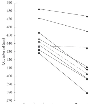
The QTc intervals were significantly increased during the episodes of severe hypoglycemia compared to the recovered stage in all patients (447.6±18.2 ms vs. 417.2±30.6 ms, P=
0.008) (Table 3, Fig. 2). In particular, the QTc intervals were significantly increased in patients taking only oral glucose Table 3. The comparison of electrocardiographic findings be- tween severe hypoglycemia and the recovered stage in all sub- jects (n=9)
Severe
hypoglycemia Recovered
stage P value Heart rate,
beats per min 74.0±14.3 74.9±18.5 1.000 PR interval, ms 167.2±13.1 161.9±18.9 0.312 QT interval, ms 408.8±40.7 380.2±39.7 0.066 QTc interval, ms 447.6±18.2 417.2±30.6 0.008 Values are presented as mean±standard deviation.
QTc, corrected QT.
Table 2. Subject characteristics Patient
no. Age,
yr Sex BMI,
kg/m2 HbA1c,
%
Dura- tion of diabetes,
yr
Treatment Before admis- sion
After admis-
sion
1 69 Male 24.9 5.5 22 S S
2 65 Female 21.9 5.9 10 M, S M, D
3 62 Male 17.9 5.4 10 M, S S
4 76 Male 27.6 5.5 20 I S
5 70 Male 23.5 8.4 30 I, M, S I, A
6 77 Male 25.0 7.5 5 S S
7 75 Female 21.4 5.4 4 M, A, S S, D
8 77 Male 20.6 4.6 3.5 S M
9 88 Male 24.0 6.3 20 M, S D
HbA1c is checked at the Emergency Department.
BMI, body mass index; HbA1c, hemoglobin A1c; S, sulfonylureas; M, metformin; D, dipeptidyl peptidase IV inhibitors; I, insulin; A, α-glucosidase inhibitors.
Fig. 2. Corrected QT (QTc) interval, individual values during severe hypoglycemia and the recovered stage in all subjects.
Only oral glucose-lowering agents users are indicated by con- tinuous lines, insulin users by dashed lines.
QTc interval (ms)
490 480 470 460 450 440 430 420 410 400 390 380
370 Severe hypoglycemia Recovered stage
lowering agents during the episodes of severe hypoglycemia compared to the recovered stage (450.0±20.2 ms vs. 417.0±
34.1 ms, P=0.018) (Table 4, Fig. 2).
However, the morphology and the amplitude of the T waves were not changed and ST-segment elevation and/or depres- sion were not found during the episodes of severe hypoglyce- mia (Fig. 1).
DISCUSSION
It has been suggested that prolonged QT intervals in patients with myocardial infarction may be associated with a worse prognosis [11]. The prolongation of QTc intervals is related to an increased risk of sudden death [12]. Tattersall and Gill [13]
reported that the cause of sudden nocturnal death in young people with type 1 diabetes was likely to be cardiac arrhythmia induced by hypoglycemia.
Marques et al. [5] demonstrated that the degree of QTc lengthening during the episodes of hypoglycemia, which was induced by intravenous (IV) insulin, was greater compared to that during the euglycemic period in type 1 diabetic patients (median [range], 156 [8 to 258] ms vs. 6 [-3 to 28] ms; P<0.02) and in type 2 diabetic patients (median [range], 128 [16 to 166]
ms vs. 4 [-3 to 169] ms; P<0.05). They suggested hypoglyce- mia could cause cardiac arrhythmias. They also found that a fall in plasma potassium and a rise in plasma adrenalin were greater during the episodes of insulin induced hypoglycemia [5].
Landstedt-Hallin et al. [6] reported that the mean QT inter- val and QT dispersion were significantly increased and serum potassium levels were significantly decreased during the epi- sodes of insulin-induced hypoglycemia in a study conducted
to thirteen patients with type 2 diabetes.
Heller conducted a study to normal adults to investigate whether the increase in both QT dispersion and QT intervals during the episodes of insulin-induced hypoglycemia could be avoided by β-blockade and the prevention of hypokalemia with IV potassium administration [7]. Many investigators have sug- gested that hypokalemia caused by hyperinsulinemia was a possible mechanism of altered cardiac repolarization during the episodes of hypoglycemia [14,15].
However, in this study, significant lengthening of QTc inter- vals during the episodes of severe hypoglycemia was observed without hypokalemia. The results of this study implied that distinct alterations in cardiac repolarization during the epi- sodes of severe hypoglycemia might not be associated with hypokalemia.
Most of the subjects in previous studies were treated with insulin while the majority of the subjects in this study were treated with only oral glucose-lowering agents (seven out of nine patients). In addition, this study investigated the signifi- cant lengthening of the QTc interval during the episodes of se- vere hypoglycemia without hypokalemia in patients treated with only oral glucose-lowering agents was investigated in this study (Table 4, Fig. 2).
It has been reported that glimepiride didn’t seem to have any effects on cardiac function in several studies [16-18]. Un- fortunately, a comparison between insulin users and glimepiri- de users could not be made in this study due to the limited num- ber of patients. Therefore, the independent effect of glimepiride on the QTc interval during the episodes of severe hypoglyce- mia cannot be established in this study.
Although the sympathetic activation response to hypogly- cemia is known to increase heart rates [19], several studies showed no increase of heart rates during hypoglycemia and they suggested that the parasympathetic activation caused by hypoglycemia itself result in this phenomenon [20,21]. It was suggested that altered neural regulation is considered as the cause of the prolongation of QTc interval during hypoglyce- mia [22]. Similar to previous studies, our study did not observe tachycardia and significant differences in their heart rates dur- ing the episodes of severe hypoglycemia compared to the re- covered stage in spite of all patients not taking alpha and beta adrenergic bloking agent.
We suggest that the probable mechanism of the prolonged QTc interval during severe hypoglycemia is altered autonomic regulation caused by hypoglycemia itself regardless of effects Table 4. The comparison of electrocardiographic findings be-
tween severe hypoglycemia and the recovered stage in only oral glucose-lowering agents users (n=7)
Severe
hypoglycemia Recovered
stage P value Heart rate,
beats per min 75.4±12.9 71.4±19.6 0.612 PR interval, ms 167.0±12.4 159.6±17.4 0.204 QT interval, ms 405.7±36.6 389.6±40.5 0.176 QTc interval, ms 450.0±20.2 417.0±34.1 0.018 Values are presented as mean±standard deviation.
QTc, corrected QT.
of insulin or hypokalemia.
Lindstrom et al. [23] demonstrated ST depression and the flattening of the T wave during the episodes of insulin-induced hypoglycemia in patients with type 2 diabetes. Ischemic T wave inversion and ST segment depression associated with hypo- glycemia without the evidence of myocardial infarction were also previously reported [24,25]. However, in this study, isch- emic changes of T wave and ST segment were not found dur- ing the episodes of severe hypoglycemia.
In summary, unlike previous studies, QTc interval prolon- gation in severe hypoglycemia without hypokalemia in type 2 diabetes was investigated in real-life conditions in this study.
Thus, the results of this study imply that QTc interval prolon- gation in severe hypoglycemia may not be associated with hy- pokalemia but be associated with hypoglycemia itself and/or others. Most of the type 2 diabetic patients in this study were treated with only oral glucose-lowering agents and ischemic changes of ECG during the episodes of severe hypoglycemia were not found in them. Further studies with larger samples are needed to determine the mechanisms of QTc interval pro- longation in severe hypoglycemia.
CONFLICTS OF INTEREST
No potential conflict of interest relevant to this article was re- ported.
REFERENCES
1. Kim JT, Oh TJ, Lee YA, Bae JH, Kim HJ, Jung HS, Cho YM, Park KS, Lim S, Jang HC, Lee HK. Increasing trend in the num- ber of severe hypoglycemia patients in Korea. Diabetes Metab J 2011;35:166-72.
2. Johnson ES, Koepsell TD, Reiber G, Stergachis A, Platt R. In- creasing incidence of serious hypoglycemia in insulin users. J Clin Epidemiol 2002;55:253-9.
3. Cryer PE, Davis SN, Shamoon H. Hypoglycemia in diabetes.
Diabetes Care 2003;26:1902-12.
4. Turchin A, Matheny ME, Shubina M, Scanlon JV, Greenwood B, Pendergrass ML. Hypoglycemia and clinical outcomes in patients with diabetes hospitalized in the general ward. Diabe- tes Care 2009;32:1153-7.
5. Marques JL, George E, Peacey SR, Harris ND, Macdonald IA, Cochrane T, Heller SR. Altered ventricular repolarization dur- ing hypoglycaemia in patients with diabetes. Diabet Med 1997;
14:648-54.
6. Landstedt-Hallin L, Englund A, Adamson U, Lins PE. Increased QT dispersion during hypoglycaemia in patients with type 2 diabetes mellitus. J Intern Med 1999;246:299-307.
7. Heller SR. Abnormalities of the electrocardiogram during hy- poglycaemia: the cause of the dead in bed syndrome? Int J Clin Pract Suppl 2002:27-32.
8. Desouza C, Salazar H, Cheong B, Murgo J, Fonseca V. Associa- tion of hypoglycemia and cardiac ischemia: a study based on continuous monitoring. Diabetes Care 2003;26:1485-9.
9. Workgroup on Hypoglycemia, American Diabetes Associa- tion. Defining and reporting hypoglycemia in diabetes: a re- port from the American Diabetes Association Workgroup on Hypoglycemia. Diabetes Care 2005;28:1245-9.
10. Wagner GS, Macfarlane P, Wellens H, Josephson M, Gorgels A, Mirvis DM, Pahlm O, Surawicz B, Kligfield P, Childers R, Gettes LS, Bailey JJ, Deal BJ, Hancock EW, Kors JA, Mason JW, Okin P, Rautaharju PM, van Herpen G; American Heart Association Electrocardiography and Arrhythmias Committee, Council on Clinical Cardiology; American College of Cardiology Foun- dation; Heart Rhythm Society. AHA/ACCF/HRS recommen- dations for the standardization and interpretation of the elec- trocardiogram: part VI: acute ischemia/infarction: a scientific statement from the American Heart Association Electrocardi- ography and Arrhythmias Committee, Council on Clinical Cardiology; the American College of Cardiology Foundation;
and the Heart Rhythm Society. Endorsed by the International Society for Computerized Electrocardiology. J Am Coll Cardi- ol 2009;53:1003-11.
11. Schwartz PJ, Wolf S. QT interval prolongation as predictor of sudden death in patients with myocardial infarction. Circula- tion 1978;57:1074-7.
12. Ewing DJ, Boland O, Neilson JM, Cho CG, Clarke BF. Auto- nomic neuropathy, QT interval lengthening, and unexpected deaths in male diabetic patients. Diabetologia 1991;34:182-5.
13. Tattersall RB, Gill GV. Unexplained deaths of type 1 diabetic patients. Diabet Med 1991;8:49-58.
14. Laitinen T, Lyyra-Laitinen T, Huopio H, Vauhkonen I, Halonen T, Hartikainen J, Niskanen L, Laakso M. Electrocardiographic alterations during hyperinsulinemic hypoglycemia in healthy subjects. Ann Noninvasive Electrocardiol 2008;13:97-105.
15. Heller SR, Robinson RT. Hypoglycaemia and associated hypo- kalaemia in diabetes: mechanisms, clinical implications and prevention. Diabetes Obes Metab 2000;2:75-82.
16. Mocanu MM, Maddock HL, Baxter GF, Lawrence CL, Standen
NB, Yellon DM. Glimepiride, a novel sulfonylurea, does not abolish myocardial protection afforded by either ischemic pre- conditioning or diazoxide. Circulation 2001;103:3111-6.
17. Nieszner E, Posa I, Kocsis E, Pogatsa G, Preda I, Koltai MZ.
Influence of diabetic state and that of different sulfonylureas on the size of myocardial infarction with and without ischemic preconditioning in rabbits. Exp Clin Endocrinol Diabetes 2002;
110:212-8.
18. Rosati B, Rocchetti M, Zaza A, Wanke E. Sulfonylureas block- ade of neural and cardiac HERG channels. FEBS Lett 1998;440:
125-30.
19. Fisher BM, Gillen G, Dargie HJ, Inglis GC, Frier BM. The ef- fects of insulin-induced hypoglycaemia on cardiovascular function in normal man: studies using radionuclide ventricu- lography. Diabetologia 1987;30:841-5.
20. Fisher BM, Gillen G, Hepburn DA, Dargie HJ, Frier BM. Car- diac responses to acute insulin-induced hypoglycemia in hu-
mans. Am J Physiol 1990;258(6 Pt 2):H1775-9.
21. Schachinger H, Port J, Brody S, Linder L, Wilhelm FH, Huber PR, Cox D, Keller U. Increased high-frequency heart rate vari- ability during insulin-induced hypoglycaemia in healthy hu- mans. Clin Sci (Lond) 2004;106:583-8.
22. Lipponen JA, Kemppainen J, Karjalainen PA, Laitinen T, Mikola H, Karki T, Tarvainen MP. Dynamic estimation of car- diac repolarization characteristics during hypoglycemia in healthy and diabetic subjects. Physiol Meas 2011;32:649-60.
23. Lindstrom T, Jorfeldt L, Tegler L, Arnqvist HJ. Hypoglycaemia and cardiac arrhythmias in patients with type 2 diabetes melli- tus. Diabet Med 1992;9:536-41.
24. Markel A, Keidar S, Yasin K. Hypoglycaemia-induced isch- aemic ECG changes. Presse Med 1994;23:78-9.
25. Skyrme-Jones RA, Gribbin B. Hypoglycaemia and electrocar- diographic changes in a subject with diabetes mellitus. Intern Med J 2001;31:368-70.