Incidental Findings on Knee Radiographs in Children and Adolescents
Sang Gyo Seo, MD, Ki Hyuk Sung, MD*, Chin Youb Chung, MD, Kyoung Min Lee, MD, Seung Yeol Lee, MD*, Young Choi, MD, Tae Gyun Kim, MD, Jeong Kook Baek, MD, Soon-Sun Kwon, PhD
†, Dae Gyu Kwon, MD
‡, In Ho Choi, MD
§, Tae-Joon Cho, MD
§,
Won Joon Yoo, MD
§, Moon Seok Park, MD
Department of Orthopedic Surgery, Seoul National University Bundang Hospital, Seongnam,
*Department of Orthopaedic Surgery, Myongji Hospital, Goyang,
†Biomedical Research Institute, Seoul National University Bundang Hospital, Seongnam,
‡Department of Orthopedic Surgery, Inha University Hospital, Incheon,
§Department of Orthopedic Surgery, Seoul National University Children’s Hospital, Seoul, Korea
Received July 1, 2013; Accepted December 2, 2013 Correspondence to: Ki Hyuk Sung, MD
Department of Orthopaedic Surgery, Myongji Hospital, 55 Hwasu-ro 14beon-gil, Deokyang-gu, Goyang 412-826, Korea
Tel: +82-31-810-5416, Fax: +82-31-969-0500 E-mail: skh1219@naver.com
Many children visit the pediatric orthopedic outpatient clinic because of knee problems such as pain and malalign-
Background: Despite the wide use of knee radiography in children and adolescent patients visiting the outpatient clinic, there has been no analysis about the prevalence and type of incidental findings yet. This study was performed to investigate the inci- dental findings on knee radiographs in children and adolescents according to age.
Methods: A total of 1,562 consecutive patients younger than 18 years of age were included. They who visited Seoul National University Bundang Hospital’s outpatient clinic with a chief complaint of knee pain or malalignment between 2010 and 2011. We reviewed the knee radiographs and analyzed the prevalence and type of incidental findings, such as metaphyseal lucent area, epiphyseal cortical irregularity, osteochondroma and Harris growth arrest line.
Results: The mean age of the patients was 10.2 years (range, 1 month to 18 years). We identified 355 incidental findings in 335 patients (21.4%) and 98 abnormal findings (6.3%). The most common incidental finding was metaphyseal lucent area (131, 8.4%), followed by epiphyseal cortical irregularity (105, 6.7%), Harris growth arrest line (75, 4.8%), and osteochondroma (44, 2.8%). An epiphyseal cortical irregularity tended to have a higher prevalence at younger age (p < 0.001) and the prevalences of metaphyseal lucent area and Harris growth arrest line were also higher at a younger age (p = 0.001 and p < 0.001, respectively). However, the osteochondroma tended to have a higher prevalence at an older age (p = 0.004).
Conclusions: This study describes the incidental findings on knee radiographs in children and adolescents and provides effective information from a viewpoint of an orthopedic doctor. The authors recommend considering those incidental findings if unfamiliar findings appear on a knee radiograph in the pediatric outpatient clinic.
Keywords: Knee radiograph, Incidental finding, Children, Adolescent
ment as one of their most common chief complaints.1,2) The radiography of the knee is widely used by clinicians for the evaluation of knee problems, and abnormal find- ings such as fracture, osteomyelitis and malignancy can be diagnosed. However, incidental findings unrelated to the patients’ problems may be observed as well. Unlike adults, children have physiologic bowing, open growth plates, and absent or only partial appearance of the ossification center.
Thus, there are some difficulties in distinguishing abnor-
Copyright © 2014 by The Korean Orthopaedic Association
This is an Open Access article distributed under the terms of the Creative Commons Attribution Non-Commercial License (http://creativecommons.org/licenses/by-nc/3.0) which permits unrestricted non-commercial use, distribution, and reproduction in any medium, provided the original work is properly cited.
Clinics in Orthopedic Surgery • pISSN 2005-291X eISSN 2005-4408
mal findings from normal findings in children.
Despite the wide use of knee radiography in child and adolescent patients visiting the outpatient clinic and the awareness of incidental findings of the knee by many researchers,3-13) there has been no analysis about the overall prevalence and the different types of these incidental find- ings. Therefore, this study was performed to investigate the prevalence and distribution of incidental findings from knee radiographs in children and adolescents according to age.
METHODS
This retrospective study was approved by the Institutional Review Board of Seoul National University Bundang Hos- pital, which is a tertiary referral hospital. Inclusion criteria were as follows: (1) consecutive patients under 18 years of age, who visited the pediatric orthopedic outpatient clinic between 2010 and 2011, (2) patients with a chief complaint of knee pain or deformity, and (3) patients who got knee anteroposterior (AP) and lateral radiographs.
Patients with inadequate knee radiographs for review were excluded from this study. All medical records and knee AP radiographic examinations were retrospectively reviewed and follow-up radiographs were analyzed for patients who underwent additional radiography. If needed, lateral ra- diographs were checked as well.
Consensus Building
The selection of incidental findings was based on a review of the literature following a PubMed (http://pubmed.gov) search. Following terms were used for the literature search on the PubMed database: (“incidental finding” [All Field]
AND “knee” [All Filed]) OR (“bone tumor” [All Field]
AND “knee” [All Field]) AND (“children” [All Field] OR
“adolescent” [All Field]) OR (“physiologic finding” [All
Field] AND “children” [All Field] OR “bone” [All Field]).
One of the orthopedic surgeons (SGS) reviewed the ab- stracts and articles and the most commonly referred in- cidental findings were pooled for consensus building. In total, 818 literatures were searched and of these, 45 litera- tures were thought to be relevant for this study. Redundant terms for incidental findings were eliminated, leaving 8 for candidacy. Relevant literatures regarding these terms were searched and reviewed for consensus building.
A consensus building session was held by five ortho- pedic surgeons (MSP, KML, KHS, SLY, and SGS) with 13, 11, 9, 8, and 4 years of orthopedic experience to select the incidental findings of knee radiographs in children and adolescents. Standardization was performed for terms for incidental findings and 6 incidental findings were finally selected and defined. Each finding was divided into three categories: incidental finding, normal finding and abnor- mal finding. Normal findings were defined as the absence of any abnormal findings on knee radiographs. Also the physiologic malalignment without pathologic finding on the knee radiograph, such as physiologic genu varum and genu valgum was defined as normal finding.
Incidental findings were defined as previously un- diagnosed medical conditions that were discovered un- intentionally and were unrelated to the current medical condition.14) Also radiographs with incidental findings of patients referred from local clinics were defined as inci- dental findings. We included the metaphyseal lucent area, epiphyseal cortical irregularity, osteochondroma and also a Harris growth arrest line in the category of incidental find- ings (Table 1). The metaphyseal lucent area was divided into cortical fibrous defect, non-ossifying fibroma and bi- lateral metaphyseal lucency. A non-ossifying fibroma was defined as an enlarged benign cortical defect occurring in the long bone of the lower extremity (Fig. 1A).15,16) A corti- cal fibrous defect was defined as well-defined, intracorti-
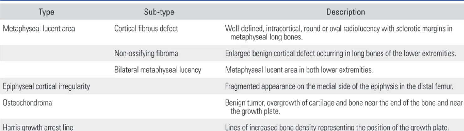
Table 1. Description of Incidental Findings
Type Sub-type Description
Metaphyseal lucent area Cortical fibrous defect Well-defined, intracortical, round or oval radiolucency with sclerotic margins in metaphyseal long bones.
Non-ossifying fibroma Enlarged benign cortical defect occurring in long bones of the lower extremities.
Bilateral metaphyseal lucency Metaphyseal lucent area in both lower extremities.
Epiphyseal cortical irregularity Fragmented appearance on the medial side of the epiphysis in the distal femur.
Osteochondroma Benign tumor, overgrowth of cartilage and bone near the end of the bone and near
the growth plate.
Harris growth arrest line Lines of increased bone density representing the position of the growth plate.
cal, round or oval radiolucency with sclerotic margins in the metaphyseal long bone (Fig. 1B).15,16) Bilateral metaph- yseal lucency was defined as the metaphyseal lucent area in both lower extremities (Fig. 1C). An epiphyseal cortical irregularity was defined as a fragmented appearance at the medial side of the epiphysis in the distal femur (Fig. 2). An osteochondroma was defined as an overgrowth of cartilage and bone near the end of the bone in close proximity to the growth plate (Fig. 3).17) And a Harris growth arrest line was defined as lines of increased bone density representing the position of the growth plate (Fig. 4).16,18)
Abnormal findings were defined as findings directly
related to the patient’s symptoms and diagnosis, subse- quently needing medical or surgical treatment. These in- cluded fractures, osteochondral lesions, tumors, infections, metabolic disorders and skeletal dysplasia. Additional studies such as magnetic resonance imaging (MRI) or computed tomography (CT) were performed to confirm the diagnosis if it was uncertain by using X-ray only.
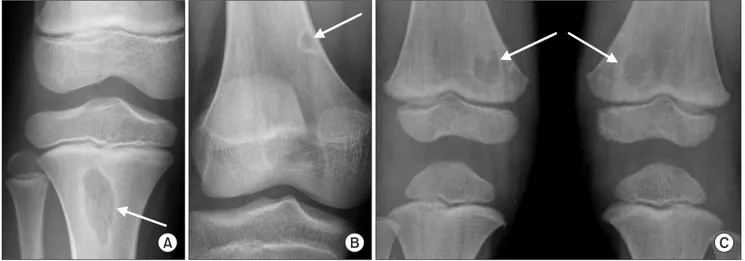
Interobserver Reliability and Review of Radiographs After building of a consensus, three orthopedic surgeons (KHS, TGK, and SGS), with 8, 7, and 4 years of orthopedic experience assessed the interobserver reliability of radio- Fig. 1. Metaphyseal lucent area. (A) The non-ossifying fibroma is an enlarged benign cortical defect occurring in long bone of the lower extremity. (B) The cortical fibrous defect is a well-defined, intracortical, less than 2 cm round or oval radiolucency with sclerotic margins in the metaphyseal long bone. (C) Bilateral metaphyseal lucency refers to the presence of metaphyseal lucent areas in both lower extremities.
Fig. 2. Epiphyseal cortical irregularity showing a fragmented appearance on the medial side of the epiphysis in the distal femur.
Fig. 3. Osteochondroma is a benign tumor characterized by an overgrowth of cartilage and bone near the end of the bone and near the growth plate.
graphic findings. Prior sample size estimation by precision analysis indicated an assessment of 36 knee radiographs as a minimum. Three examiners independently assessed the radiographic findings without knowledge of the patients’
clinical information and the other examiners’ measure- ments. All measurements were collected by a research as- sistant (HMK), who did not otherwise participate in the study.
All radiographic studies were assessed by two ex- aminers (SGS and KHS) with 4 and 8 years of orthopedic experience. AP radiographs of both knees in standing or supine positions were reviewed to search for incidental findings. If there was a controversy, 3 authors (SGS, KHS, and MSP) had a consensus process until agreement was reached. Each radiograph was classified into incidental finding, normal finding or abnormal finding related to the diagnosis and the type of abnormal findings were documented. Periodic radiographs and patients’ medi- cal records were followed for radiographs with incidental findings.
Statistical Analysis
Prior precision analysis was performed to identify the minimal sample size required for reliability testing. Intra- class correlation coefficients (ICCs) were used for reliabil- ity testing at a target value of 0.80 and a 95% confidence interval (CI) width of 0.2 for three observers. The minimal sample size needed was 36 by Bonett’s approximation.19) ICCs and their 95% CIs were used to summarize the in- terobserver reliability of the radiographic measurements
and were calculated in a setting using a two-way random effect model assuming a single measurement and absolute agreement.20) An ICC value of 1 indicated perfect reliabil- ity and an ICC greater than 0.8 indicated excellent reliabil- ity.
Descriptive statistics were used to provide demo- graphic data and radiographic findings. Chi-square tests were performed to compare the prevalence of findings by age. Statistical analyses were conducted using SPSS ver.
18.0 (SPSS Inc., Chicago, IL, USA) and the level of signifi- cance was set at p < 0.05.
RESULTS
A total of 1,572 patients were reviewed with knee radio- graphs between 2010 and 2011. Ten patients were excluded due to inadequate knee radiographs. Thus, a total of 1,562 consecutive patients were enrolled in this study. There were 975 males and 587 females with mean age of 10.2 years (range, 1 month to 18 years) at the time of outpatient clinic visit. Three hundred and seventy-seven patients were under 5 years of age, 445 patients were between 5 and 10 years, and 740 patients were over 10 years old. Of 1,562 patients, 355 incidental findings were identified in 335 pa- tients (21.4%). Abnormal findings were seen in 98 radio- graphs (6.3%) and normal findings in 1,129 radiographs (72.3%) (Table 2). Radiographic assessments showed an excellent reliability (ICC, 0.811; 95% CI, 0.700 to 0.891) in terms of the interobserver reliability for overall incidental findings.
Incidental Findings
Of 355 incidental findings, there were 131 patients (8.4%) with metaphyseal lucent area, 105 patients (6.7%) with epiphyseal cortical irregularity, 75 patients (4.8%) with Harris growth arrest line, and 44 patients (2.8%) with os- teochondroma (Table 3).
In patients under 5 years of age we identified 173
Fig. 4. The Harris growth arrest line is an increased bone density representing the position of the growth plate.
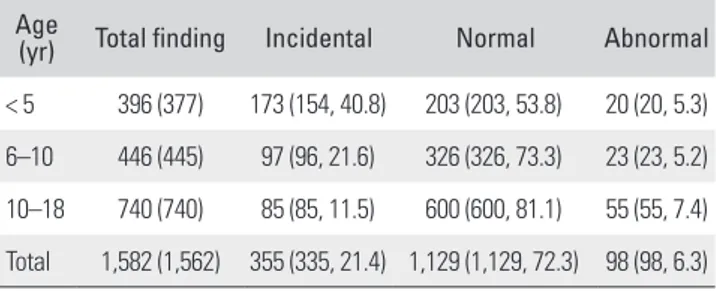
Table 2. Findings from Knee Radiographs
Age (yr) Total finding Incidental Normal Abnormal
< 5 396 (377) 173 (154, 40.8) 203 (203, 53.8) 20 (20, 5.3) 6–10 446 (445) 97 (96, 21.6) 326 (326, 73.3) 23 (23, 5.2) 10–18 740 (740) 85 (85, 11.5) 600 (600, 81.1) 55 (55, 7.4) Total 1,582 (1,562) 355 (335, 21.4) 1,129 (1,129, 72.3) 98 (98, 6.3) Values are presented as number of findings (patients, %).
incidental findings (40.8%), including 99 cases (26.3%) of epiphyseal cortical irregularity. In patients between 5 and 10 years of age, we found 97 incidental findings (21.6%) and 47 cases (10.6%) with metaphyseal lucent area, while we found 85 incidental findings (11.5%) with 30 cases of osteochondroma (4.1%) in patients over 10 years of age (Table 3).
There was a higher prevalence of epiphyseal corti- cal irregularity in patients with young age (p < 0.001).
Also the prevalence of metaphyseal lucent area and Harris growth arrest line was higher in patients with young age (p
= 0.001, p < 0.001). However, osteochondroma was found to have a higher prevalence in patients with higher age (p
= 0.004) (Table 3).
Abnormal Findings Related to the Diagnosis
Ninety-eight abnormal findings (6.3%) were identified in knee radiographs. Findings of tumors were seen in 41 knee radiographs, fractures in 31, osteochondral lesions in 10, skeletal dysplasias in 6, infections in 5 and metabolic disorders in 5.
DISCUSSION
In our cohort, we identified incidental findings in knee radiographs of 21% of patients. A metaphyseal lucent area was the most commonly seen incidental finding, followed by epiphyseal cortical irregularity, Harris growth arrest line and osteochondroma. An epiphyseal cortical irregu- larity, metaphyseal lucent area as well as a Harris growth arrest line was found with higher prevalence in young age while osteochondroma was more prevalent in older age.
Our study has its limitation. The reading of the radiographs may depend on the examiner’s decision al- though we had several consensus meetings. There have been some studies reported on malignant tumors mim- icking benign lesions, such as a parosteal osteosarcoma
mimicking an osteochondroma and an osteogenic sar- coma mimicking a non-ossifying fibroma.21,22) Therefore, additional studies such as bone scan, MRI or biopsy may be needed to confirm the diagnosis in selected cases with an uncertain diagnosis on radiography.
Knee pain is one of the most common complaints in an orthopedic outpatient clinic1,2) and radiography of the knee is the most simple and widely used test in those patients. However, knee radiography findings in children and adolescents have quite different characteristics com- pared to adults. Those characteristics include the physi- ologic bowing, open growth plate and an absent or partial appearance of the ossification center. In addition to these normal characteristics, we studied incidental findings of the knee in children and adolescents. The incidental find- ings were unexpectedly high in knee radiography. Under- standing the prevalence and results of incidental findings in knee radiography may be helpful in the decision mak- ing for pediatric orthopedic patients. Also age is a very im- portant factor in the clinical history of a patient with knee problems as certain benign tumors are age dependent.23,24)
To date, there has been no study investigating on the incidence of epiphyseal cortical irregularity in children.
In our study, an epiphyseal cortical irregularity occurred in 105 out of 1,562 patients. A cortical irregularity was observed with a much higher proportion than in other age groups in patients under 5 years of age. An epiphyseal cortical irregularity usually appeared in patients under 4 years of age and there was a tendency that it is to be found at an earlier age in girls than in boys. Such findings tended to disappear on follow-up radiographs as the patients grew older. It may be inferred due to the fact that a cortical ir- regularity is related to a delayed ossification. Therefore, an epiphyseal cortical irregularity is thought to be an in- cidental finding which needs no further evaluation and treatment. Some infants may show a minimal beaking or fragmentation at the edge of the metaphyses. This finding Table 3. Common Incidental Findings According to Age
Incidental finding Total Age (yr)
p-value
< 5 6–10 10–18
Metaphyseal lucent area 131 (8.4) 41 (10.9) 47 (10.6) 43 (5.8) 0.001
Epiphyseal cortical irregularity 105 (6.7) 99 (26.3) 6 (1.3) 0 < 0.001
Harris growth arrest line 75 (4.8) 32 (8.5) 31 (7.0) 12 (1.6) < 0.001
Osteochondroma 44 (2.8) 1 (0.3) 13 (2.9) 30 (4.1) 0.004
Values are presented as number (%).
is considered as a normal variation.12) It appears as spurs, discrete linear mineralized sections at the periphery of the physis or as a beak on an osseous projection.25)
The metaphyseal lucent area is classified among non-ossifying fibromas, cortical desmoids and bicorti- cal multiple lucencies.11) A cortical desmoid is known as a benign, fibrous or fibro-osseous lesion9,10) and has been reported to most commonly occur between the age of 10 and 15 years.7,8) Its prevalence is reported to be 11.5% for males and 3.6% for females.6) However, in our study, the prevalence of cortical desmoid was 9.1% for males and 7.2% for females. The prevalence in patients under 5 years of age and between 5 and 10 years of age was higher than in patients over 10 years of age. Bilateral metaphyseal lu- cency, which symmetrically involves both extremities, is thought to be an incidental finding that needs no further evaluation and treatment. However, further studies such as bone scan and MRI can be performed if needed in case of cortical desmoid and non-ossifying fibroma to rule out malignancy.
The Harris growth arrest line has been well de- scribed in the literature and is classically described as radiographic dense lines found in long bones.16,18,26) Major causes for physeal injury and temporary growth arrest are a systemic illness, nutritional deficit, physeal injury and a fracture in the growing period.16,18,23,27,28) A previous study reported that the Harris line was found in 7% of boys and 3% of girls.29) Another study showed the highest frequency of Harris line in boys between 1 and 4 years of age, with another peak at 5 years, while the highest frequency of the lines in girls was observed between 1 and 3.5 years of age.30) Our results showed an overall prevalence of Har- ris growth arrest line with 4.8% for both gender. It was more common in younger patients. This is because Har-
ris growth arrest line is obvious around the growth plate.
At higher age, we found it far from the growth plate and tapered. Considering our hospital as a tertiary institution, the prevalence of Harris growth arrest line may have had a higher appearance than in the general population.
Osteochondroma is the most common benign bony lesion. It usually appears as a single lesion, but it can also be part of osteochondromatosis (multiple hereditary ex- ostoses).16,17) A previous study reported the frequency of a single osteochondroma was 2% as an incidental finding during the course of a radiographic examination for other lesions.4) In our study the prevalence was 2.8%, (by gender 2.7% for boys and 3.1% for girls) which is in accordance with the results of previous studies. Osteochondroma tended to be more prevalent in patients over 10 years of age. In summary, we were able to demonstrate the prevalence of different types of incidental findings in knee radiographs of children and adolescents, such as metaphy- seal lucent area, epiphyseal cortical irregularity, Harris growth arrest line and osteochondroma. As a result, the authors recommend to consider those incidental findings if unfamiliar findings appear in knee radiographs of chil- dren and adolescents.
CONFLICT OF INTEREST
No potential conflict of interest relevant to this article was reported.
ACKNOWLEDGEMENTS
We thank Mi Sun Ryu, BS and Hyun Mi Kim, BS for data collection.
REFERENCES
Paediatr Oncol. 2011;32(4):187-91.
5. Walden MJ, Murphey MD, Vidal JA. Incidental enchondro- mas of the knee. AJR Am J Roentgenol. 2008;190(6):1611-5.
6. Simon H. Medial distal metaphyseal femoral irregularity in children. Radiology. 1968;90(2):258-60.
7. Vieira RL, Bencardino JT, Rosenberg ZS, Nomikos G. MRI features of cortical desmoid in acute knee trauma. AJR Am J Roentgenol. 2011;196(2):424-8.
8. Johnson LC, Genner BA III, Engh CA, Brown RH. Cortical desmoids. J Bone Joint Surg Am. 1968;50(4):828-9.
9. Bufkin WJ. The avulsive cortical irregularity. Am J Roent- 1. Perquin CW, Hazebroek-Kampschreur AA, Hunfeld JA, et
al. Pain in children and adolescents: a common experience.
Pain. 2000;87(1):51-8.
2. Goodman JE, McGrath PJ. The epidemiology of pain in children and adolescents: a review. Pain. 1991;46(3):247-64.
3. Baena-Ocampo Ldel C, Ramirez-Perez E, Linares-Gonzalez LM, Delgado-Chavez R. Epidemiology of bone tumors in Mexico City: retrospective clinicopathologic study of 566 patients at a referral institution. Ann Diagn Pathol. 2009;
13(1):16-21.
4. Solooki S, Vosoughi AR, Masoomi V. Epidemiology of mus- culoskeletal tumors in Shiraz, south of Iran. Indian J Med
genol Radium Ther Nucl Med. 1971;112(3):487-92.
10. Sklar DH, Phillips JJ, Lachman RS. Case report 683: distal metaphyseal femoral defect (cortical desmoid; distal femoral cortical irregularity). Skeletal Radiol. 1991;20(5):394-6.
11. Betsy M, Kupersmith LM, Springfield DS. Metaphyseal fi- brous defects. J Am Acad Orthop Surg. 2004;12(2):89-95.
12. Swischuk LE. Imaging of the new bone, infant and young child. 5th ed. Philadelphia, PA: Lippincott Williams &
Wikins; 2003. 725-34.
13. Verdonk PC, Verstraete K, Verdonk R. Distal femoral corti- cal irregularity in a 13-year old boy: a case report. Acta Or- thop Belg. 2003;69(4):377-81.
14. Wolf SM, Paradise J, Caga-anan C. The law of incidental findings in human subjects research: establishing research- ers' duties. J Law Med Ethics. 2008;36(2):361-83.
15. Kumar R, Madewell JE, Lindell MM, Swischuk LE. Fibrous lesions of bones. Radiographics. 1990;10(2):237-56.
16. Resnick D, Kransdorf MJ. Bone and joint imaging. 3rd ed.
Philadelphia, PA: Elsevier Saunders; 2005.
17. Lange RH, Lange TA, Rao BK. Correlative radiographic, scintigraphic, and histological evaluation of exostoses. J Bone Joint Surg Am. 1984;66(9):1454-9.
18. Ecklund K, Jaramillo D. Imaging of growth disturbance in children. Radiol Clin North Am. 2001;39(4):823-41.
19. Bonett DG. Sample size requirements for estimating intra- class correlations with desired precision. Stat Med. 2002;
21(9):1331-5.
20. Lee KM, Lee J, Chung CY, et al. Pitfalls and important issues in testing reliability using intraclass correlation coefficients in orthopaedic research. Clin Orthop Surg. 2012;4(2):149- 55.
21. Choong PF, Pritchard DJ, Rock MG, Sim FH, McLeod RA,
Unni KK. Low grade central osteogenic sarcoma: a long- term followup of 20 patients. Clin Orthop Relat Res. 1996;
(322):198-206.
22. Papathanassiou ZG, Alberghini M, Thiesse P, et al. Parosteal osteosarcoma mimicking osteochondroma: a radio-histo- logic approach on two cases. Clin Sarcoma Res. 2011;1(1):2.
23. Kristy LW, Mary IO. Benign cartilage tumors. In: Herbert SS, ed. Orthopaedic knowledge update: musculoskeletal tu- mors 2. Rosemont, IL: American Academy of Orthopaedic Surgeons; 2007. 103-20.
24. Subhas N, Bui KL, Sundaram M, Ilaslan H, Recht MP. In- cidental tumor and tumor-like lesions around the knee.
Semin Musculoskelet Radiol. 2009;13(4):353-70.
25. Kleinman PK, Belanger PL, Karellas A, Spevak MR. Normal metaphyseal radiologic variants not to be confused with findings of infant abuse. AJR Am J Roentgenol. 1991;156(4):
781-3.
26. Tang CW, Kay RM, Skaggs DL. Growth arrest of the distal radius following a metaphyseal fracture: case report and re- view of the literature. J Pediatr Orthop B. 2002;11(1):89-92.
27. Carey J, Spence L, Blickman H, Eustace S. MRI of pediatric growth plate injury: correlation with plain film radiographs and clinical outcome. Skeletal Radiol. 1998;27(5):250-5.
28. Havranek P, Lizler J. Magnetic resonance imaging in the evaluation of partial growth arrest after physeal injuries in children. J Bone Joint Surg Am. 1991;73(8):1234-41.
29. Sontag LW, Harris LM. Evidences of disturbed prenatal and neonatal growth in bones of infants aged one month. II.
contributing factors. Am J Dis Child. 1938;56(6):1248-55.
30. Gindhart PS. The frequency of appearance of transverse lines in the tibia in relation to childhood illnesses. Am J Phys Anthropol. 1969;31(1):17-22.