*Corresponding Author. Tel: 82-42-860-4225; Fax: 82-42-860-4593; E-mail: ipchoi@kribb.re.kr This work was supported by a grant from GRL project, KICOS, Korea.
Expression of Gpnmb in NK Cell Development from Hematopoietic Stem Cells
Nara Shin, Jiwon Lee, Jiwon Lee, Mira Jeong, Mi Sun Kim, Suk Hyung Lee, Suk Ran Yoon, Jin Woong Chung, Tae-Don Kim and Inpyo Choi*
Stem Cell Research Center, Korea Research Institute of Bioscience and Biotechnology, Yusong, Daejeon, Korea
Background: Molecular mechanisms of natural killer (NK) cell development from hematopoietic stem cells (HSCs) have not been clearly elucidated, although the roles of some genes in NK cell development have been reported previously.
Thus, searching for molecules and genes related NK cell de- velopmental stage is important to understand the molecular events of NK cell development. Methods: From our previous SAGE data-base, Gpnmb (Glycoprotein non-metastatic mel- anoma protein B) was selected for further analysis. We con- firmed the level of mRNA and protein of Gpnmb through RT-PCR, quantitative PCR, and FACS analysis. Then we per- formed cell-based ELISA and FACS analysis, to know wheth- er there are some molecules which can bind to Gpnmb. Using neutralizing antibody, we blocked the interaction between NK cells and OP9 cells, and checked IFN-γ production by ELISA kit. Results: Gpnmb expression was elevated during in vitro developmental stage and bound to OP9 cells, but not to NK precursor cells. In addition, we confirmed that the lev- els of Gpnmb were increased at NK precursor stage in vivo.
We confirmed syndecan4 as a candidate of Gpnmb's binding molecule. When the interaction between NK cells and OP9 cells were inhibited in vitro, IFN-γ production from NK cells were reduced. Conclusion: Based on these observations, it is concluded that Gpnmb has a potential role in NK cell de- velopment from HSCs.
[Immune Network 2008;8(2):53-58]
INTRODUCTION
Natural killer (NK) cells are derived from hematopoietic stem cells (HSCs) which can be differentiated into several hema-
topoietic lineages. It has been reported that NK cells are mainly matured in bone marrow as they express several re- ceptors (1). These receptors endow NK cells to advance into the late stages of maturation. Although the outline of NK cell development from hematopoietic stem cells was reported pre- viously (2,3), it is far from full understanding of NK cell development. Thus, the identification of other receptors re- lated differentiation of NK cells is a critical issue to under- stand NK cell development.
Based on our published SAGE database (4), we selected some genes that may have potential to affect the differ- entiation of NK cells. Among these candidates, Gpnmb (glycoprotein non-metastatic melanoma protein B) showed a pattern of elevated expression in precursor NK (pNK) stage, compared to HSCs. Gpnmb is a typical trans-membrane gly- coprotein composed with 574 amino acids. It is found on the plasma membrane and membranes of the endosome com- partments. Gpnmb contains heparin binding motif and in- tegrin recognition RGD domain in its extracellular region, while in the cytoplasmic region Gpnmb contains ITAM (Immunoreceptor tyrosine-based activation motif) and lysoso- mal targeting motif. It was previously reported that it is in- volved in osteoblast differentiation and participates in mela- noma subsistence (5,6). In breast cancer, Gpnmb has tumor suppressor functions or metastatic functions (7). Gpnmb is al- so known as osteoactivin, dendritic cell-associated heparin sulfate proteoglycan-integrin ligand, and hematopoietic growth factor inducible neurokinin-1 type (7).
In this report, we evaluated the potential role of Gpnmb in NK cell developmental from HSCs. Expression of Gpnmb was increased during developmental stage of NK cells and
maintained until they got full maturation. During this stage, pNK cells have chance to interact with stromal cells for the full maturation (8). Thus, we checked the roles of Gpnmb in interacting between these cells. Finally we identified that Gpnmb induces NK cells differentiation in aspect of IFN-γ production through in vitro co-culture system of NK cells and stromal cells.
MATERIALS AND METHODS Mice
C57BL/6 mice were purchased from DaeHan Bio-Link (Korea). Whenever we have the necessity of fostering mice, we housed mice under specific pathogen-free (SPF) con- ditions as recommended by institutional regulation, and used between 6 and 10 weeks of age.
RT PCR and Quantitative real time PCR
To verify the expression of NK cell associated genes from each developmental stage, we conducted isolation of mRNA classically using TRIzol (Invitrogen). After quantitative nor- malization for each gene using β-actin expression, for Gpnmb detection, we accomplished RT-PCR using 3' primer- TCCCATCTCGAAGGTGAAAG and 5' primer-CAATTGTGATG- GTGGCTCTG. To detection of SD1, SD2, SD3, and SD4, we designed 3' primer- GAAGTGCTGGGAGGTGTCAT and 5' pri- mer- GCTTGGTGGGTTTCTGGTAG, 3' primer- CCTCAGCAA- ACCCAAAGATG and 5' primer- TAACACCAGTCCGCAACAAG, 3' primer- CTGGCTCCTCTAACCATCCA, and 5' primer- ACTGTGTGGGCTTGTGACTG, and 3' primer- ATCCTCTTTG- CCGTTTTCCT and 5' primer- CACAACCCTCCACTCCTCTC, respectively. Levels of mRNA expression were normalized rel- ative to β-actin gene expression levels.
Antibodies and Flow cytometric Analysis
All antibodies for flow cytometric analysis were purchased from Becton Dickinson (BD) and PharMingen (SanDiego, CA). Mouse IgG Fc conjugated recombinant Gpnmb, and an- ti-Gpnmb antibody were purchased from R&D systems and used as manufacturer's guide line and reported (9). Cells were stained with the indicated antibodies in a staining buffer for 15min in 4oC, washed away, and analyzed by flow cy- tometry (FACScalibur: BD Biosciences) using CellQuest Pro (BD Biosciences) program.
in vitro differentiation of NK cells from HSCs
NK cell differentiation from HSCs was performed as described previously (3). In brief, after removing erythrocytes in extract from mouse bone marrow by ACK solution, c-Kit+, Lin− (B cells, T/NK cells, granulocytes, monocytes, NK/NKT cells, and erythrocytes were depleted by B220, CD2, Gr-1, CD11b, NK1.1, and TER-119, respectively) were purified by MACS Cell Separation kit (Miltenyi Biotec) according to the manu- facturer's protocol. Antibodies used for MACS purification were purchased from Becton-Dickinson (BD) and Phar- Mingen (San Diego, CA).
Co-culture with OP9
To perform a co-culture with OP9 stromal cells, OP9 cells were plated at a density of 5×10³ cells/ml in a 24-well plate with alpha-MEM (Invitrogen) supplemented with 20% fetal bovine serum. Attached OP9 cells were irradiated with 2000 rads for 24 h prior to the procedure. After removing al- pha-MEM medium, pNK cells which were cultured for 7 days from HSC stage were plate onto OP9 cells at a density of 2×10⁶ cells/ml in the presence IL-15 (50 ng/ml), and in- cubated for another 5 days.
Cell-based ELISA
OP9 cells were seeded at near 70% confluence in a 96-well plate containing alpha-MEM supplemented with 20% fetal bo- vine serum on 1 day prior to cell fixation. After removing residual liquid, cells were fixed by 3.7% formaldehyde diluted in PBS for 10 min at room temperature, washed 3 times with PBST, and blocked with 10% FBS diluted PBS for 1 h at room temperature. After washing, recombinant Gpnmb and control IgG were added into 96-well for 2 h at room temperature.
Cells were washed 3 times, added with anti-mouse HRP as a secondary antibody at room temperature for 1h, washed 3 times again, added with substrate and stop solution, and then scanned at 450 nm.
Mouse IFN-γ ELISA
To detect IFN-γ, we collected the supernatants of NK cell culture on 12 day after, diluted, and used mouse IFN-γ ELISA kit from R&D systems. Then we followed manufactur- er's guideline.
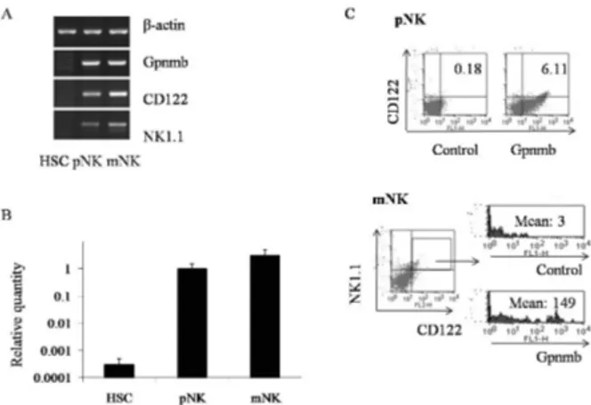
Figure 1. Gpnmb expression was increased during NK differentiation.
(A) HSCs, pNK, and mNK cells were collected during in vitro NK differentiation, and isolated total RNAs. The expression of Gpnmb, CD122, and NK1.1 were analyzed by RT-PCR. The results shown are representative of at least three individual experiments with similar results. (B) Using cDNA from Fig. 1A, the expressions of Gpnmb were examined by real time quantitative PCR. A vertical axis shows the relative quantity. Data are representative of three independent experiments. (C) During in vitro culture, pNK and mNK cells were
Figure 2. Gpnmb expression was elevated at pNK stage in vivo.
Single cell suspensions of bone marrow were stained with several markers. G1, G2, G3, and G4 represented lineage-/c-kit+, lineage-/
CD122+, CD122+/NK1.1-, and CD122+/NK1.1+, respectively. The
RESULTS
Gpnmb expression was increased during in vitro NK cell differentiation
To confirm the expression pattern of Gpnmb during NK cell differentiation, we checked mRNA and protein level of Gpnmb in in vitro culture system (Fig. 1). HSCs isolated from wild type mice were cultured in RPMI media containing SCF, Flt3L, and IL-7 for 7 days (pNK). After cultivation, we changed RPMI media containing IL-15 and cultured for anoth- er 5 days (mature NK, mNK). As shown in Fig. 1A, the ex- pression of Gpnmb was not detected at HSC, but it was in- creased from 7 days of cultivation. To know relative quantity of Gpnmb mRNA, we performed real-time PCR (Fig. 1B). The dramatic change of Gpnmb mRNA suggests that Gpnmb may have some roles in NK cell differentiation. To know the ex- pression level of Gpnmb protein in vitro, we checked its ex- pression in the cells at each in vitro stage. On the 7 day, when pNK cells express CD122 (10), Gpnmb expression was increased in CD122 positive cells (Fig. 1C, top). On the 12 day when mNK cells express both CD122 and NK1.1, more Gpnmb expression was observed in CD122 and NK1.1 dou- ble positive populations (Fig. 1C, bottom). Overall, these re- sults demonstrate that Gpnmb expression was increased dur-
ing in vitro NK cell developmental stage.
Level of Gpnmb expression was elevated at pNK stage in vivo
To confirm whether each in vivo developmental stage of NK cells has the same expression pattern with in vitro devel- opmental stage, we checked the expression of Gpnmb in bone marrow cells. After removing erythrocytes, lymphocytes from bone marrow were stained with several markers indicat- ing each developmental status. HSC represented by c-kit pos- itive and lineage negative showed that mean value of Gpnmb was 12.6 (Fig. 2, G1). In contrast to HSC, pNK cells stained as CD122 positive and lineage/NK1.1 negative displayed higher mean value of Gpnmb (Fig. 2, G2 and G3). CD122 and NK1.1 double positive mNK cells from bone marrow showed the reduced expression of Gpnmb (Fig. 2, G4), sim- ilar to human mNK cells, which CD56 positive or CD94 pos- itive cells showed the low expression level of Gpnmb in vivo (data not shown). It seems that Gpnmb may function mainly at pNK stage rather than mNK stage at least in vivo system.
Meanwhile, comparing Gpnmb expression in vitro or in vivo, mNK cells have different expression pattern of Gpnmb. This
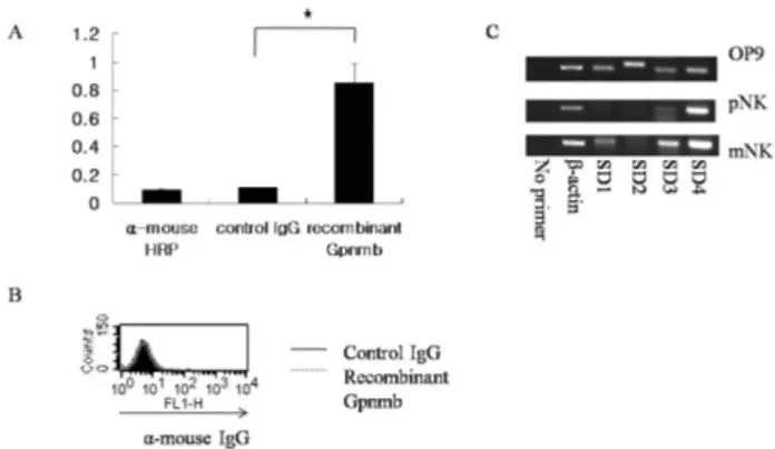
Figure 3. Gpnmb binds to the ligand of stromal cells. (A) OP9 cells were plated at about 70% confluence. After one day cultivation, cell-based ELISA was performed. For conjugation, control IgG and recombinant Gpnmb protein were used. A vertical axis shows optical density value. The values represent the means of 3 or 4 individual experiments. *p<0.05. (B) pNK cells were collected and stained with control IgG and recombinant Gpnmb to know whether Gpnmb is involved in binding of NK cells by themselves. As a secondary antibody, anti-mouse IgG conjugated FITC was used. The results represent at least three individual experiments. (C) OP9, pNK cells, and mNK cells were collected, and total RNAs were isolated. As a negative control, no primer was used, and the expressions of SD family were examined.
Figure 4. Neutralizing anti-Gpnmb antibody induced the reduction of IFN-γ production. After irradiation of OP9 cells, pNK cells were co-cultured with OP9 cells or OP9 cells with Fc Blocker (FcBl) (1ug/ml) alone or both FcBl and anti-Gpnmb antibody (1.5 ug/ml).
On 12 day, supernatants were collected and essayed for mouse IFN- γ ELISA. A vertical axis shows IFN-γ concentration (pg/ml). The results shown are mean values which are representative of at least three individual experiments. *p<0.01, **p<0.1.
may be due to the different microenvironment conditions of in vitro and in vivo systems or uneven maturation of NK cells in in vitro culture system. Collectively, Gpnmb may be related to CD122 expression at pNK stage where NK cell develop- ment is accelerated by expressing CD122 molecule.
Gpnmb binds ligand of stromal cells
During differentiation, it is already reported that bone marrow environment is an essential factor for NK cell maturation (11).
To know the effect of Gpnmb expression on NK cell differ- entiation, we tested whether Gpnmb can bind to bone mar- row stromal cells. For this purpose, we checked interaction of Gpnmb with OP9 stromal cells using recombinant Gpnmb conjugated Fc region. Recombinant Gpnmb conjugated Fc re- gion or control IgG were added on plated OP9 cells, and their optical density value were analyzed by cell-based ELISA using mouse HRP (Fig. 3A). Compared to control IgG or without Gpnmb, recombinant Gpnmb showed high level of optical density value, indicating that Gpnmb can bind to unidentified component of OP9 cells. Meanwhile, we also checked the in- teraction of pNK cells by themselves, because it is possible that pNK cell might be a binding partner each other.
However, pNK cells showed unchanged levels of anti-mouse IgG in the presence of recombinant Gpnmb or control IgG
(Fig. 3B). These results show that pNK cell interacts with stro- mal cell, but not with pNK cell itself.
It has been reported that Gpnmb binds to heparin heparan sulfate proteoglycan (12). In addition, one of the binding partner of Gpnmb is syndecan4 (SD4) on activated T cell which functions as a negative regulator on T cell activation, although SD4 on B cell did not bind to Gpnmb (12,13). Thus, we determined the expression of SD4 and other SD family in OP9 cells to determine a candidate which enables to bind Gpnmb on cell surface (Fig. 3C). As shown in Fig. 3C, OP9 cells showed SD1, SD2, SD3, and SD4 mRNA expression. In case of pNK cells, they expressed SD4 mRNA, but not SD1, SD2, and SD3 mRNA. However, mNK cells showed elevated SD family expressions. A previous report suggested that there is discordance between expression and binding activity, be- cause SD4 can form diverse heparan sulfate structures (13).
Considering all of these results, it seems that Gpnmb on NK cells binds to SD4 on OP9 cells directly, but not to SD4 on NK cells which might have different heparin sulfate structure at pNK stage. More study will be required to resolve this issue.
Gpnmb is involved in IFN-γ production
To know the meaning of Gpnmb binding to stromal cells dur-
ing NK cell differentiation, we test the inhibition effects of anti- Gpnmb antibody on IFN-γ production which is a key molecule for NK cell function and development (14). Using neutralizing anti-Gpnmb antibody, we found that inhibition of interaction between pNK cells and OP9 cells reduced IFN-γ production (Fig. 4). Compared with IFN-γ production of other control groups, NK cells treated with anti-Gpnmb anti- body (lane 4) produced low level of IFN-γ, about 50%
reduction. These results indicate that Gpnmb has an im- portant role in IFN-γ production and NK cell development.
DISCUSSION
Our results demonstrate that Gpnmb elevated at pNK stage has a positive role in NK cell differentiation. Its expression was increased during developmental stage, bound to OP9 cells through unfound mechanism, and blocking interaction with OP9 cells by anti- Gpnmb antibody reduced IFN-γ production.
Gpnmb has a heparin biding motif (15), and SD family and glypican family are major candidates of cell surface-heparan sulfate (16). Thus, Gpnmb has a possibility to bind to SD family and glypican family. As it was reported that SD4 is a binding molecule of Gpnmb (13), thus we examined SD family as binding candidates including SD4. Because SD4 is expressed constructively in endothelial cells and B cells (16,17), other cells might able to bind to Gpnmb. Based on expression pattern of SD4 and binding pattern, it seems that SD4 is a candidate for Gpnmb's binding partner in this case.
In blocking experiments between OP9 cells and Gpnmb of NK cells, we scrutinized several developmental markers such as CD122, NK1.1, DX5, NKG2D, and IFN-γ. Although there were no consistent changes in all markers, IFN-γ production was remarkably reduced. IFN-γ production has been consid- ered as an essential marker in NK cell development (14).
Furthermore, IFN-γ production has been used as a key mol- ecule to monitoring NK cell activity (18,19). It is very interest- ing that Gpnmb can modulate IFN-γ production during NK cell development. It seems that Gpnmb regulates NK cell de- velopment by controlling NK cell and stromal cell interaction via IFN-γ production. The direct effects of Gpnmb on NK cell development in connection with stromal cell will be required.
In conclusion, the expression of Gpnmb was increased as NK cells were matured. It bound to stromal cells directly
production. Taken together, Gpnmb has a potential value as a positive regulator in NK cell development.
REFERENCES
1. Freud AG and Caligiuri MA: Human natural killer cell development. Immunol Rev 214;56-72, 2006
2. Mrozek E, Anderson P, Caligiuri MA: Role of interleukin-15 in the development of human CD56+ natural killer cells from CD34+ hematopoietic progenitor cells. Blood 87;2632-2640, 1996
3. Williams NS, Klem J, Puzanov IJ, Sivakumar PV, Bennett M, Kumar V: Differentiation of NK1.1+, Ly49+ NK cells from flt3+ multipotent marrow progenitor cells. J Immunol 163;2648-2656, 1999
4. Kang HS, Kim EM, Lee S, Yoon SR, Kawamura T, Lee YC, Kim S, Myung PK, Wang SM, Choi I: Stage-dependent gene expression profiles during natural killer cell development.
Genomics 86;551-565, 2005
5. Rich JN, Shi Q, Hjelmeland M, Cummings TJ, Kuan CT, Bigner DD, Counter CM, Wang XF: Bone-related genes ex- pressed in advanced malignancies induce invasion and metastasis in a genetically defined human cancer model.
J Biol Chem 278;15951-15957, 2003
6. Selim AA, Abdelmagid SM, Kanaan RA, Smock SL, Owen TA, Popoff SN, Safadi FF: Anti-osteoactivin antibody in- hibits osteoblast differentiation and function in vitro. Crit Rev Eukaryot Gene Expr 13;265-275, 2003
7. Rose AA, Siegel PM: Osteoactivin/HGFIN: is it a tumor sup- pressor or mediator of metastasis in breast cancer? Breast Cancer Res 9;403, 2007
8. Ogasawara K, Hida S, Azimi N, Tagaya Y, Sato T, Yokochi-Fukuda T, Waldmann TA, Taniguchi T, Taki S:
Requirement for IRF-1 in the microenvironment supporting development of natural killer cells. Nature 391;700-703, 1998
9. Nakamura A, Ishii A, Ohata C, Komurasaki T: Early in- duction of osteoactivin expression in rat renal tubular epi- thelial cells after unilateral ureteral obstruction. Exp Toxicol Pathol 59;53-59, 2007
10. Rosmaraki EE, Douagi I, Roth C, Colucci F, Cumano A, Di Santo JP: Identification of committed NK cell progenitors in adult murine bone marrow. Eur J Immunol 31;1900- 1909, 2001
11. Roth C, Rothlin C, Riou S, Raulet DH, Lemke G: Stromal- cell regulation of natural killer cell differentiation. J Mol Med 85;1047-1056, 2007
12. Chung JS, Sato K, Dougherty II, Cruz PD Jr, Ariizumi K:
DC-HIL is a negative regulator of T lymphocyte activation.
Blood 109;4320-4327, 2007
13. Chung JS, Dougherty I, Cruz PD Jr, Ariizumi K: Syndecan-4 mediates the coinhibitory function of DC-HIL on T cell activation. J Immunol 179;5778-5784, 2007
14. Vosshenrich CA, Samson-Villeger SI, Di Santo JP: Distin- guishing features of developing natural killer cells. Curr Opin Immunol 17;151-158, 2005
cloning of a dendritic cell-associated transmembrane pro- tein, DC-HIL, that promotes RGD-dependent adhesion of endothelial cells through recognition of heparan sulfate proteoglycans. J Biol Chem 276;8125-8134, 2001 16. Bernfield M, Gotte M, Park PW, Reizes O, Fitzgerald ML,
Lincecum J, Zako M: Functions of cell surface heparan sul- fate proteoglycans. Annu Rev Biochem 68;729-777, 1999 17. Yamashita Y, Oritani K, Miyoshi EK, Wall R, Bernfield M
and Kincade PW: Syndecan-4 is expressed by B lineage lymphocytes and can transmit a signal for formation of den- dritic processes. J Immunol 162;5940-5948, 1999
18. Kim S, Poursine-Laurent J, Truscott SM, Lybarger L, Song YJ, Yang L, French AR, Sunwoo JB, Lemieux S, Hansen TH, Yokoyama WM: Licensing of natural killer cells by host ma- jor histocompatibility complex class I molecules. Nature 436;709-713, 2005
19. Samson SI, Richard O, Tavian M, Ranson T, Vosshenrich CA, Colucci F, Buer J, Grosveld F, Godin I, Di Santo JP:
GATA-3 promotes maturation, IFN-gamma production, and liver-specific homing of NK cells. Immunity 19;701-711, 2003