─ 249 ─
슬관절 관절 내・외에 동시 발생한 국소형 색소성 융모 결절성 활액막염
- 1례 보고 -
한양대학교 의과대학 구리병원 정형외과학교실
박 태 수・최 용 현
= 국문 초록=
슬관절 상 슬개낭 및 비복근 내측두에 동시에 발생한 국소형 색소성 융모 결절성 활액막염 1례에 대하여 관절경적 및 관혈적 절제술을 통하여 완전히 제거한 후 만족스러운 치료 결과를 얻었기에 보고하고자 한다.
색인 단어 : 슬관절, 색소성 융모 결절성 활액막염, 관절 내・외 병변
Corresponding Author : Tae-Soo Park, M.D.
Department of Orthopaedic Surgery, Kuri Hospital, Hanyang University College of Medicine
#249-1 Kyomoon-dong, Kuri 471-020, Korea
Tel : 031-560-2314, Fax : 031-557-8781, E-mail : parkts@hanyang.ac.kr
大韓膝關節學會誌・第 1 3卷・第2號 Volume 13, Number 2, December 2001
색소성 융모 결절성 활액막염은 관절 활액막, 점액 낭 및 건초에서 기시하여 증식을 일으키는 병인 불명 의 활액막 질환으로서 슬관절에서 가장 호발한다9 ). 슬관절에서 발견되는 색소성 융모 결절성 활액막염은 미만형(diffused form)과 국소형(localized form) 의 두 형태로 나타나며, 전자는 관절 활액막의 전반 에 걸쳐 여러 부위를 침범하는 반면, 후자는 결절이 나 자루형 종물 형태로 활액막의 일부에 국한되어 나 타나고 국소 활막 절제술 만으로도 높은 치유 결과를 보인다9 ).
저자들은 상 슬개낭 및 비복근의 내측두 주위 등 슬관절 내・외에 동시에 발생한 색소성 융모 결절성 활액막염 1례에 대하여 수술적 절제술을 통하여 만 족스러운 치료 결과를 얻었기에 문헌고찰과 함께 보 고하고자 한다.
증례 보고
5 9세 남자 환자가 2년 전부터 발생한 우측 슬관절 의 부종, 동통 및 관절운동 제한을 주소로 내원하였 다. 외상력은 없었으며, 슬관절 종창과 잠김 증상으 로 타병원에서 수 차례에 걸쳐 관절 천자를 시행한
결과 혈 슬관절증 소견을 보였으며 증상이 자주 재발 하는 과거력이 있었다. 초진 시 이학적 검사 상 각각 1 0도 씩의 굴곡 구축 및 굴곡 감소를 보이는 관절운 동 제한과 중등도의 관절 부종을 보였다. 슬관절의 최대 굴곡 시 슬개골의 직상방 및 슬관절 후내측에 압통을 보였으며, 대퇴 원위부 전방에 부종 소견과 함께 슬관절 후내측부에서는 종물이 촉지되었다.
단순 방사선 소견상 전반적으로 경도의 퇴행성 및 연부 조직의 종창 소견을 보였으며, 자기 공명 영상 소견에서는 관절액 증가와 함께 후방 십자 인대의 후 상방으로 관절 낭에 인접한 관절외 종물과 슬개골 상 부에 위치한 관절내 종물이 관찰되었다. 이 종물들은 T1 강조 영상에서는 비교적 일정한 중등도의 신호 강도의 소견을, T2 강조 영상에서는 슬개골 상부에 위치한 관절내 종물은 증등도 및 저신호 강도 소견 을, 슬관절 후내측부에 위치한 관절외 종물은 고신호 및 저신호 강도가 혼재된 소견을 각각 보였으며, 증 식된 활액막 내에 관찰되는 다수의 국소 침착물은 T1 및 T2 강조 영상 모두에서 저신호 강도를 보여 혈철소( h e m o s i d e r i n )로 사료되었다(Fig. 1, 2). 치 료는 관절내 상 슬개낭 종물에 대해서는 관절경적 절 제술을, 슬관절 후내측의 관절외 종물에 대해서는 관
혈적 절제술을 각각 시행하였다. 먼저 환자를 앙와 위에서 시행한 관절경 소견상 상 슬개낭 근위부에서 상방 관절 낭 쪽으로 염전되어 붙어 있는 종물은 2 . 5
×2 . 5×1 . 5㎝ 크기의 자루형으로서 적갈색을 띄고 있었으며(Fig. 3), 이는 절단 겸자(basket forcep) 및 동력화된 흡입 절단기( s h a v e r )를 이용하여 모두 제거하였다. 그 후 체위를 복와 위로 바꾼 후 후방 접근법의 피부 절개를 통하여 종물을 노출시켰으며, 이는 적갈색의 종물로서 비복근의 내측두를 U자 모 양으로 후방에서부터 내측을 거쳐 전방으로 둘러싸면 서 비복근의 내측두(Fig. 4), 반막양근의 근막과 후 내측 관절낭에 부착되어 있었다. 종물은 반막양건을 일부 절개한 후 모두 제거할 수 있었으며(Fig. 5) 이후 절개한 반막양건 봉합술을 시행하였다.
관절 내・외에서 절제한 종물의 병리 조직 소견은 서로 일치하였으며, 연질( s o f t e n )의 결절성으로 다 방성이며 회색의 색소 침착 소견을 보였고 다면형의 조직구 기질과 혈철소 과립을 함유한 다핵 거대세포 와 포말 세포의 응집을 보였다(Fig. 6).
술 후 2주간 장하지 석고 부목 고정 후 관절 운동 및 체중 부하 운동을 시행하였으며, 15개월 추시에 서 증상은 완전 소실되었으며 더 이상의 종물은 촉진 되지 않았고 정상적인 일상생활을 하고 있다.
─ 250 ─
— 대한슬관절학회지 제 13 권 제 2 호 —
FIGURE 1. T1-weighted sagittal Magnetic Resonance Image of right knee shows the homogenous intermediate signal masses in the suprapatellar pouch(wedge) and medial head of the Gastrocnemius(arrow).
FIGURE 2. T2-weighted sagittal Magnetic Resonance Image of right knee shows the inhomogenous intermedi- ate and low signal mass in the suprapatellar pouch(wedge), and the inhomogenous high and low signal mass in the medial head of the Gas- trocnemius(arrow).
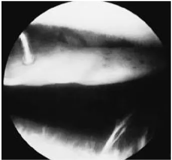
FIGURE 3. Arthroscopic view shows the pedunculated mass in the suprapatellar pouch. below : sulcus of the femoral condyle, above : inferior aspect of the patella
고 찰
색소성 융모 결절성 활액막염은 Jaffe 등3 )에 의하 여 명명된 활액막의 증식성 질환으로서 연간 인구 백 만명 당 1 . 8례가 발생할 정도로 드문 질환이며6 ) 활 액막 전체를 침범하는 미만형과 결절형, 자루형으로 침범 부위가 국한된 국소형으로 구분된다. 빈도는 남 녀 동일하게 발생하고, 국소형의 경우 2 0 ~ 3 0대의 젊은 성인에서 호발하며2 , 7 ) 슬관절과 수지의 건초에 서 가장 흔히 발생한다고 알려져 있지만2 ) 본 증례에
서는 5 9세 남자의 슬관절 관절 내・외에서 동시에 발 생하였다.
단순 방사선 검사는 국소형에서 경우에 따라 나타 나는 연부 조직 종창을 제외하고는 미만형에서의 변 화를 보이지 않으므로 진단에 도움이 되지 않고1 , 1 0 ), 종물의 촉지와 함께 종창, 압통, 발적, 기계적 잠김 및 관절 운동 제한 등의 증세와 함께 자기 공명 영상 이 진단에 매우 효과적이다4 , 5 ).
자기 공명 영상 소견상 T1 및 T2 영상에서는 혈철 소로 인한 저신호 강도를 보이며4 ) 종물의 크기와 침 범된 정도, 위치 등의 정보를 얻을 수 있다5 ).
조직학적 소견은 활액막에 증식이 왕성한 다각형의 섬유 아세포와 다핵 거대세포의 산재가 특징적 소견 이며 혈철소와 포말 세포의 침착이 있지만 염증성 질 환이 아니므로 임파구와 형질세포들과 섞여서 분포되 어 있다.
이 병변의 일반적인 재발율은 9 %에서 4 4 %까지 보 고되고 있으나9 ) 국소형은 병변의 국소 절제술로써 만 족한 결과를 얻을 수 있으며9 ), 특히 병변이 연골에 침 투하기 전 가능하면 조기에 수술을 시행하는 것이 필
요하다1 , 4 , 7 ). 관절내 종물로 인한 잠김 등의 기계적 증상
을 나타낼 때는 반월상 연골 손상 증세를 동반한 추벽 증후군이나 다른 공간 점유 병변 등에 의한 슬내장과 의 감별을 요하며8 ), 이를 위한 진단 수단으로 관절경 수술은 자기 공명 영상과 함께 유용하게 사용될 수 있 다. 후방 구획을 포함한 관절내 국소형 병변은 관절경 적 절제술로서 충분한 완전 제거가 가능하기 때문에 술 후 낮은 재발율을 기대할 수 있으며, 본 증례에서도 슬관절 내・외에 동시에 존재하는 자루형 종물들에 대 하여 관절경 및 관혈적 피부 절개를 이용한 절제술로
─ 251 ─
— 박태수 외 : 슬관절 관절 내・외에 동시 발생한 국소형 색소성 융모 결절성 활액막염 —
FIGURE 4. Operative field shows the mass(M) surrounding the medial head of the Gastrocnemius muscle(G).
FIGURE 6. On high-power view, the lesion shows the ten- dency to increased dissociation of the individual cells, the hemosiderin(wedge) and the numerous mutinucleated giant cells(arrow)(H-E, ×400).
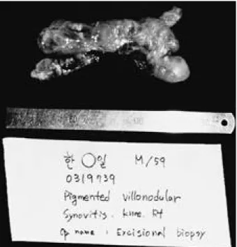
FIGURE 5. This picture shows the gross morphology of the excised mass located in popliteal area of the knee.
완전히 제거할 수 있었고, 술 후 1 5개월 추시에서 종 물은 재발하지 않았으며 슬관절 증상도 없었다.
슬관절 동통 및 혈 관절증이 동반된 슬관절 내・외 에서 동시에 병발한 국소형 색소성 융모 결정성 활액 막액염 1례에 대하여 관절경적 및 관혈적 절제술을 통하여 증상이 소실되고 재발이 없는 만족스러운 치 료 결과를 얻었으며, 이때 자기 공명 영상 및 관절경 검사는 진단과 치료에 매우 유용하게 사용되었다.
REFERENCES
01) Breimer CW and Freiberger RH : Bone lesions associated with villonodular synovitis. Am J Roent- genol, 79:618-629, 1969.
02) Docken WP : Pigmented villonodular synovitis. A review with illustrative case reports. Seminars Arthri - tis Rheumatism, 9:1-22, 1979.
03) Jaffe HL, Lichtenstein L and Sutro CJ : Pigmented villonodular synovitis, bursitis and tenosynovitis. A discussion of the synovial and bursal equivalent of the tenosynovial leision commonly denoted as xanthoma, xanthogranuloma, gaint cell tumor, or myelodyplaxo- ma of the tendon sheath lesion itself. Arch Pathol Lab M e d , 31:731-765, 1941.
04) Kaneko K, Naknhara D, Tobe M, et al : Pigment- ed villonodular synovitis of the ankle in an adoles- cent. Int Orthop, 24:234-237, 2000.
05) Mandelbaum BR, Grant TT and Hartzmann S, et a l : The use of MRI to assist in diagnosis of pig- mented villonodular synovitis of the knee joint. Clin Orthop, 23:135-139, 1988.
06) Mayer BW, Masi AT and Feigenbaum SL : Pig- mentd villonodular synovitis and tenosynovitis: A cli- nical epidemiological study of 166 cases and literature review. M e i d i c i n e , 59:223-238, 1980.
07) Nilsonne U, Moberger G : Pigmented villonodular synovitis of joints: Histological and clinical problems in diagnosis. Acta Orthop Scand, 40:448-460, 1969.
08) Palumbo RC, Matthews LS and Reuben JM : Lo- calized pigmented villonodular synovitis of the pa- tella fat pad: A report of two cases. Arthroscopy, 10:
400-403, 1994.
09) Schwartz HS, Unni KK and Pritchard DJ : Pigment- ed villonodular synovitis. A retrospective review of affected large joints. Clin Orthop, 247:243-255, 1989.
10) Van Meter CD and Rowdon GA : Localized pig- mented villonodular synovitis presenting as a locked lateral meniscal bucket tear: A case report and review of the literature. A r t h r o s c o p y , 10:309-312, 1994.
─ 252 ─
— 대한슬관절학회지 제 13 권 제 2 호 —
─ Abstract ─
Localized Pigmented Villonodular Synovitis of the Knee with Concomitant Intra- and Extra-Articular Lesion
– A Case Report –
Tae-Soo Park, M.D., Yong-Hyun Choi, M.D.
Department of Orthopaedic Surgery, Kuri Hospital, Hanyang University College of Medicine, Kuri, Korea
We report a case of localized pigmented villonodular synovitis of the knee with intra- and extra-articu- lar involvement simultaneously, which was resected completely by the arthroscopic and open methods.
Key Words : Knee, Pigmented villonodular synovitis, Intra- and extra-articular lesion