A Decline in Renal Function is Associated With Loss of Bone Mass in Korean Postmenopausal Women With Mild Renal Dysfunction
This study was conducted to assess the relationship between estimated glomerular filtration rate (eGFR) and bone mineral density (BMD) in Korean postmenopausal women with mild renal dysfunction. A total of 328 postmenopausal women who underwent BMD measurement during health check-up was investigated. BMD was measured in lumbar spine (L1-L4), femoral neck, total proximal femur and femoral trochanteric areas by dual energy radiography absorptiometry and renal function was estimated by eGFR using Cockcroft-Gault equation. Of the 328 subjects, 317 (96.6%) had an eGFR ≥60 mL/
min/1.73 m2. By using simple linear regression analysis, age, height, weight and eGFR were significantly associated with BMD for the 4 aforementioned anatomic sites, while serum levels of creatinine and blood urea nitrogen did not influence BMD. When multiple regression analyses were applied, age and body weight still had significant associations with BMD at 4 different anatomic sites (P < 0.001). A significant association of eGFR with BMD remained in the lumbar spine, femoral neck and proximal total femur (P < 0.05) but not in the trochanteric area (P = 0.300). Our study suggests that a decline of renal function is associated with lower BMD in the lumbar spine, femoral neck and total proximal femur areas in Korean menopausal women with mild renal dysfunction.
Key Words: Association; Bone Density; Koreans; Postmenopause; Renal Insufficiency Hack-Lyoung Kim1,2, In Young Park1,
Jin Man Choi1, Se-Min Hwang3, Hyo Sang Kim2, Jae-Sung Lim1, Min Kim1, and Min-Jeong Son4
1Armed Forces Seoul Hospital, Seoul; 2Department of Internal Medicine, Seoul National University Hospital, Seoul; 3Department of Preventive Medicine, The Armed Forces Medical Commands, Seongnam; 4Division of Nephrology, Department of Internal Medicine, The Armed Forces Capital Hospital, Seongnam, Korea
Received: 26 July 2010 Accepted: 14 December 2010 Address for Correspondence:
Min-Jeong Son, MD
Division of Nephrology, Department of Internal Medicine, The Armed Forces Capital Hospital, 66 Yul-dong-2-gil, Bundang-gu, Seongnam 463-040, Korea
Tel: +82.31-725-6285, Fax: +82.31-706-0987 E-mail: [email protected]
DOI: 10.3346/jkms.2011.26.3.392 • J Korean Med Sci 2011; 26: 392-398 Nephrology
INTRODUCTION
It has been well established that patients with end stage renal disease (ESRD) have reduced bone mineral density (BMD) and are at risk of osteoporosis and fragility fractures (1, 2). Multiple mechanisms including dysregulation of bone mineral metabo- lism, vitamin D deficiency, hyperparathyroidism and chronic acidosis are involved in the development of uremic bone dis- ease (3). Accumulating evidence suggests that abnormal phos- phate retention and subsequent elevation in parathyroid hor- mone (PTH) may occur from the early stage of renal impairment and negatively affect BMD (3-5). However, data on BMD in the population with mild renal dysfunction are scarce. Although sev- eral studies have demonstrated the association between renal function and BMD in the population without significant renal disease, their results are inconsistent and even conflicting (6-16).
Some studies showed that decreased renal function is associat- ed with lower BMD (6, 7, 9-11, 15, 16), whereas, others failed to show the relationship between them (8, 12, 14). Besides the dif- ferences in the study population and the methods for calculat- ing renal function and BMD measurements, the methods of ad- justment for potential confounders such as age, sex and body
weight affected the study results and caused discrepancies (11, 12). Since age, sex and body weight are strongly associated with both BMD and renal function and usually included in the for- mulas for estimating renal function such as the Cockcroft-Gault (CG) and Modification of Diet in Renal Disease (MDRD) equa- tions (17), correcting these potential confounders may be the most important in accurately assessing the relationship between renal function and BMD.
Considering that osteoporosis and subsequent fractures are major clinical burden to the public health care system in old age (18), and that the number of the subjects with early stage of re- nal dysfunction is increasing (19), assessing the association be- tween renal function and BMD in elderly without significant renal impairment is of great importance.
This study was conducted to evaluate the association between renal function and BMD in Korean postmenopausal women with mild renal dysfunction by adjusting for potential confounders.
MATERIALS AND METHODS
Over a 5-yr period, between January 2005 and December 2009, 328 postmenopausal women who visited the Armed Forces
Seoul Hospital (Seoul, Korea) for their health check-ups and also underwent BMD measurements were selected on the ba- sis of the following criteria. All subjects 1) were Korean; 2) were a minimum of 2 yr postmenopausal; 3) showed no evidence of any chronic diseases or alcoholism; 4) showed no abnormality in thyroid function test; and 5) had not taken calcium, vitamin D, estrogen, calcitonin, bisphosphonate, furosemide, thiazide diurectics, steroid, anticonvulsant, or any other medications known to influence bone metabolism or renal function.
A family medicine doctor performed the health check-ups which included the recording of medical and medication histo- ry as well as social habits including alcohol intake, cigarette smoking and regular exercise. Alcohol intake was considered when it consisted of at least 2 drinks per week and regular exer- cise was considered when it consisted of at least 3 times per week and at least 30 min each time. A gynecologist recorded history of menstruation and hormonal replacement therapy or that of medication for osteoporosis. Participants were studied in the morning following an overnight fast. A trained nurse mea- sured systolic and diastolic blood pressure (SBP and DBP), body weight and height, and performed blood sampling. In addition to serum creatinine and blood urea nitrogen (BUN), serum lev- els of glucose and total cholesterol were measured on the same day (20). Renal function was estimated by estimated glomeru- lar filtration rate (eGFR), which was calculated using the CG equation: eGFR (mL/min/1.73 m2) = (140-age) × weight (kg)/
serum creatinine (mg/dL)/72 × (0.85 for females) × 1.73 m2/ body surface area (BSA), BSA and body mass index (BMI) were
calculated as follows: BSA = weight (kg)0.425 × height (cm)0.725 × 0.007184, BMI = weight (kg)/height2 (m) (17).
BMD was measured at the lumbar spine (L1-L4), femoral neck, total proximal femur and femoral trochanteric area by Dual energy radiography absorptiometry (DXA) using a Prodi- gy Advance (GE Lunar Health Care, Madison, WI, USA), which was calibrated daily with a standard phantom which had been provided by the manufacturer (20). For the lumbar spine, the mean BMD for L1 to L4 was obtained unless individual values for one more of these vertebrae were spuriously elevated by os- teophytes or sclerotic degenerative changes (7). All BMD was expressed as exact values in g/cm2. The measurements were within an accuracy of ≤ 1.0%. DXA was performed on all sub- jects with the same machine by the same examiner and ana- lyzed with the same software.
Statistical analysis
Data are presented as mean values with standard deviation or percentage. The Pearson’s correlation method was used to de- termine the correlation between eGFR and BMD. Simple and multiple linear regression analyses were used to estimate the association of variables of interest with BMD. A two-tailed P value of < 0.05 was considered statistically significant. All data were analyzed using SPSS for Windows 13.0 (Chicago, IL, USA).
Ethics statement
This cross-sectional study was approved by the institutional re- view board of the Armed Forces Medical Command (Seongnam, Korea, research number AFMC-10-IRB-018). Informed consent was exempted due to anonymous information collection.
RESULTS
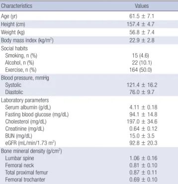
The baseline characteristics of the study participants are shown in Table 1. The participants were aged 43-84 yr, 60% of whom Table 1. Baseline characteristics of study participants (n = 328)
Characteristics Values
Age (yr) 61.5 ± 7.1
Height (cm) 157.4 ± 4.7
Weight (kg) 56.8 ± 7.4
Body mass index (kg/m2) 22.9 ± 2.8
Social habits Smoking, n (%) Alcohol, n (%) Exercise, n (%)
15 (4.6) 22 (10.1) 164 (50.0) Blood pressure, mmHg
Systolic Diastolic
121.4 ± 16.2 76.0 ± 9.7 Laboratory parameters
Serum albumin (g/dL) Fasting blood glucose (mg/dL) Cholesterol (mg/dL) Creatinine (mg/dL) BUN (mg/dL) eGFR (mL/min/1.73 m2)
4.11 ± 0.18 94.1 ± 14.8 197.0 ± 34.6 0.64 ± 0.12 15.0 ± 3.5 92.8 ± 20.3 Bone mineral density (g/cm2)
Lumbar spine Femoral neck Total proximal femur Femoral trochanter
1.06 ± 0.16 0.81 ± 0.10 0.87 ± 0.11 0.69 ± 0.10 Values are expressed as mean ± standard deviation or number. BUN, blood urea nitrogen; eGFR, estimated glomerular filtration rate.
Proportion (%)
eGFR (mL/min/1.73 m2)
3.4
45.7
50.9
30-59 60-89 ≥ 90
60 50 40 30 20 10 0
Fig. 1. Proportion of study participants according to their eGFR values. Participants who had eGFR ≥ 90, 69-89, and 30-59 mL/min/1.73 m2 were 50.9%, 45.7%, and 3.4% of all participants, respectively. eGFR, estimated glomerular filtration rate.
were aged > 60 yr. Most subjects were not overweight and did not smoke, and half of the subjects exercised regularly. The mean eGFR was 92.8 ± 20.3 mL/min/1.73 m2 and mean serum creati- nine level was 0.64 ± 0.12 mg/dL. The BMD for the lumbar spine (L1-L4), femoral neck, total proximal femur and trochanteric
areas were 1.06 ± 0.16, 0.81 ± 0.10, 0.87 ± 0.11, 0.69 ± 0.10 g/cm2, respectively.
Fig. 1 displays the distribution of the participants according to their eGFR values. About half (50.9%) had an eGFR of ≥ 90 mL/min/1.73 m2, the other half (45.7%) had an eGFR between
Table 2. Simple linear regression analyses showing the associations between variables and BMD
Variables Lumbar spine Femoral neck Total proximal femur Trochanter
Age -0.281 (0.001)/0.079* -0.384 (0.001)/0.148* -0.340 (0.001)/0.115* -0.285 (0.001)/0.081*
Height 0.253 (0.002)/0.064* 0.306 (0.001)/0.093* 0.216 (0.001)/0.216* 0.217 (0.001)/0.047*
Weight 0.279 (0.001)/0.078* 0.267 (0.001)/0.071* 0.305 (0.001)/0.093* 0.298 (0.001)/0.089*
BMI 0.176 (0.003)/0.031* 0.141 (0.002)/0.020* 0.223 (0.002)/0.050* 0.217 (0.002)/0.047*
Smoking 0.032 (0.042)/0.001 0.060 (0.028)/0.004 0.040 (0.030)/0.002 -0.058 (0.028)/0.003
Alcohol -0.008 (0.029)/0.000 -0.007 (0.020)/0.000 0.023 (0.021)/0.001 -0.003 (0.020)/0.000
Exercise 0.172 (0.017)/0.030 0.067 (0.012)/0.004 0.057 (0.013)/0.003 0.050 (0.012)/0.003
SBP -0.106 (0.001)/0.011 -0.120 (0.000)/0.014‡ -0.079 (0.000)/0.006 -0.083 (0.000)/0.007
DBP 0.040 (0.001)/0.002 0.019 (0.001)/0.000 0.050 (0.001)/0.002 0.043 (0.001)/0.002
Albumin 0.055 (0.050)/0.003 0.095 (0.033)/0.009 0.159 (0.035)/0.025‡ 0.122 (0.034)/0.015‡
FBG 0.034 (0.001)/0.001 -0.054 (0.000)/0.003 -0.016 (0.000)/0.000 -0.0074 (0.000)/0.000
Cholesterol 0.047 (0.000)/0.002 0.052 (0.000)/0.003 0.083 (0.000)/0.007 0.050 (0.000)/0.003
BUN -0.102 (0.003)/0.010 -0.036 (0.002)/0.001 -0.047 (0.002)/0.002 -0.076 (0.002)/0.006
Creatinine 0.101 (0.070)/0.010 0.066 (0.047)/0.004 0.061 (0.050)/0.004 0.038 (0.047)/0.001
eGFR 0.122 (0.000)/0.015‡ 0.157 (0.000)/0.025† 0.176 (0.000)/0.031† 0.178 (0.000)/0.032†
Values are expressed as β (SE)/R2. *P < 0.001, †P < 0.01, ‡P < 0.05. β, standardized regression coefficient; SE, standard error; R2, percent variance explained by each variable.
BMD, bone mineral density; BMI, body mass index; SBP, systolic blood pressure; DBP, diastolic blood pressure; FBG, fasting blood glucose; BUN, blood urea nitrogen; eGFR, estimated glomerular filtration rate.
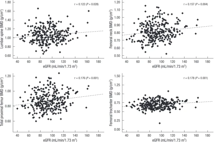
Fig. 2. Linear associations between eGFR and BMD. Significant positive linear associations between eGFR and BMD are shown in scatterplots. r, Pearson’s correlation coefficient.
eGFR, estimated glomerular filtration rate; BMD, bone mineral density.
Lumbar spine BMD (g/cm2)Total proximal femur BMD (g/cm2) Femoral neck BMD (g/cm2)Femoral trochanter BMD (g/cm2)
eGFR (mL/min/1.73 m2)
eGFR (mL/min/1.73 m2)
eGFR (mL/min/1.73 m2)
eGFR (mL/min/1.73 m2)
r = 0.122 (P = 0.028)
r = 0.176 (P = 0.001)
r = 0.157 (P = 0.004)
r = 0.178 (P = 0.001)
40 60 80 100 120 140 160 180
40 60 80 100 120 140 160 180
40 60 80 100 120 140 160 180
40 60 80 100 120 140 160 180
1.80 1.60 1.40 1.20 1.00 0.80 0.60
1.20
1.00
0.80
0.60
1.20 1.10 1.00 0.90 0.80 0.70 0.60 0.50
1.50 1.25 1.00 0.75 0.50 0.25 0.00
60 and 89 mL/min/1.73 m2 and only 3.4% of the participants had an eGFR of < 60 mL/min/1.73 m2.
Fig. 2 demonstrates a significant linear association between eGFR and BMD for the 4 different sites (P < 0.05 for each). The Pearson’s correlation coefficients between eGFR and lumbar spine, femoral neck, total proximal femur and femoral trochan- teric BMD were 0.122, 0.157, 0.176, and 0.178, respectively.
We performed simple linear regression analysis to assess as- sociation of each variable with BMD (Table 2). Age, height, body weight and BMI were all significantly associated with BMD at all 4 sites (P < 0.001 for each). SBP was associated with femoral neck BMD; and serum albumin was associated with total proxi- mal femur and trochanteric BMDs (P < 0.05 for each). In addi- tion, eGFR was associated with BMD for the 4 sites (P < 0.01 each versus femoral neck, total proximal femur and trochanter- ic BMDs; P < 0.05 each versus lumbar spine BMD). However, serum levels of creatinine and BUN were not associated with BMD (P > 0.05 for each). We performed multiple linear regres- sion analyses to identify the independent variables affecting BMD (Table 3). We used variables that were significant (P < 0.05) in a simple linear regression model (Table 2) as confounding factors in this multiple regression analyses. Height and body mass index were not taken into account in the analyses because of the eventual problems with multicollinearity with body weight (21). Age and body weight still had a significant association with BMD from four different skeletal sites (P < 0.001 for each). A sig- nificant association of eGFR with BMD was remained in lum- bar spine, femoral neck and proximal total femur (P < 0.05 for each) but not for trochanteric BMD (P = 0.300).
When the MDRD equation for estimating GFR was applied,
similar results were obtained demonstrating positive relation- ship between renal function and bone density. In multiple linear regression analyses, femoral neck BMD had a significant posi- tive association with eGFR (P = 0.012), lumbar spine and proxi- mal total femur BMD had a marginal significance (P = 0.060 and P = 0.079, respectively), whereas, trochanteric BMD was not as- sociated with eGFR (P = 0.422) (data not shown).
DISCUSSION
The kidney plays a pivotal role in regulating calcium and phos- phorous metabolism. The kidney is not only the target organ for various bone regulating hormones such as PTH, but also a prin- cipal site for the production of calcitriol (1,25-dihydroxyvitamin D). It has been known that decrease in calcitriol and abnormal retention of phosphorus occur from a relatively early stage of chronic kidney disease (CKD) followed by a subsequent increase in PTH (3-5). Although these changes contribute to the mainte- nance of serum levels of calcium and phosphorous within the normal range until advanced stage of CKD, the results adverse- ly affect bone formation (22, 23).
In this study, we found independent association between eGFR and BMD for the lumbar spine, femoral neck and proxi- mal total femur in 328 Korean menopausal women with mild renal dysfunction. No significant relationships of serum levels of creatinine and BUN with BMD were observed in our study, suggesting that eGFR is more important than serum creatinine or BUN in estimating the BMD in postmenopausal women.
CKD is a worldwide health problem. The prevalence of CKD is reported to be about 11% among the USA (19) and Australian adults (24). In a recent Korean population-based study with el- derly aged > 65 yr living in a satellite city of Korea, 52% of the sub- jects showed an eGFR values of < 60 mL/min/1.73 m2 as mea- sured by the MDRD equation (25). In our study, only 3.4% had an eGFR of < 60 mL/min/1.73 m2, which is much lower than those in previous reports (19, 24, 25). These differences may be due to a different study population. We selected postmenopaus- al women who had no medical problems such as hypertension and diabetes, whereas the previous study (25) included 71.1%
of patients with hypertension and 20.9% of patients with diabe- tes, which may have negatively affected renal function. In addi- tion, the subjects were relatively younger in our study and the methods for eGFR estimation were different: we used the CG while the previous study used the MDRD equation (25). It has been reported that the GFR calculated by the CG equation is likely to be overestimated compared to that by the MDRD equa- tion (26).
Our study results are consistent with those of previous stud- ies showing that the decline in renal function was associated with decreased BMD (6, 7, 11, 16). Yendt et al. (6, 7) reported a strong positive correlation between creatinine clearance (Ccr) and the Table 3. Multiple linear regression analyses showing the independent association
between variables and BMD
β (SE) t P VIF
Lumbar spine BMD (Durbin-Watson = 1.853; adjusted R2 = 0.169) Age
Weight eGFR
-0.342 (0.001) 0.317 (0.001) 0.131 (0.000)
-5.889 5.901 2.163
< 0.001
< 0.001 0.031
1.317 1.122 1.439 Femoral neck BMD (Durbin-Watson = 2.050; adjusted R2 = 0.231)
Age Weight SBP eGFR
-0.435 (0.001) 0.322 (0.001) -0.086 (0.000) 0.122 (0.000)
-7.211 5.872 -1.581 1.997
< 0.001
< 0.001 0.102 0.047
1.423 1.171 1.155 1.465 Total hip BMD (Durbin-Watson = 2.609; adjusted R2 = 0.227)
Age Weight Albumin eGFR
-0.445 (0.001) 0.315 (0.001) 0.051 (0.030) 0.145 (0.000)
-7.775 5.988 1.027 2.429
< 0.001
< 0.001 0.305 0.016
1.333 1.128 1.013 1.446 Trochanter BMD (Durbin-Watson = 1.968; adjusted R2 = 0.202)
Age Weight Albumin eGFR
-0.308 (0.008) 0.359 (0.007) 0.098 (0.263) 0.063 (0.003)
-5.300 6.715 1.942 1.038
< 0.001
< 0.001 0.053 0.300
1.333 1.128 1.013 1.446 β, standardized regression coefficient; SE, standard error; t, corresponding t values;
VIF, variance inflation factor. BMD, bone mineral density; eGFR, estimated glomerular filtration rate; SBP, systolic blood pressure.
lumbar spine BMD, which was primarily independent on age and body surface area in healthy aging women with and with- out osteoporosis. They calculated Ccr using 24-hr urine sam- ples, however, we did not include a 24-hr urine samples for the estimation of renal function in our study. Jassal et al. (11) also showed a significant positive linear association between calcu- lated renal function (eGFR by using the CG and MDRD equa- tions) and hip BMD in elderly. However, they did not adjust for age and body weight during the multiple regression analysis.
Kaji et al. (16) showed significant positive correlations between eGFR and BMD values at the femoral neck and radius among Japanese postmenopausal women with stage 2 CKD. In the same study, they found that postmenopausal women with ver- tebral fractures have lower eGFR than those without vertebral fractures in stage 2 CKD (60 ≤ eGFR < 90 mL/min/1.73 m2). Con- sidering the study population of postmenopausal women at an early stage of CKD, the study results reported by Kaji et al. (16) are very similar to those of our study. When subgroup analysis was performed an 317 participants (96.6%) with stage 1 or 2 CKD (eGFR ≥ 60 mL/min/1.73 m2), eGFR was significantly associat- ed with femoral neck BMD in multiple regression analysis after adjustment for confounders (P = 0.014). However, the associa- tions between eGFR and BMDs at lumbar spine, total proximal femur or trochanteric area were not observed in our study pop- ulation (P > 0.05 for each).
Although simple linear regression analysis showed a positive association between eGFR and femoral trochanteric BMD in our study, this association disappeared after adjustment for con- founding factors including age and body weight. This result is similar to those of previous studies showing the importance of confounding factors in assessing the association between renal function and BMD (8, 12-14). Ccr had a significant relationship with BMD in old man and women, however, this relationship was due to the inter-correlation of both variables with BMD and lost its significance after adjustment for age (8, 14). A recent epi- demiological study with 13,848 adults (12) also showed that the association between renal impairment and lower BMD, which was shown to be significant in unadjusted analysis, disappeared after adjustment for age and body weight in the multivariate models. Cystatin-C, a new marker for kidney function, was as- sociated with BMD in the linear regression model, however, this association disappeared after adjustment for age and body fat in 885 elderly women (13).
There is accumulating evidence that old age and lower body weight are strongly associated with low BMD (27), and these potential confounders are included when renal function is cal- culated using the CG or MDRD equations (17). Therefore, it is mandatory to properly adjust these confounders for accurate assessment of the relationship between BMD and renal func- tion if renal function is estimated by the CG or MDRD equations as in this study. Some investigators have tried to avoid the effect
of these strong confounders by not adjusting for age and body weight because these confounders are included in the CG equa- tion (11) or by using other markers that are independent of age and body weight (13). In our study, we attempted to overcome the multicollinearity problem by using the variance influence factor (VIF) (21). The maximal VIF value did not exceed 1.5 in each multiple regression model, which indicated that the de- gree of multicollinearity between the independent variables was not serious in our study.
Sometimes serum levels of creatinine and BUN can be useful markers for estimating renal function because they are less af- fected by age and body weight than using the eGFR by the CG or MDRD equation (12). However, serum creatinine levels do not predict renal function precisely, especially in old individu- als because they usually have a normal serum creatinine levels in spite of impaired renal function (28). Our finding that serum levels of both creatinine and BUN were not associated with BMD suggest that renal function should be assessed by eGFR rather than either the serum creatinine or BUN level in elderly subjects because renal impairment can be masked by an apparently nor- mal serum creatinine or BUN levels (14).
Proteinuria is an important marker for renal damage and progression to ESRD (29). In our study population, 10 subjects (3.0%) had proteinuria above the normal limit in the spot urine dipstick test. Among them, 9 (90.0%) had an eGFR of ≥ 60 mL/
min/1.73 m2 (data not shown). These subjects with proteinuria had the potential of more advanced stage of renal dysfunction, however, we did not consider proteinuria as an additional fac- tor for estimating renal function in this study.
This study has several limitations. First, because our data were obtained from a cross-sectional study, there is possibility of a temporal association between renal function and BMD which makes it difficult to ascertain our conclusion. Longitudinal stud- ies are needed to better characterize this relationship. Second, since only Korean postmenopausal women were included in the study, our results may not apply to the general population.
Third, other potential confounders, including the history of frac- tures, childbearing, income level, education background and calcium intake, which are known to affect BMD in Korean adults (30), were not considered in this study. Fourth, we did not ana- lyze serum levels of vitamin D and PTH, which may be impor- tant in the association between renal function and BMD (3). Fi- nally, we did not use gold standard measures of GFR such as inulin, radioisotope clearance or 24-hr urine samples, which would have helped accurately assess the association between renal function and BMD (13).
In conclusion, the results of this study demonstrate that a de- cline in renal function may be associated with low BMD in Ko- rean menopausal women with mild renal dysfunction. Further studies with a larger sample size and various assessment meth- ods are needed to confirm our results.
ACKNOWLEDGMENTS
The authors thank Mr. Damien Lim (Richmond Hill, ON, Cana- da) for his skillful editing of the text.
REFERENCES
1. Alem AM, Sherrard DJ, Gillen DL, Weiss NS, Beresford SA, Heckbert SR, Wong C, Stehman-Breen C. Increased risk of hip fracture among patients with end-stage renal disease. Kidney Int 2000; 58: 396-9.
2. Stehman-Breen C. Bone mineral density measurements in dialysis pa- tients. Semin Dial 2001; 14: 228-9.
3. Elder G. Pathophysiology and recent advances in the management of re- nal osteodystrophy. J Bone Miner Res 2002; 17: 2094-105.
4. Martinez I, Saracho R, Montenegro J, Llach F. The importance of dietary calcium and phosphorous in the secondary hyperparathyroidism of pa- tients with early renal failure. Am J Kidney Dis 1997; 29: 496-502.
5. Fajtova VT, Sayegh MH, Hickey N, Aliabadi P, Lazarus JM, LeBoff MS.
Intact parathyroid hormone levels in renal insufficiency. Calcif Tissue Int 1995; 57: 329-35.
6. Yendt ER, Cohanim M, Jarzylo S, Jones G, Rosenberg G. Reduced creati- nine clearance in primary osteoporosis in women. J Bone Miner Res 1993;
8: 1045-52.
7. Yendt ER, Cohanim M, Jarzylo S, Jones G, Rosenberg G. Bone mass is related to creatinine clearance in normal elderly women. J Bone Miner Res 1991; 6: 1043-50.
8. Sherman SS, Tobin JD, Hollis BW, Gundberg CM, Roy TA, Plato CC. Bio- chemical parameters associated with low bone density in healthy men and women. J Bone Miner Res 1992; 7: 1123-30.
9. Rix M, Andreassen H, Eskildsen P, Langdahl B, Olgaard K. Bone miner- al density and biochemical markers of bone turnover in patients with predialysis chronic renal failure. Kidney Int 1999; 56: 1084-93.
10. Klawansky S, Komaroff E, Cavanaugh PF Jr, Mitchell DY, Gordon MJ, Connelly JE, Ross SD. Relationship between age, renal function and bone mineral density in the US population. Osteoporos Int 2003; 14: 570-6.
11. Jassal SK, von Muhlen D, Barrett-Connor E. Measures of renal function, BMD, bone loss, and osteoporotic fracture in older adults: the Rancho Bernardo study. J Bone Miner Res 2007; 22: 203-10.
12. Hsu CY, Cummings SR, McCulloch CE, Chertow GM. Bone mineral density is not diminished by mild to moderate chronic renal insufficien- cy. Kidney Int 2002; 61: 1814-20.
13. Fried LF, Shlipak MG, Stehman-Breen C, Mittalhenkle A, Seliger S, Sar- nak M, Robbins J, Siscovick D, Harris TB, Newman AB, Cauley JA. Kid- ney function predicts the rate of bone loss in older individuals: the Car- diovascular Health Study. J Gerontol A Biol Sci Med Sci 2006; 61: 743-8.
14. Buchanan JR, Myers CA, Greer RB 3rd. Effect of declining renal function on bone density in aging women. Calcif Tissue Int 1988; 43: 1-6.
15. Bianchi ML, Colantonio G, Montesano A, Trevisan C, Ortolani S, Rossi R, Buccianti G. Bone mass status in different degrees of chronic renal fail- ure. Bone 1992; 13: 225-8.
16. Kaji H, Yamauchi M, Yamaguchi T, Shigematsu T, Sugimoto T. Mild re-
nal dysfunction is a risk factor for a decrease in bone mineral density and vertebral fractures in Japanese postmenopausal women. J Clin En- docrinol Metab 2010; 95: 4635-42.
17. Botev R, Mallié JP, Couchoud C, Schück O, Fauvel JP, Wetzels JF, Lee N, De Santo NG, Cirillo M. Estimating glomerular filtration rate: Cockcroft- Gault and Modification of Diet in Renal Disease formulas compared to renal inulin clearance. Clin J Am Soc Nephrol 2009; 4: 899-906.
18. Keene GS, Parker MJ, Pryor GA. Mortality and morbidity after hip frac- tures. BMJ 1993; 307: 1248-50.
19. Coresh J, Astor BC, Greene T, Eknoyan G, Levey AS. Prevalence of chron- ic kidney disease and decreased kidney function in the adult US popula- tion: Third National Health and Nutrition Examination Survey. Am J Kidney Dis 2003; 41: 1-12.
20. Park KK, Kim SJ, Moon ES. Association between bone mineral density and metabolic syndrome in postmenopausal Korean women. Gynecol Obstet Invest 2010; 69: 145-52.
21. Taaffe DR, Cauley JA, Danielson M, Nevitt MC, Lang TF, Bauer DC, Har- ris TB. Race and sex effects on the association between muscle strength, soft tissue, and bone mineral density in healthy elders: the Health, Aging, and Body Composition Study. J Bone Miner Res 2001; 16: 1343-52.
22. Bacchetta J, Boutroy S, Vilayphiou N, Juillard L, Guebre-Egziabher F, Rognant N, Sornay-Rendu E, Szulc P, Laville M, Delmas PD, Fouque D, Chapurlat R. Early impairment of trabecular microarchitecture assessed with HR-pQCT in patients with stage II-IV chronic kidney disease. J Bone Miner Res 2010; 25: 849-57.
23. Kovesdy CP, Kalantar-Zadeh K. Bone and mineral disorders in pre-dial- ysis CKD. Int Urol Nephrol 2008; 40: 427-40.
24. Chadban SJ, Briganti EM, Kerr PG, Dunstan DW, Welborn TA, Zimmet PZ, Atkins RC. Prevalence of kidney damage in Australian adults: The AusDiab kidney study. J Am Soc Nephrol 2003; 14 (7 Suppl 2): S131-8.
25. Chin HJ, Song YR, Lee JJ, Lee SB, Kim KW, Na KY, Kim S, Chae DW.
Moderately decreased renal function negatively affects the health-related quality of life among the elderly Korean population: a population-based study. Nephrol Dial Transplant 2008; 23: 2810-7.
26. Lin J, Knight EL, Hogan ML, Singh AK. A comparison of prediction equa- tions for estimating glomerular filtration rate in adults without kidney disease. J Am Soc Nephrol 2003; 14: 2573-80.
27. Mazess RB, Barden HS, Drinka PJ, Bauwens SF, Orwoll ES, Bell NH. In- fluence of age and body weight on spine and femur bone mineral density in U.S. white men. J Bone Miner Res 1990; 5: 645-52.
28. Swedko PJ, Clark HD, Paramsothy K, Akbari A. Serum creatinine is an inadequate screening test for renal failure in elderly patients. Arch Intern Med 2003; 163: 356-60.
29. Ishani A, Grandits GA, Grimm RH, Svendsen KH, Collins AJ, Prineas RJ, Neaton JD. Association of single measurements of dipstick proteinuria, estimated glomerular filtration rate, and hematocrit with 25-year inci- dence of end-stage renal disease in the multiple risk factor intervention trial. J Am Soc Nephrol 2006; 17: 1444-52.
30. Shin CS, Choi HJ, Kim MJ, Kim JT, Yu SH, Koo BK, Cho HY, Cho SW, Kim SW, Park YJ, Jang HC, Kim SY, Cho NH. Prevalence and risk factors of osteoporosis in Korea: a community-based cohort study with lumbar spine and hip bone mineral density. Bone 2010; 47: 378-87.
AUTHOR SUMMARY
A Decline in Renal Function is Associated With Loss of Bone Mass in Korean Postmenopausal Women With Mild Renal Dysfunction
Hack-Lyoung Kim, In Young Park, Jin Man Choi, Se-Min Hwang, Hyo Sang Kim, Jae-Sung Lim, Min Kim, and Min-Jeong Son Kidney function is intimately associated with bone health. We assessed the relationship between estimated glomerular filtration rate (eGFR) and bone mineral density (BMD) in Korean postmenopausal women with mild renal dysfunction. BMD was measured in lumbar spine and femur regions by dual energy radiography absorptiometry, and eGFR was estimated by using Cockcroft-Gault equation. Of the 328 study subjects, 317 (96.6%) had an eGFR ≥ 60 mL/min/1.73 m2. Significant positive association between eGFR and BMD was observed in Pearson’s correlation. In multiple linear regression analyses, eGFR significantly associated with BMD in the lumbar spine, femoral neck and proximal total femur (P < 0.05) but not in the trochanteric area (P = 0.300) after controlling confounders including age and body weight. Our study demonstrated that, even with mild renal dysfunction, a decline of renal function is associated with lower BMD in Korean menopausal women.