INTRODUCTION
Because early complications such as anastomosis site leak- age can be disastrous and cause significant morbidity and mortality to patients undergoing esophageal reconstruction, the successful esophagogastric anastomosis is closely related to early patient outcome (1). Cervical esophagogastric anas- tomosis (CEGA) is a widely accepted procedure despite crit- icisms from some authors (2-4). Many surgeons prefer this technique due to better tumor eradication, and reduced mor- bidity and mortality associated with anastomotic breakdown (1, 5, 6) despite known risks of leakage, stricture formation and recurrent laryngeal nerve injury (3, 7, 8). Currently, CEGA is becoming an increasingly common procedure (9).
CEGA consists partly of a hand-sewn procedure and partly of a method that employs mechanical suturing devices. Al- though there has been one prospective randomized trial advo- cating the hand-sewn technique in fashioning cervical esoph- agogastric anastomoses (10), there are disagreements in the literature (11). Kim and colleagues (12) and Orringer and associates (9) have reported the usefulness of endo-GIA sta- pler devices in esophago-enteric anastomosis. Orringer and associates (9) introduced the CEGA technique incorporating both the hand sewn technique and that using endo-GIA (side- to-side stapled anastomosis); this method has been reported
to reduce the incidence of anastomosis related complications.
In this study, we evaluated the mid-term results of CEGA using side-to-side stapled anastomosis in patients with eso- phageal squamous cell carcinoma.
MATERIALS AND METHODS
Retrospective medical records review was conducted for 13 of 18 patients who underwent esophagogastrostomy from January 2001 to November 2005. All 13 patients were diag- nosed with esophageal squamous cell carcinoma and under- went curative CEGA. Patients diagnosed with non-squamous cell carcinoma, who underwent palliative surgery, or thoracic esophagogastrostomy were excluded from the study. The male to female ratio was 11:2 with an average age of 62.6 yr (47- 79 yr). The most common presenting symptom was dyspha- gia, and chest pain. Tumor location was diverse; 8 patients had midthoracic esophageal tumors, 2 patients had tumors in the upper thoracic esophagus, 1 patient had lower thoracic esophageal carcinoma, and 2 patients had dual locations (up- per and mid thoracic esophagus, and mid and lower thoracic esophagus, respectively) The two patients diagnosed with dual esophageal cancer had celiac lymph node metastasis and was diagnosed as stage IVB (Table 1).
Won-Min Jo, Jae Seung Shin, In Sung Lee
Department of Thoracic & Cardiovascular Surgery, Ansan Hospital, Korea University School of Medicine, Seoul, Korea
Address for correspondence Won-Min Jo, M.D.
Department of Thoracic & Cardiovascular Surgery, Ansan Hospital, Korea University, Gojan-1-dong, Ansan 425-707, Korea
Tel : +82.31-412-5060, Fax : +82.31-414-3249 E-mail : jowonmin@korea.ac.kr
1033 J Korean Med Sci 2006; 21: 1033-6
ISSN 1011-8934
Copyright � The Korean Academy of Medical Sciences
Mid-term Outcomes of Side-to-Side Stapled Anastomosis in Cervical Esophagogastrostomy
This study was conducted to evaluate the mid-term results of cervical esophago- gastric anastomosis using a side-to-side stapled anastomosis method for treatment of patients with malignant esophageal disease. A total of 13 patients were reviewed retrospectively from January 2001 to November 2005 who underwent total esopha- gectomy through a right thoracotomy, gastric tube formation through a midline lapa- rotomy and finally a cervical esophagogastric anastomosis. Average patient age was 62.6 yr old and the male to female ratio was 11:2. The mean anastomosis time was measured to be about 32.5 min; all patients were followed for about 22.8±±9.9 months postoperatively. There were no early or late mortalities. There were no com- plications of anastomosis site leakage or conduit necrosis. A mild anastomotic stric- ture was noted in one patient, and required two endoscopic bougination procedures at postoperative 4th month. Construction of a cervical esophagogastric anastomo- sis by side-to-side stapled anastomosis is relatively easy to apply and can be per- formed in a timely manner. Follow up outcomes are very good. We, therefore, sug- gest that the side-to-side stapled anastomosis could be used as a safe and effec- tive option for cervical esophagogastric anastomosis.
Key Words : Esophageal Neoplasms; Carcinoma, Squamous Cell; Side-to-Side Stapled Anastomosis; Anas- tomosis, Surgical; Suture Techniques
Received : 23 February 2006 Accepted : 23 April 2006
1034 W.M. Jo, J.S. Shin, I.S. Lee
All patients underwent diagnostic and metastatic workup under a preset protocol including endoscopy with endoscopic ultrasonography, upper gastrointestinal series with or without colon studies, CT scans (neck, chest, and abdomen), bone scan, optional brain CT, and optional PET-CT. After such evalua- tion, the patients were grouped into according to the pres- ence or absence of distant metastasis; M0 group-immediate
surgery, M1 group-neoadjuvant chemo- or radiotherapy before surgery. The two pati- ents in the M1 group received neoad- juvant chemotherapy before operation.
The operative technique consisted of a total thoracic esopha- gectomy with mediastinal lymphadenectomy via a right tho- racotomy and gastric tube construction via a midline laparo- tomy incision with preservation of the right gastroepiploic artery. Celiac lymph node dissection was also conducted dur- ing the laparotomy phase. Gastric drainage procedures were performed in all patients; Heller’s pyloromyotomy (4 patients), pylorus fracture by manipulation (9 patients). The gastric tube was pulled up through the posterior mediastinum to the left cervical area for side-to-side CEGA. CEGA was per- formed according to the method introduced by Orringer and colleagues (9). Their technique recommends a side-to-side anastomosis with an endo-GIA 30 mm stapler on the ante- rior stomach wall. The endo-GIA II 30-3.5 mm stapler (Au- tosuture�, Norwalk, CT, U.S.A.) was used in the first phase of the CEGA, with care taken to avoid involvement of the conduit tip. The second phase of the CEGA, the anterior clo- sure of the anastomosis site contained within the hood of the overlying esophagus, was performed by a hand-sewn method using Vicryl 5-0 (Ethicon�, Piscateway, NJ, U.S.A.) in inter- rupted sutures. In patients showing no cervical lymph node
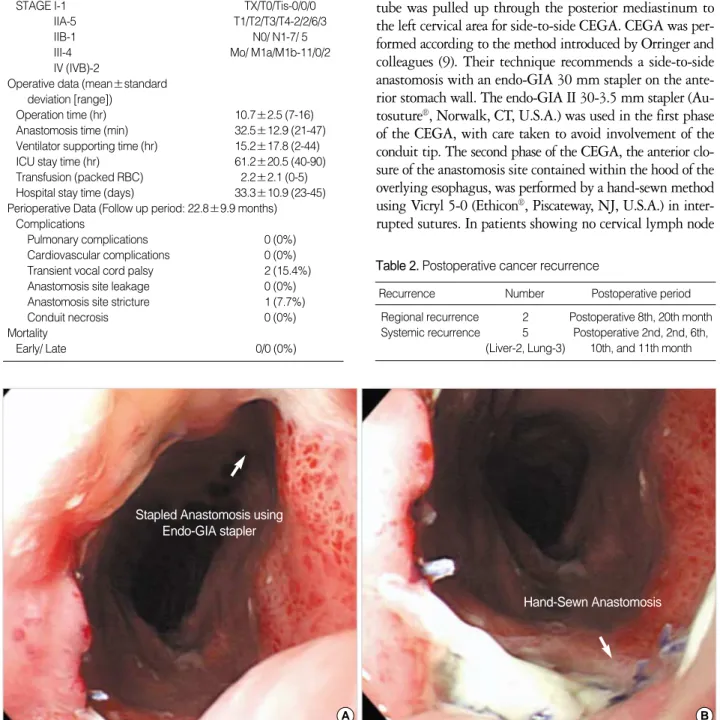
Fig. 1.Endoscopic findings of CEGA at postoperative 2nd month conducted by stapled anastomosis using Endo-GIA stapler (A) and hand- sewn anastomosis (B).
A B
Stapled Anastomosis using Endo-GIA stapler
Hand-Sewn Anastomosis Operative staging (AJCC)
STAGE I-1 TX/T0/Tis-0/0/0
IIA-5 T1/T2/T3/T4-2/2/6/3
IIB-1 N0/ N1-7/ 5
III-4 Mo/ M1a/M1b-11/0/2
IV (IVB)-2
Operative data (mean±standard deviation [range])
Operation time (hr) 10.7±2.5 (7-16)
Anastomosis time (min) 32.5±12.9 (21-47) Ventilator supporting time (hr) 15.2±17.8 (2-44)
ICU stay time (hr) 61.2±20.5 (40-90)
Transfusion (packed RBC) 2.2±2.1 (0-5) Hospital stay time (days) 33.3±10.9 (23-45) Perioperative Data (Follow up period: 22.8±9.9 months)
Complications
Pulmonary complications 0 (0%)
Cardiovascular complications 0 (0%)
Transient vocal cord palsy 2 (15.4%)
Anastomosis site leakage 0 (0%)
Anastomosis site stricture 1 (7.7%)
Conduit necrosis 0 (0%)
Mortality
Early/ Late 0/0 (0%)
Table 1.Operative and perioperative data
Regional recurrence 2 Postoperative 8th, 20th month Systemic recurrence 5 Postoperative 2nd, 2nd, 6th,
(Liver-2, Lung-3) 10th, and 11th month Table 2.Postoperative cancer recurrence
Recurrence Number Postoperative period
Side-to-side Stapled Anastomosis in Cervical Esophagogastrostomy 1035
metastasis on neck CT (all of the patients in this study), cer- vical lymph node dissection was not performed.
Postoperative esophagography was performed on the 7th postoperative day to evaluate functional results and to iden- tify anastomosis site complications. All patients received en- doscopic examination 2 months after surgery (Fig. 1); neck, chest, and abdominal CT scans were performed routinely 6 months after the operation.
RESULTS
The mean operation time was 10.7 hr with a mean anas- tomosis tome of 32.5 min. All patients received mechanical ventilation during the immediate postoperative period for an average of 15.2 hr to reduce pulmonary complications. Post- operative intensive care unit stay was 61.2±20.5 hr.
The mean hospital stay was 33.3 days and patients were followed up for 22.8±9.9 months postoperatively. There were no early or late mortalities related to the procedure. We experienced no respiratory or cardiovascular complications throughout this study. Two patients complained of postop- erative voice hoarseness and intermittent episodes of aspira- tion. Laryngoscopic evaluation revealed vocal cord palsies, but these were transient symptoms and both patients recovered fully by 2 months after surgery.
There were no cases of anastomosis site leakage or conduit necrosis. A mild anastomosis site stricture was noted in one patient and he received two endoscopic bougienations four months after surgery. Postoperative staging and operative data are summarized in Table 1.
Cancer recurrences were noted in 7 patients (Table 2) dur- ing follow up. Two patients confirmed with regional recur- rences had mediastinal and celiac lymph node metastases.
DISCUSSION
Esophageal anastomosis site complications are important causes of postoperative morbidity and mortality after eso- phagectomy. Cervical esophagogastric anastomosis is pre- ferred by many surgeons because of larger resection margins and less dangerous anastomosis leakage (5, 6), although it has an increased risk for recurrent laryngeal nerve injury (8).
Anastomosis techniques, both the hand-sewn and mechani- cal stapling procedures, have been evaluated by many inves- tigators. Gandhi and Naunheim (13) reported a 5-26% inci- dence of anastomosis leak, and 10-15% incidence of benign anastomosis site strictures after hand-sewn CEGA. In another study, Fok and associates (14) reported a 5% incidence of anastomosis leakage in hand-sewn CEGA versus a 3.8% inci- dence in a group using a circular stapler for gatro-enteric anastomosis. However, a greater number of stapled anasto- moses resulted in strictures. In general, stapled anastomosis
is a safe and effective technique (15-18).
Although Hsu and colleagues (19) reported that the circu- lar stapler is a feasible option for CEGA, the application of these devices in the cervical region is technically complicated (10, 11). Many investigator, therefore, discourage the use of circular stapling devices (10). Kim et al. (12) and Orringer et al. (9) have reported on the usefulness of Endo-GIA 30 mm stapler in CEGA. They found that Endo-GIA was easy to handle in the cervical region and with a generous 3 cm anas- tomosis there was a reduction in anastomosis site stricture and postoperative dysphagia compared to circular staplers.
Also, anastomosis leak was uncommon.
Collard and associates (20) have reported on a side-to-side stapled CEGA with an Endo-GIA stapler using the tip of the mobilized stomach. This, in effect, creates a functional end- to-end esophagogastric connection. Orringer and colleagues report that gastroesophageal refulx was less common when the anastomosis is performed using the anterior wall of the stomach. We conducted CEGA according to Orringer’s me- thod and confirmed the benefits of this approach. The CEGA technique was associated with gastric conduit tip necrosis, a rare but very serious problem (4, 9). Fortunately, we did not experience any cases of graft failure or conduit tip necrosis in our study.
Although Orringer’s technique requires manual sewing in the final anterior closure of the CEGA, this did not increase leakage rates after esophageal resection of squamous cell carci- nomas. The benign stricture rate, surgical outcome and long- term results were satisfactory. In conclusion, side-to-side sta- pled anastomosis according to the technique introduced by Orringer and colleagues is the preferred procedure for CEGA because it is relatively easy to perform and therefore less oper- ator dependent, and requires less time to perform hand sewn method (11).
REFERENCES
1. Urschel JD. Esophagogastrostomy anastomotic leaks complicating esophagectomy: a review. Am J Surg 1995; 169: 634-40.
2. Lam TC, Fok M, Cheng SW, Wong J. Anastomotic complications after esophagectomy for cancer. A comparison of neck and chest anas- tomoses. J Thorac Cardiovasc Surg 1992; 104: 395-400.
3. Chasseray VM, Kiroff GK, Buard JL, Launois B. Cervical or thoracic anastomosis for esophagectomy for carcinoma. Surg Gynecol Obstet 1989; 169: 55-62.
4. Iannettoni MD, Whyte RI, Orringer MB. Catastrophic complications of the cervical esophagogastric anastomosis. J Thorac Cardiovasc Surg 1995; 110: 1493-500.
5. Muller JM, Erasmi H, Stelzner M, Zieren U, Pichlmaier H. Surgical therapy of oesophageal carcinoma. Br J Surg 1990; 77: 845-57.
6. Patil PK, Patel SG, Mistry RC, Deshpande RK, Desai PB. Cancer of the esophagus: esophagogastric anastomotic leak--a retrospective study of predisposing factors. J Surg Oncol 1992; 49: 163-7.
1036 W.M. Jo, J.S. Shin, I.S. Lee
7. Dewar L, Gelfand G, Finley RJ, Evans K, Inculet R, Nelems B. Fac- tors affecting cervical anastomotic leak and stricture formation fol- lowing esophagogastrectomy and gastric tube interposition. Am J Surg 1992; 163: 484-9.
8. Fok M, Law S, Stipa F, Cheng S, Wong J. A comparison of trans- hiatal and transthoracic resection for oesophageal carcinoma. Endo- scopy 1993; 25: 660-3.
9. Orringer MB, Marshall B, Iannettoni MD. Eliminating the cervical esophagogastric anastomotic leak with a side-to-side stapled anas- tomosis. J Thorac Cardiovasc Surg 2000; 119: 277-88.
10. Laterza E, de’ Manzoni G, Veraldi GF, Guglielmi A, Tedesco P, Cordiano C. Manual compared with mechanical cervical oesopha- gogastric anastomosis: a randomised trial. Eur J Surg 1999; 165:
1051-4.
11. Urschel JD, Blewett CJ, Bennett WF, Miller JD, Young JE. Hand- sewn or stapled esophagogastric anastomoses after esophagectomy for cancer: meta-analysis of randomized controlled trials. Dis Esoph- agus 2001; 14: 212-7.
12. Kim IH, Kim KT, Park SM, Lee SY, Baek MJ, Sun K, Kim HM, Lee IS. Cervical esophago-enteric anastomosis with straight endo- stapler. Korean J Thorac Cardiovasc Surg 1999; 32: 924-9.
13. Gandhi SK, Naunheim KS. Complications of transhiatal esophagec- tomy. Chest Surg Clin N Am 1997; 7: 601-10.
14. Fok M, Ah-Chong AK, Cheng SW, Wong J. Comparison of a single
layer continuous hand-sewn method and circular stapling in 580 oe- sophageal anastomoses. Br J Surg 1991; 78: 342-5.
15. Law S, Fok M, Chu KM, Wong J. Comparison of hand-sewn and stapled esophagogastric anastomosis after esophageal resection for cancer: a prospective randomized controlled trial. Ann Surg 1997;
226: 169-73.
16. Beitler AL, Urschel JD. Comparison of stapled and hand-sewn eso- phagogastric anastomoses. Am J Surg 1998; 175: 337-40.
17. Honkoop P, Siersema PD, Tilanus HW, Stassen LP, Hop WC, van Blankenstein M. Benign anastomotic strictures after transhiatal eso- phagectomy and cervical esophagogastrostomy: risk factors and man- agement. J Thorac Cardiovasc Surg 1996; 111: 1141-6.
18. Wong J, Cheung H, Lui R, Fan YW, Smith A, Siu KF. Esophagogas- tric anastomosis performed with a stapler: the occurrence of leakage and stricture. Surgery 1987; 101: 408-15.
19. Hsu HH, Chen JS, Huang PM, Lee JM, Lee YC. Comparison of man- ual and mechanical cervical esophagogastric anastomosis after eso- phageal resection for squamous cell carcinoma: a prospective ran- domized controlled trial. Eur J Cardiothorac Surg 2004; 25: 1097- 101.
20. Collard JM, Romagnoli R, Goncette L, Otte JB, Kestens PJ. Termi- nalized semimechanical side-to-side suture technique for cervical esophagogastrostomy. Ann Thorac Surg 1998; 65: 814-7.