Copyrights © 2016 by The Korean Gastric Cancer Association www.jgc-online.org This is an open-access article distributed under the terms of the Creative Commons Attribution Non-Commercial License (http://creativecommons.org/
licenses/by-nc/4.0) which permits unrestricted noncommercial use, distribution, and reproduction in any medium, provided the original work is properly cited.
Introduction
Gastric cancer is the fourth most common cancer and one of the leading causes of cancer-related deaths in the world1 (and the second most common cancer and the third most com- mon cause of cancer deaths2 in Korea). Patients’ survival and
prognosis are largely determined by the stage of gastric cancer.3 Owing to national cancer screening programs, the detection of early gastric cancer has improved in Korea. However, mass en- doscopic screening, regardless of risk factors, may not be cost- effective and can cause procedure-related complications. There- fore, it is necessary to develop better diagnostic biomarkers of early-stage gastric cancer.
MicroRNAs (miRNAs) are small noncoding RNAs that regu- late gene expression through post-transcriptional silencing of target genes.4 The dysregulation of miRNAs is related to cancer development through abnormal proliferation of cells, apoptosis, and differentiation.5 In gastric cancer, various miRNAs have been shown to have tumor suppressor or oncogenic functions.6 Correspondence to: Hyun Yong Jeong
Department of Internal Medicine, Chungnam National University Hospital, Chungnam National University School of Medicine, 282 Munhwa-ro, Jung-gu, Daejeon 35015, Korea
Tel: +82-42-280-7164, Fax: +82-42-254-4553 E-mail: jeonghy@cnuh.co.kr
Received July 29, 2016 Revised September 9, 2016 Accepted September 22, 2016
Dysregulation of MicroRNA-196b-5p and MicroRNA-375 in Gastric Cancer
Seung Woo Lee, Ki Cheol Park1, Jeong Goo Kim2, Sung Jin Moon, Sang Bum Kang, Dong Soo Lee, Hae Joung Sul3, Jeong Seon Ji4, and Hyun Yong Jeong5
Division of Gastroenterology, Department of Internal Medicine, 1Clinical Research Institute, 2Department of General Surgery,
3Department of Pathology, Daejeon St. Mary’s Hospital, School of Medicine, The Catholic University of Korea, Daejeon,
4Division of Gastroenterology, Department of Internal Medicine, Incheon St. Mary’s Hospital, School of Medicine,
The Catholic University of Korea, Incheon, 5Department of Internal Medicine, Chungnam National University School of Medicine, Daejeon, Korea
Purpose: Dysregulated microRNAs (miRNAs) can contribute to cancer development by leading to abnormal proliferation of cells, apoptosis, and differentiation. Although several miRNAs that are related to gastric cancer have been identified, the reported results have been inconsis- tent. The aim of this study was to determine miRNA expression profiles and validate miRNAs up- and down-regulated in gastric cancer.
Materials and Methods: We evaluated 34 primary gastric cancer tissues and paired adjacent nontumorous gastric tissues. Total RNA was extracted, and low-molecular-weight RNAs (<200 nucleotides) were isolated for further analysis. Two pairs of tissues were pro- cessed for GeneChip microarray analysis, and the identified up- and down-regulated miRNAs were validated by real-time quantitative polymerase chain reaction (qPCR).
Results: In the set of differentially expressed miRNAs, 5 were overexpressed by more than 2 fold, and 5 were reduced by 2 fold or less in gastric cancer tissues compared with normal gastric tissues. Four of these miRNAs (miR-196b-5p, miR-375, miR-483-5p, and miR- 486-5p) were then validated by qPCR, and the relative expression levels of 2 miRNAs (miR-196b-5p and miR-375) were significantly different between cancer and normal tissues.
Conclusions: Our results revealed that the expression of miR-196b-5p and miR-375 significantly correlates with gastric cancer. These miRNAs could therefore serve as diagnostic biomarkers of gastric cancer.
Key Words: MicroRNAs; Stomach; Neoplasms; Microarray analysis; Polymerase chain reaction
Although many gastric cancer-related miRNAs have been identified, the reported results have been inconsistent and they require further validation. The aim of this study was to deter- mine the expression profiles of miRNAs and to validate up- and down-regulated miRNAs in gastric adenocarcinoma.
Materials and Methods
1. Materials
We included patients undergoing gastrectomy for potentially curable gastric cancer in Daejeon St. Mary’s Hospital in 2013 and 2014. The inclusion criterion in this study was histologically confirmed adenocarcinoma of the stomach, and all patients re- ceived complete resections, with an attempt at complete tumor removal. We collected information on clinical characteristics of the patients retrospectively. Cancer staging was performed ac- cording to the 7th edition of the American Joint Committee on Cancer TNM (tumor-node-metastasis) criteria.7
All of the samples were obtained from surgical specimens of patients with gastric adenocarcinoma, and all of the patients pro- vided written informed consent for the use of these tissues for research purposes. We obtained 34 gastric cancer tissues and 34 paired adjacent nontumorous tissues (1 pair from each patient).
Among the 34 paired samples, we used 2 paired samples for GeneChip microarray analysis. The basic patient demographic characteristics are summarized in Table 1. The study protocol was approved by the Institutional Review Board of Daejeon St.
Mary’s Hospital.
2. RNA extraction
In total, 34 gastric cancer tissues and paired adjacent normal gastric tissues were homogenized with Tissue Lyser2 (Qiagen, Germantown, MD, USA). TRIzol Reagent (Invitrogen, Carlsbad, CA, USA) was used for total RNA extraction. Low-molecular- weight RNAs (<200 nucleotides [nt]) were separated from total RNA using mirVana miRNA purification columns (Ambion, Austin, TX, USA) for microarray analysis and quantitative poly- merase chain reaction (qPCR). The quality and quantity of each RNA sample were determined using a NanoDrop ND-1000 spectrophotometer (Agilent Technologies UK, Ltd., West Lo- thian, UK).
3. Microarray analysis of microRNA expression Each total RNA sample (700 ng) was labeled and hybridized
using the FlashTag Biotin HSR RNA Labeling Kit (Genisphere LLC, Hatfield, PA, USA). Total RNA was labeled using polyA polymerase. Biotin-labeled RNA was hybridized to the Affyme- trix miRNA v3.0 array for 16 to 18 hours at 45oC. GeneChips were washed and stained in an Affymetrix Fluidics Station 450 (Affymetrix, Santa Clara, CA, USA) and scanned using the Af- fymetrix GeneChip scanner 3000 7G (Affymetrix). We analyzed the data with RMA-DABG using the normalization method.
The normalized and log-transformed intensity values were analyzed using Expression Console (Affymetrix). Fold-change filters included the requirements that the miRNAs be expressed at greater than or equal to 2 fold of control levels for upregulated miRNAs and less than 2 fold of control levels for downregulated miRNAs.
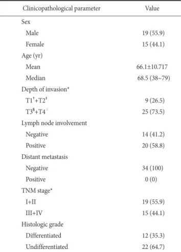
Table 1. Basic characteristics and clinical characteristics (n=34) Clinicopathological parameter Value Sex
Male 19 (55.9)
Female 15 (44.1)
Age (yr)
Mean 66.1±10.717
Median 68.5 (38~79)
Depth of invasion*
T1†+T2‡ 9 (26.5)
T3§+T4∥ 25 (73.5)
Lymph node involvement
Negative 14 (41.2)
Positive 20 (58.8)
Distant metastasis
Negative 34 (100)
Positive 0 (0)
TNM stage*
I+II 19 (55.9)
III+IV 15 (44.1)
Histologic grade
Differentiated 12 (35.3)
Undifferentiated 22 (64.7)
Values are presented as number (%), mean±standard deviation, or median (range). *Classification according to the American Joint Committee on Cancer 7th edition. †Lamina propria, muscularis mucosae, or submucosa. ‡Muscularis propria. §Penetrates subserosal connective tissue without invasion of visceral peritoneum or adjacent structures. ∥Serosa (visceral peritoneum) or adjacent structures.
4. Reverse transcription and real-time quantitative polymerase chain reaction
To validate the fold change results of the miRNA arrays, we used real-time qPCR to assess the expression of four randomly selected miRNAs including two upregulated (miR-196b-5p and miR-375) and two downregulated (miR-483-5p and miR-486- 5p) miRNAs (Table 2). Total RNA was extracted from 34 paired cancer and nontumor tissues using NucleoSpin RNA II Kit (Macherey-Nagel, Düren, Germany). Reverse transcription was performed using mature miRNA-specific primer sets and the miRNA Reverse Transcription Kit (Applied Biosystems, Foster City, CA, USA). We purchased miRNA-specific TaqMan-based probes from Applied Biosystems, and real-time qPCR was per- formed on a 7500 Fast Real-Time PCR System (Applied Bio- systems). The fold change for each miRNA was calculated using the comparative Ct (2–∆∆Ct) method, and RNU48 (a small nuclear RNA) served as an endogenous control. We performed all of the reactions in triplicate for each sample.
5. Gene ontology analysis
We used this analysis to predict target gene function and to conduct pathway analysis of the differentially expressed mi- RNAs identified in GeneChip microarray analysis (Table 3). The DAVID bioinformatics resources tool was used for this purpose.
6. Statistical analysis
This analysis was performed in the PASW Statistics ver. 18.0 (IBM Co., Armonk, NY, USA). Student’s t-test was used for evaluation of differences in miRNA expression between cancer and normal tissues and for measuring significance in clinico- pathological parameters. Data were considered statistically sig- nificant at P<0.05.
Results
1. MicroRNA expression profiles in gastric cancer and normal gastric tissues
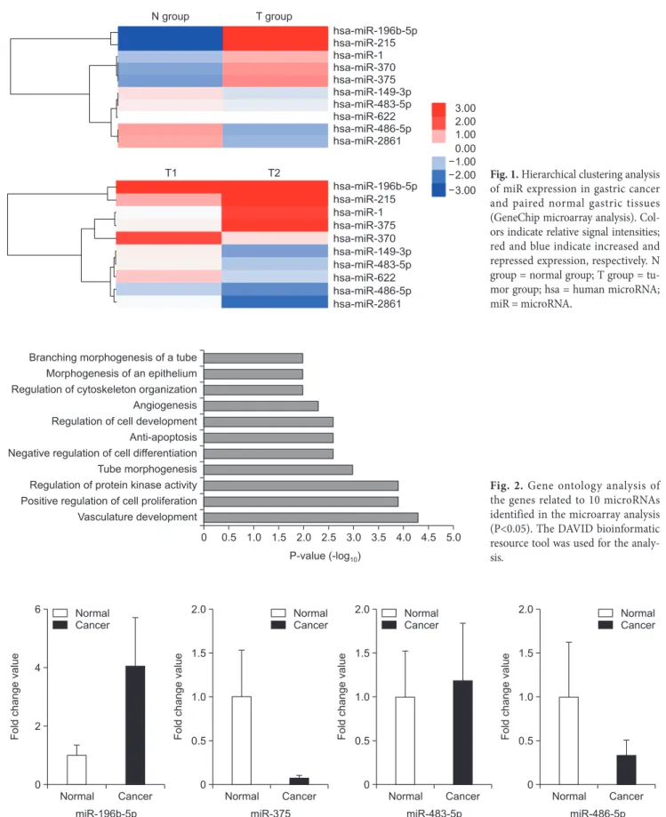
miRNA expression patterns significantly differed between gastric cancer and normal gastric tissues (Fig. 1). Fold-change filters included the requirements that the miRNAs be expressed at greater than or equal to 2 fold of control levels for upregu- lated miRNAs and less than 2 fold of control levels for down- regulated miRNAs (Table 3).
Ten miRNAs satisfied the above requirements. Among these differentially expressed miRNAs, 5 (miR-196b-5p, miR-215, miR-375, miR-1, and miR-370) were significantly overex- pressed, and 5 (miR-2861, miR-483-5p, miR-486-5p, miR-622, and miR-149-3p) were significantly underexpressed in gastric cancer tissues compared with normal gastric tissues.
Table 3. MicroRNAs (miRNA) with altered expression in gastric cancer tissues compared (GeneChip microarray analysis)
miRNA Fold-change
Upregulated miRNAs
hsa-miR-215 4.734756
hsa-miR-375 4.603461
hsa-miR-1 2.505603
hsa-miR-370 2.390836
hsa-miR-196b-5p 5.503479
Downregulated miRNAs
hsa-miR-2861 –2.6286
hsa-miR-483-5p –2.481
hsa-miR-622 –2.2829
hsa-miR-149-3p –2.2675
hsa-miR-486-5p –2.4789
hsa = human microRNA; miR = microRNA.
Table 2. The information on primers for real-time polymerase chain reaction
MicroRNA Company Country Product number
miR-196b-5p Applied Biosystems Foster City, CA, USA 002215
miR-375 Applied Biosystems Foster City, CA, USA 000564
miR-483-5p Applied Biosystems Foster City, CA, USA 002338
miR-486-5p Applied Biosystems Foster City, CA, USA 001278
miR = microRNA.
0 Branching morphogenesis of a tube
Morphogenesis of an epithelium Regulation of cytoskeleton organization Angiogenesis Regulation of cell development Anti-apoptosis Negative regulation of cell differentiation Tube morphogenesis Regulation of protein kinase activity Positive regulation of cell proliferation Vasculature development
0.5 1.0 1.5 2.0 2.5 3.0 3.5 4.0 4.5 5.0 P-value -log( 10)
Fig. 2. Gene ontology analysis of the genes related to 10 microRNAs identified in the microarray analysis (P<0.05). The DAVID bioinformatic resource tool was used for the analy- sis.
Foldchangevalue
Normal 0
Cancer miR-196b-5p 6
4
2
Foldchangevalue
Normal 0
Cancer miR-375 2.0
1.5
1.0
0.5 Foldchangevalue
Normal 0
Cancer miR-483-5p 2.0
1.5
1.0
0.5 Foldchangevalue
Normal 0
Cancer miR-486-5p 2.0
1.5
1.0
0.5 Normal
Cancer
Normal Cancer
Normal Cancer
Normal Cancer
Fig. 3. Summary of real-time quantitative polymerase chain reaction analysis. The increased (miR-196b-5p and miR-375) and the repressed (miR- 483-5p and miR-486-5p) miRNAs are presented. The miR-483-5p and miR-375 results were contrary to the microarray results. Four selected miR- NAs are listed on the X-axis, and relative expression levels are indicated on the Y-axis. Two miRNAs (miR-196b-5p and miR-375) demonstrated significantly different expression levels between gastric cancer tissues and adjacent non-tumorous tissues (P<0.05). miR = microRNA.
3.00 2.00 1.00 0.00 1.00 2.00 3.00
N group T group
hsa-miR-196b-5p hsa-miR-215 hsa-miR-1 hsa-miR-370 hsa-miR-375 hsa-miR-149-3p hsa-miR-483-5p hsa-miR-622 hsa-miR-486-5p hsa-miR-2861
hsa-miR-196b-5p hsa-miR-215 hsa-miR-1 hsa-miR-375 hsa-miR-370 hsa-miR-149-3p hsa-miR-483-5p hsa-miR-622 hsa-miR-486-5p hsa-miR-2861
T1 T2 Fig. 1. Hierarchical clustering analysis
of miR expression in gastric cancer and paired normal gastric tissues (GeneChip microarray analysis). Col- ors indicate relative signal intensities;
red and blue indicate increased and repressed expression, respectively. N group = normal group; T group = tu- mor group; hsa = human micro RNA;
miR = microRNA.
2. Gene ontology analysis
The pathways for the 10 miRNAs identified in the microar- ray analysis included vascular development, positive regulation of cell proliferation, regulation of protein kinase activity, tube morphogenesis, negative regulation of cell differentiation, anti- apoptosis, regulation of cell development, angiogenesis, regula- tion of cytoskeleton organization, morphogenesis of an epithe- lium, and branching morphogenesis of a tube (listed in the order of decreasing frequency; Fig. 2).
3. Validation of microarray data by real-time quantitative polymerase chain reaction
miR-196b-5p and miR-486-5p were upregulated 4.04- and 1.17-fold, respectively, in the gastric cancer tissues compared with the adjacent nontumorous gastric tissues. miR-375 and miR-483-5p were downregulated 14.2- and 3.1-fold, respec- tively, in the gastric cancer tissues compared with the non- tumorous gastric tissues. The relative expression changes of 2 of these miRNAs (miR-196b-5p and miR-486-5p) according to qPCR analysis were consistent with the microarray analysis, but two miRNAs (miR-375 and miR-483-5p) yielded the opposite results, as noted in the histogram (P<0.05) in Fig. 3. miR-196b- 5p and miR-375 showed statistically significant differences in the qPCR analysis.
4. Relations between miRNA expression and clinical features of the patients with gastric cancer We analyzed the relations between clinical characteristics and the qPCR results for the four miRNAs (miR-196b-5p, miR- 375, miR-483-5p, and miR-486-5p). miR-375 was found to be associated with low T-stage cancers (T1+T2) and differentiated histologic grade (Table 4); the other miRNAs had no significant associations.
Discussion
Recently, aberrant miRNA expression was reported in vari- ous solid cancers, including breast, lung, pancreas, and colon cancer.8-12 Altered miRNA expression contributes to cancer initiation and progression. The relations between miRNAs and tumors have thus become the focus of many cancer studies. Nu- merous research articles describe the utility of miRNAs as bio- markers for early detection and for progression, recurrence, and theranostics.13 However, few researchers have assessed miRNAs in gastric cancer in Korea.
In gastric cancer, various miRNAs have been reported to be associated with tumor development, progression, metastasis, and prognosis. Overexpressed miR-296b-5p significantly accelerates gastric cancer cell growth through attenuation of Caudal-related homeobox 1-induced antigrowth effects.14 miR-223 provokes Table 4. Correlation between microRNA (miR) expression and clinical characteristics of patients
Clinicopathological parameter
miR-196b-5p miR-375 miR-483-5p miR-486-5p
Mean fold change P-value Mean fold change P-value Mean fold change P-value Mean fold change P-value Depth of invasion*
T1†+T2‡ 1.582 0.122 0.191 0.020¶ 3.074 0.109 0.134 0.334
T3§+T4∥ 5.820 0.005 0.808 0.215
Lymph node involvement
Negative 5.480 0.304 0.155 0.097 1.671 0.318 0.106 0.242
Positive 3.517 0.041 0.963 0.227
TNM stage*
I+II 4.228 0.484 0.118 0.163 1.240 0.489 0.083 0.076
III+IV 4.083 0.033 1.200 0.319
Histologic grade
Differentiated 2.235 0.194 0.170 0.032¶ 2.460 0.174 0.135 0.341
Undifferentiated 5.404 0.006 0.856 0.208
*Classification according to the American Joint Committee on Cancer 7th edition. †Lamina propria, muscularis mucosae, or submucosa.
‡Muscularis propria. §Penetrates subserosal connective tissue without invasion of visceral peritoneum or adjacent structures. ∥Serosa (visceral peritoneum) or adjacent structures. ¶Statistically significant (P<0.05).
migration of nonmetastatic gastric cancer cells and invasion in vitro and in vivo by suppressing erythrocyte membrane pro- tein band 4.1-like 3 expression.15 Li et al.16 demonstrated that a 7-miRNA signature (miR-10b, miR-21, miR-223, miR-338, let-7a, miR-30a-5p, and miR-126) can predict the clinical outcome of gastric cancer. There have been studies showing that circulating miRNA in blood could serve as noninvasive biomarkers. The plasma concentrations of miR-17-5p, miR- 21, miR-106a, miR-106b, and miR-18a have been reported to be significantly higher in patients with gastric cancer than in healthy volunteers.17,18 Another study showed that miR-21 has clinical value as a noninvasive biomarker and potential diag- nostic value with good specificity and moderate sensitivity.19 To date, definitive noninvasive biomarkers have not been found, and larger-scale studies are required.
In this study, we identified miRNAs with significantly up- or down-regulated expression in gastric cancer tissues versus adjacent nontumorous tissues. We identified 5 upregulated and 5 downregulated miRNAs in gastric cancer tissues by GeneChip microarray analysis. Of these 10, we validated 2 upregulated and 2 downregulated miRNAs by qPCR. The 2 upregulated miRNAs are miR-196b-5p and miR-375, and the 2 downregulated mi- RNAs are miR-483-5p and miR-486-5p. We also determined that miR-196b-5p and miR-375 are significantly associated with gastric cancer.
The miR-196 family is encoded at three paralogous loci in the mammalian homeobox (HOX) clusters, and several HOX genes are regulated via targeting of their 3′untranslated region.20 The miR-196 family is composed of miR-196a and miR-196b, and mature miR-196b differs from miR-196a by 1 nt.21 miR- 196 is upregulated in pancreatic adenocarcinoma, breast cancer, leukemia, esophageal adenocarcinoma, and colonic cancer,22-26 and downregulated in melanoma.27 Several reports on miR-196 in gastric cancer have been published. Tsai et al.28 noticed that the lack of promoter methylation in miR-196b may explain its overexpression in the majority of gastric cancers and that miR- 196b overexpression may be a tumor marker. This group later reported that miR-196b expression is significantly repressed by the transcription factor E26 transformation-specific sequence-2 (ETS2), whose expression is implicated in a reduced incidence of solid tumors.29 A knockdown of ETS2 significantly promotes migration and invasiveness of gastric cancer cells.29 Alterations in ETS2 and miR-196b expression in gastric cancer cell lines influence the expression of epithelial-mesenchymal-transition-
related genes.29 Sun et al.30 reported that aberrant miR-196a overexpression and consequent downregulation of p27kip1 may contribute to gastric carcinogenesis. In another study, miR- 196a/-196b levels were found to be frequently upregulated in human gastric cancer and significantly associated with clinically advanced stages and lymph node metastasis. In that experiment, miR-196a and miR-196b promoted gastric cancer cell migration and invasion in vitro and metastasis in vivo, and radixin was iden- tified as a direct functional target of miR-196a and miR-196b.31
Our study revealed that miR-196b-5p expression is increased in gastric cancer tissues compared with nontumorous tissues. To date, there have been a few studies on miR-196b-5p in gastric cancer. Xie et al.32 and Liu et al.33 reported that miR-196b-5p is downregulated in a gastric cancer cell line. This result contradicts ours, but those studies were conducted on a gastric cancer cell line rather than primary tumor tissues. Another microarray study revealed that miR-196b-5p is upregulated in human gastric can- cer tissues, in agreement with our results, but no validation was performed there.34
miR-375 is predominantly expressed in islets of the pan- creas.35 miR-375 has an important function in the formation of insulin-secreting pancreatic islets; this result was obtained by inhibition of miR-375 maturation with morpholinos in zebraf- ish.36 miR-375 is downregulated in multiple types of cancer and it inhibits the hallmarks of cancer by targeting several key onco- genes, including Yes associated protein 1 (YAP1), astrocyte el- evated gene-1 protein (AEG-1), 3-phosphoinositide dependent protein kinase-1 (PDK1), and insulin-like growth factor (IGF) 1R.37 Ding et al.38 reported that miR-375 is repressed in more than 90% of stomach cancer samples compared with their non- tumorous counterparts and suppresses proliferation of malignant stomach cells partially by targeting Janus kinase 2 (JAK2). Tsu- kamoto et al.39 also reported that reduced miR-375 expression may enhance survival of gastric carcinoma tumors by activation of the PDK1/Akt survival pathway; therefore, miR-375 may be a tumor suppressor miRNA in stomach cancer. Helicobacter pylori infection is known to be a risk factor of gastric cancer.
In one study, miR-375 was found to be downregulated in re- sponse to H. pylori infection and to target JAK2. JAK2 activates signal transducer and activator of transcription 3 and promotes neoplastic transformation by changing the expression of B cell lymphoma 2 (BCL-2) and twist related protein 1 (TWIST1).40 Conversely, Zhang et al.41 reported that expression of miR-375 is increased and may predict recurrence risk in patients with
recurrent stomach cancer after surgical resection. Because of these discrepancies, further studies involving clinical data are needed to confirm the function of miR-375 in gastric cancer. In the present study, miR-375 was downregulated according to the qPCR analysis but upregulated in the microarray analysis.
The miR-483-5p gene is located in the chromosomal 11p15.5 region in the second intron of the IGF2 gene and expresses two mature forms (miR-483-5p and miR-483-3p).42 To date, few studies have been conducted to reveal the function of miR-483- 5p in gastric cancer, and its relation to carcinogenesis remains uncertain. Wang et al.42 reported that miR-483-5p is downregu- lated in human gliomas compared with normal brain tissues and that miR-483-5p can repress glioma cell proliferation by di- rectly targeting extracellular signal regulated kinase 1, which is a core controller of cell proliferation and cell differentiation. Qiao et al.43 suggested that miR-483-5p plays a role of an angiogen- esis inhibitor. In contrast, miR-483-5p is reportedly upregulated in plasma samples of patients with hepatocellular carcinoma.44 miR-483-5p expression was found to be downregulated in the microarray analysis in the present study. We were unable to confirm the association of miR-483-5p with gastric cancer by qPCR analysis.
miR-486 is located in chromosomal 8p11 region, the site undergoing frequent genomic loss in various carcinomas.45 Solo- mides et al.46 and Tan et al.47 found that miR-486-5p is down- regulated in lung cancer tissue and may serve as a new diagnos- tic biomarker of lung cancer. Oh et al.45 reported that miR-486 expression is decreased in gastric cancer, and that olfactomedin4 is a direct target gene. Recently, Zhu et al.48 reported that miR- 486-5p concentration is consistently elevated in plasma from patients with gastric cancer. The exact roles of this miRNA in human cancers have not been fully characterized to date. In the present study, miR-486-5p expression was decreased in the mi- croarray, but the qPCR results were not significant.
We also analyzed the relation between these miRNAs and clinical features. A lower T stage (T1+T2) and a differentiated histo- logic type were associated with high miR-375 expression. This result supports the previously reported function of miR-375 as a tumor suppressor.38,39 Other miRNAs yielded no significant correlations.
We predicted possible genetic pathways related to the 10 above-mentioned miRNAs using bioinformatics resource tool analysis. Among the pathways involved, regulation of protein kinase activity was related to both miR-375 and miR-483-5p, and angiogenesis was related to miR-483-5p.
The interaction between dysregulated miRNAs and their po- tential target genes is complex, and miRNAs can be influenced by various factors, such as pathology, hypoxia, infection, and cytotoxictreatment.49 This complexity may explain the inconsis- tent results on miR-375 and miR-483-5p in our study.
There are a few limitations to this study. First, our study in- cludes a relatively small number of patients. Thus, further large- scale research is required to clarify the exact roles of miRNAs in gastric cancer. Second, we could not study the functional roles of the significantly expressed miRNAs in gastric cancer.
However, our study does provide basic data for future research on mi RNAs in gastric cancer. The data from this study should facilitate further research on gastric cancer aimed at elucidation of the functional roles and clinical significance of miRNAs or at the development of miRNA-based noninvasive biomarkers.
In conclusion, our results revealed that several miRNAs are significantly differentially expressed between gastric cancer tis- sues and nontumorous tissues. We used real-time qPCR analy- sis to validate the expression of 4 randomly selected miRNAs, including 2 downregulated and 2 upregulated miRNAs. Two mi RNAs (miR-196b-5p and miR-375) were significantly ex- pressed in the qPCR validation analysis. miR-196b-5p expres- sion was consistent with the microarray analysis, but miR-375 yielded the opposite results. We propose that these 2 validated miRNAs may serve as diagnostic biomarkers of gastric cancer.
Conflicts of Interest
No potential conflict of interest relevant to this article was reported.
References
1. Danaei G, Vander Hoorn S, Lopez AD, Murray CJ, Ezzati M;
Comparative Risk Assessment collaborating group (Cancers).
Causes of cancer in the world: comparative risk assessment of nine behavioural and environmental risk factors. Lancet 2005;366:1784-1793.
2. Jung KW, Park S, Kong HJ, Won YJ, Lee JY, Seo HG, et al.
Cancer statistics in Korea: incidence, mortality, survival, and prevalence in 2009. Cancer Res Treat 2012;44:11-24.
3. Hartgrink HH, Jansen EP, van Grieken NC, van de Velde CJ.
Gastric cancer. Lancet 2009;374:477-490.
4. Calin GA, Croce CM. MicroRNA signatures in human can-
cers. Nat Rev Cancer 2006;6:857-866.
5. Iorio MV, Croce CM. MicroRNAs in cancer: small molecules with a huge impact. J Clin Oncol 2009;27:5848-5856.
6. Link A, Kupcinskas J, Wex T, Malfertheiner P. Macro-role of microRNA in gastric cancer. Dig Dis 2012;30:255-267.
7. Edge SB, Compton CC. The American Joint Committee on Cancer: the 7th edition of the AJCC cancer staging manual and the future of TNM. Ann Surg Oncol 2010;17:1471-1474.
8. Melo S, Villanueva A, Moutinho C, Davalos V, Spizzo R, Ivan C, et al. Small molecule enoxacin is a cancer-specific growth inhibitor that acts by enhancing TAR RNA-binding protein 2-mediated microRNA processing. Proc Natl Acad Sci U S A 2011;108:4394-4399.
9. Volinia S, Calin GA, Liu CG, Ambs S, Cimmino A, Petrocca F, et al. A microRNA expression signature of human solid tumors defines cancer gene targets. Proc Natl Acad Sci U S A 2006;103:2257-2261.
10. Iorio MV, Ferracin M, Liu CG, Veronese A, Spizzo R, Sabbioni S, et al. MicroRNA gene expression deregulation in human breast cancer. Cancer Res 2005;65:7065-7070.
11. Yanaihara N, Caplen N, Bowman E, Seike M, Kumamoto K, Yi M, et al. Unique microRNA molecular profiles in lung cancer diagnosis and prognosis. Cancer Cell 2006;9:189-198.
12. Bloomston M, Frankel WL, Petrocca F, Volinia S, Alder H, Hagan JP, et al. MicroRNA expression patterns to differentiate pancreatic adenocarcinoma from normal pancreas and chronic pancreatitis. JAMA 2007;297:1901-1908.
13. Wang F, Sun GP, Zou YF, Hao JQ, Zhong F, Ren WJ. Micro- RNAs as promising biomarkers for gastric cancer. Cancer Bio- mark 2012;11:259-267.
14. Li T, Lu YY, Zhao XD, Guo HQ, Liu CH, Li H, et al. Mi- croRNA-296-5p increases proliferation in gastric cancer through repression of Caudal-related homeobox 1. Oncogene 2014;33:783-793.
15. Li X, Zhang Y, Zhang H, Liu X, Gong T, Li M, et al. miRNA- 223 promotes gastric cancer invasion and metastasis by target- ing tumor suppressor EPB41L3. Mol Cancer Res 2011;9:824- 833.
16. Li X, Zhang Y, Zhang Y, Ding J, Wu K, Fan D. Survival predic- tion of gastric cancer by a seven-microRNA signature. Gut 2010;59:579-585.
17. Tsujiura M, Komatsu S, Ichikawa D, Shiozaki A, Konishi H, Takeshita H, et al. Circulating miR-18a in plasma contributes to cancer detection and monitoring in patients with gastric
cancer. Gastric Cancer 2015;18:271-279.
18. Tsujiura M, Ichikawa D, Komatsu S, Shiozaki A, Takeshita H, Kosuga T, et al. Circulating microRNAs in plasma of patients with gastric cancers. Br J Cancer 2010;102:1174-1179.
19. Zeng Z, Wang J, Zhao L, Hu P, Zhang H, Tang X, et al. Poten- tial role of microRNA-21 in the diagnosis of gastric cancer: a meta-analysis. PLoS One 2013;8:e73278.
20. McGlinn E, Yekta S, Mansfield JH, Soutschek J, Bartel DP, Tabin CJ. In ovo application of antagomiRs indicates a role for miR-196 in patterning the chick axial skeleton through Hox gene regulation. Proc Natl Acad Sci U S A 2009;106:18610- 18615.
21. Chen C, Zhang Y, Zhang L, Weakley SM, Yao Q. MicroRNA- 196: critical roles and clinical applications in development and cancer. J Cell Mol Med 2011;15:14-23.
22. Hui AB, Shi W, Boutros PC, Miller N, Pintilie M, Fyles T, et al.
Robust global micro-RNA profiling with formalin-fixed par- affin-embedded breast cancer tissues. Lab Invest 2009;89:597- 606.
23. Zhang Y, Li M, Wang H, Fisher WE, Lin PH, Yao Q, et al.
Profiling of 95 microRNAs in pancreatic cancer cell lines and surgical specimens by real-time PCR analysis. World J Surg 2009;33:698-709.
24. Schotte D, Chau JC, Sylvester G, Liu G, Chen C, van der Velden VH, et al. Identification of new microRNA genes and aberrant microRNA profiles in childhood acute lymphoblastic leukemia. Leukemia 2009;23:313-322.
25. Maru DM, Singh RR, Hannah C, Albarracin CT, Li YX, Abra- ham R, et al. MicroRNA-196a is a potential marker of progres- sion during Barrett's metaplasia-dysplasia-invasive adenocar- cinoma sequence in esophagus. Am J Pathol 2009;174:1940- 1948.
26. Schimanski CC, Frerichs K, Rahman F, Berger M, Lang H, Galle PR, et al. High miR-196a levels promote the oncogenic phenotype of colorectal cancer cells. World J Gastroenterol 2009;15:2089-2096.
27. Braig S, Mueller DW, Rothhammer T, Bosserhoff AK. Mi- croRNA miR-196a is a central regulator of HOX-B7 and BMP4 expression in malignant melanoma. Cell Mol Life Sci 2010;67:3535-3548.
28. Tsai KW, Hu LY, Wu CW, Li SC, Lai CH, Kao HW, et al. Epi- genetic regulation of miR-196b expression in gastric cancer.
Genes Chromosomes Cancer 2010;49:969-980.
29. Liao YL, Hu LY, Tsai KW, Wu CW, Chan WC, Li SC, et al.
Transcriptional regulation of miR-196b by ETS2 in gastric can- cer cells. Carcinogenesis 2012;33:760-769.
30. Sun M, Liu XH, Li JH, Yang JS, Zhang EB, Yin DD, et al.
MiR-196a is upregulated in gastric cancer and promotes cell proliferation by downregulating p27(kip1). Mol Cancer Ther 2012;11:842-852.
31. Tsai MM, Wang CS, Tsai CY, Chen CY, Chi HC, Tseng YH, et al. MicroRNA-196a/-196b promote cell metastasis via nega- tive regulation of radixin in human gastric cancer. Cancer Lett 2014;351:222-231.
32. Xie J, Tan ZH, Tang X, Mo MS, Liu YP, Gan RL, et al. MiR- 374b-5p suppresses RECK expression and promotes gastric cancer cell invasion and metastasis. World J Gastroenterol 2014;20:17439-17447.
33. Liu J, Ma L, Wang Z, Wang L, Liu C, Chen R, et al. MicroRNA expression profile of gastric cancer stem cells in the MKN- 45 cancer cell line. Acta Biochim Biophys Sin (Shanghai) 2014;46:92-99.
34. Chang H, Kim N, Park JH, Nam RH, Choi YJ, Lee HS, et al. Different microRNA expression levels in gastric can- cer depending on Helicobacter pylori infection. Gut Liver 2015;9:188-196.
35. Poy MN, Eliasson L, Krutzfeldt J, Kuwajima S, Ma X, Macdon- ald PE, et al. A pancreatic islet-specific microRNA regulates insulin secretion. Nature 2004;432:226-230.
36. Kloosterman WP, Lagendijk AK, Ketting RF, Moulton JD, Plasterk RH. Targeted inhibition of miRNA maturation with morpholinos reveals a role for miR-375 in pancreatic islet de- velopment. PLoS Biol 2007;5:e203.
37. Yan JW, Lin JS, He XX. The emerging role of miR-375 in can- cer. Int J Cancer 2014;135:1011-1018.
38. Ding L, Xu Y, Zhang W, Deng Y, Si M, Du Y, et al. MiR-375 fre- quently downregulated in gastric cancer inhibits cell prolifera- tion by targeting JAK2. Cell Res 2010;20:784-793.
39. Tsukamoto Y, Nakada C, Noguchi T, Tanigawa M, Nguyen LT, Uchida T, et al. MicroRNA-375 is downregulated in gastric
carcinomas and regulates cell survival by targeting PDK1 and 14-3-3zeta. Cancer Res 2010;70:2339-2349.
40. Miao L, Liu K, Xie M, Xing Y, Xi T. miR-375 inhibits Helico- bacter pylori-induced gastric carcinogenesis by blocking JAK2- STAT3 signaling. Cancer Immunol Immunother 2014;63:699- 711.
41. Zhang X, Yan Z, Zhang J, Gong L, Li W, Cui J, et al. Combina- tion of hsa-miR-375 and hsa-miR-142-5p as a predictor for recurrence risk in gastric cancer patients following surgical resection. Ann Oncol 2011;22:2257-2266.
42. Wang L, Shi M, Hou S, Ding B, Liu L, Ji X, et al. MiR-483-5p suppresses the proliferation of glioma cells via directly target- ing ERK1. FEBS Lett 2012;586:1312-1327.
43. Qiao Y, Ma N, Wang X, Hui Y, Li F, Xiang Y, et al. MiR-483-5p controls angiogenesis in vitro and targets serum response fac- tor. FEBS Lett 2011;585:3095-3100.
44. Shen J, Wang A, Wang Q, Gurvich I, Siegel AB, Remotti H, et al. Exploration of genome-wide circulating microRNA in he- patocellular carcinoma: MiR-483-5p as a potential biomarker.
Cancer Epidemiol Biomarkers Prev 2013;22:2364-2373.
45. Oh HK, Tan AL, Das K, Ooi CH, Deng NT, Tan IB, et al. Ge- nomic loss of miR-486 regulates tumor progression and the OLFM4 antiapoptotic factor in gastric cancer. Clin Cancer Res 2011;17:2657-2667.
46. Solomides CC, Evans BJ, Navenot JM, Vadigepalli R, Peiper SC, Wang ZX. MicroRNA profiling in lung cancer reveals new molecular markers for diagnosis. Acta Cytol 2012;56:645-654.
47. Tan X, Qin W, Zhang L, Hang J, Li B, Zhang C, et al. A 5-micro RNA signature for lung squamous cell carcinoma diagnosis and hsa-miR-31 for prognosis. Clin Cancer Res 2011;17:6802-6811.
48. Zhu C, Ren C, Han J, Ding Y, Du J, Dai N, et al. A five-microRNA panel in plasma was identified as potential biomarker for early detection of gastric cancer. Br J Cancer 2014;110:2291-2299.
49. Wang J, Wang Q, Liu H, Hu B, Zhou W, Cheng Y. MicroRNA expression and its implication for the diagnosis and therapeu- tic strategies of gastric cancer. Cancer Lett 2010;297:137-143.