Introduction
The increased left ventricular diastolic filling pressure evolves in left ventricular (LV) diastolic dysfunction.1)2) This haemody- namic condition usually is demonstrated by the impairment of E/A mitral inflow ratio (E/A < 1) or by the change of nor- mal pattern of pulmonary veins’ flow. The combination of early inflow velocity curve and tissue Doppler imaging of the mitral annulus (E/E’ ratio) better estimates this condition.
But, in the absence of any mitral valve derangement, LV dia- stolic dysfunction directly affects Left Atrial Volume (LAV).
This parameter can be easily measured by two-dimensional echocardiography and indexed to the body surface area (BSA)
as left atrial volume index (LAVI).3)4) Therefore, LAVI also may be used as faithful indicator of LV diastolic dysfunction.5) On the other hand, LV function can be adequately evaluated by myocardial performance index (MPI).6)7) This (also called Tei in- dex) can be measured either with conventional Doppler method or tissue Doppler echocardiography (TDE).8) This last method has the advantage to directly assess transmural myocardial ve- locities.9) In addition, TDE-MPI is more sensitive than the con- ventional Doppler MPI in to define LV function, especially in the presence of regional wall motion abnormality.9)
In this study, we evaluated the relationship between LAVI and diastolic LV function defined with TDE-MPI in a group ORIGINAL ARTICLE J Cardiovasc Ultrasound 2012;20(1):25-29
Left Atrial Volume Index as Indicator of Left Ventricular Diastolic
Dysfunction: Comparation
between Left Atrial Volume Index and Tissue Myocardial Performance Index
Fulvio Cacciapuoti, MD, Anna Scognamiglio, MD, Venere Delli Paoli, MD, Concetta Romano, MD and Federico Cacciapuoti, MD
Echocardiographic Laboratory, Department of Internal Medicine and Geriatry, Second University of Naples, Naples, Italy
Background: To point out a possible correlation between left atrial volume index (LAVI) and left ventricular (LV) diastolic time interval to better define LV diastolic dysfunction, this study was performed.
Methods: In 62 hypertensive-hypertrophic patients without LV systolic dysfunction, LV volumes, myocardial mass index, ejection fraction% (EF%) and LAVI were measured by two-dimensional echocardiography. Instead, tissue Doppler echocardiography (TDE) was used to measure myocardial performance index (MPI) and its systo-diastolic time intervals, such as:
iso-volumetric contraction time (IVCT); iso-volumetric relaxation time (IVRT); ejection time. LAVI, TDE-MPI and time intervals where also measured in 15 healthy controls, to obtain the reference values.
Results: Results shown a significant increase of LV volumes in hypertensives in comparison to the control group (p < 0.05). LV mass index also augmented (p < 0.001). Instead, EF% not significantly changed in hypertrophic patients in comparison with healthy controls. LAVI raised in hypertensives wih left ventricular hypertrophy, whereas IVCT resulted within the normal limits.
On the contrary, IVRT significantly raised. Accordingly, MPI resulted higher in controls.
Conclusion: LAVI, MPI and its time intervals appear as reliable tools to non-invasively individualize LV diastolic dysfunction in systemic hypertension, in absence of mitral valve disease.
KEY WORDS: Left atrial volume index · Left ventricular hypertrophy · Myocardial performance index · LV diastolic dysfunction · Ejection fraction%.
• Received: September 27, 2011 • Revised: February 16, 2012 • Accepted: February 17, 2012
• Address for Correspondence: Federico Cacciapuoti, Department of Internal Medicine, Faculty of Medicine, Second University of Naples, 2 Piazza L. Miraglia, Naples 80138, Italy Tel: +39-81-566-5022, Fax: +39-81-566-5022, E-mail: [email protected]
• This is an Open Access article distributed under the terms of the Creative Commons Attribution Non-Commercial License (http://creativecommons.org/licenses/by-nc/3.0) which permits unrestricted non-commercial use, distribution, and reproduction in any medium, provided the original work is properly cited.
of hypertensive patients with LV diastolic dysfunction and ejection fraction% (EF%) > 50%.
Methods
Since October 2009 to February 2011, 62 hypertensive pa- tients (43 males and 29 females) aged from 45 to 61 years (mean age = 55 ± 6 years) and without any valvular heart dis- eases were examined. The leading epidemiological, metabolic and echocardiographic characteristics of controls and hyper- tensive patients (group II) were shown in Table 1. These were in sinus rhythm and have an echocardiographic finding of left ventricular hypertrophy (LVH).10) Coronary artery disease was excluded by coronary angiography in 24 of these, and by rest and effort myocardial SPECT in the remaining 38. Cumulative anti-hypertensive treatments given in patients of group II were shown in Table 2. In accordance with the recommenda- tions for the evaluation of LV function by echocardiography,11) the patients were diagnosed as affected by LV diastolic dys- function, with EF% > 50% (group II).12)
Fifteen (8 males and 7 females) healthy subjects (M and F;
mean age = 54 ± 3 years) was also enrolled, as controls. In these, echocardiographic left ventricular function was defined too (group I).
Conventional and tissue Doppler echocardiography
Two groups were echocardiographically examined in the left-lateral position by using a iE33 machine (Philips, Am- sterdam, the Netherlands). According to the biplane Simp- son’s rule, LV end-diastolic volume (LVEDV) and LV end-sys- tolic volume (LVESV) in mL and EF% were defined.13) Inter ventricular septum (IVS) thickness was measured (in mm) during systole. LV wall thickness was also measured (in mm).
Left ventricular mass and its indexed value was assessed by the method proposed by Devereux et al.10) Left atrial (LA) volume in systole was also measured just before the mitral valve open- ing, using the biplane Simpson’s method, as a mean between the values recorded in apical four- and two-chamber approach- es. Subsequently, LAV was indexed for BSA, such as LAVI in mL/m2.14) Finally, MPI was evaluated by using TDE method.
Pulsed-wave TDE was performed by activating the tissue Doppler function. Sample volume was placed at the lateral an- nular mitral site in apical four chamber view, in order to record the following cardiac time intervals: iso-volumetric contraction time (IVCT) in ms; iso-volumetric relaxation time (IVRT) in ms; and ejection time (ET) in ms. Images were acquired with a variable frequency phased-array transducer. The filter settings were kept low, and gains were adjusted to the minimal opti- mal level to minimize noise and eliminate the signals pro- duced by the transmitral flow. Three consecutive beats were measured and averaged for each parameter at a sweep speed of 100 mm/s. MPI was defined as the sum of IVCT and IVRT divided by ET.15) LAVI; MPI; IVCT; IVRT; and ET were de-
fined in controls (group I) too, as reference values (Table 3).
Statistical analysis
Echocardiographic data are presented as a mean values ± SD. Statistical analyses were performed with SPSS statistical software (SPSS, Chicago, IL, USA). Differences between two groups were examined by an unpaired t test. A p value < 0.05 was considered significant. Finally, LAVI was compared to MPI, IVCT, IVRT and ET between controls (group I) and hy- pertensive patients (group II) by unpaired t-test.
Results
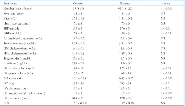
Mean values of LVEDV and LVESV were 95 ± 18 mL and 39 ± 17 mL respectively in controls (group I). These resulted 125 ± 15 mL (LVEDV), and 48 ± 11 mL (LVESV) in hyper- tensive patients (group II). Differences were significant (p <
0.05). IVS thickness was = 10 ± 0.4 mm in controls, it result- ed 135 ± 0.5 mm in hypertensive-patients. Mean value of E/A waves ratio was 1.21 ± 0.38 in normal and 0.85 ± 0.27 in hy- pertensives (p < 0.01). Mitral deceleration time (DT) resulted
= 135 ± 3.4 ms in healthy adults and 245 ± 31 in hyperten- sive patients (p < 0.01). On the other hand, LV walls’ thick- ness was 9 ± 0.3 mm in controls (group I) and 17 ± 0.2 mm in hypertensives (group II). Differences were significant (p <
0.001). The mean value of LV mass index was 90 ± 21 g/m2 in control group. It increased to 178 ± 29 g/m2 in hyperten- sive group (p < 0.001). EF% resulted of 61 ± 0.8% in controls (group I) and of 57 ± 0.9% in hypertrophic patients (group II). Differences between two groups weren’t significant (NS) (Table 1).
With reference to LAVI, a mean of 47 ± 5 mL/m2 was found in hypertensive-hypertrophic patients (group II). This value was significantly higher (p < 0.001) than that recorded in controls (group I) (23 ± 4 mL/m2). Normal values of TDE- MPI (0.34 ± 0.05) obtained in control-group significantly in- creased (p < 0.01) in patients with LV hypertrophy (0.46 ± 0.09). Particularly, IVCT resulted 28 ± 7 ms in healthy indi- viduals, almost similar to that obtained in hypertensive pa- tients (30 ± 8 ms), without significant differences (NS). On the contrary, IVRT was significantly (p < 0.001) prolonged (107 ± 9 ms) in hypertensives in comparison to healthy sub- jects (79 ± 6 ms). ET was within the limits both in normals (315 ± 10 ms) and in hypertensive patients (312 ± 10 ms) (NS) (Table 3).
Discussion
LAV may be calculated by three different methods: the bi- plane area lengh; the biplane modified Simpson’s, and the prolate ellipse method.16) Significant differences among three diverse methods exist, even through all three shown highly satisfactory reproducibility. In this study, we used biplane Simpson’s method indexed for BSA, to obtain LAVI mesaured in mL/m2. Mean val- ue of LAVI reported by several AA is 22 ± 6 mL/m2.17-21) In our
healthy controls, a mean value of 23 ± 4 mL/m2 was found. This was reported as reference value for our laboratory.
It is known that mechanical function of LA has described in three phases: reservoir; conduit, and contractile phase. The
“reservoir” corresponds to the difference between maximal and minimum LA volumes occurring in the interval-just before the opening mitral valve and just before the aortic valve open- ing. “Conduit” is the early phase of ventricular diastole. The blood is passively transferred to left ventricle just after mitral valve opening. “Contractile” phase or “booster pump” is calcu- lated as the difference between minimum and pre-atrial con- traction. It serves to augment the stroke volume. The contri- bution of three phases of LA function changes according to the diastolic properties of LV. In normal conditions, the con- tribution of reservoir, conduit and contractile function of the LA to the LV filling is 40%, 35%, and 25% respectively. As LV relaxation worsens, the contribution of different LA phases gradually increases,22) in accordance with recent experiences performed in patients with LV diastolic dysfunction.23)24)
In the present study, we evaluated the relationship between LAVI and LV diastolic dysfunction due to LV hypertrophy. LV diastolic impairment was demonstrated by the increase of IVRT and TDE-MPI. Achieved results indicate that LAVI significantly raised in comparison to the contol values in hy- pertensives with LV hypertrophy. This faithfullly certainly re- flects LV diastolic dyfunction consequent to LVH. The in-
crease of IVRT and TDE-MPI (with normal values of IVCT) can be considered as an useful and reliable tool to identify LV diastolic LV dysfunction.25)26) Several research groups previous- ly have shown that MPI and IVRT reflect LV diastolic dysfunc-
Table 1. Epidemiological, metabolic, and echocardiographic characteristics of controls and enrolled patients
Parameters Controls Patients p value
Number (male : female) 15 (8 : 7) 62 (43 : 29) p < 0.001
Mean age (years) 54 ± 3 55 ± 6 NS
BSA (m2) 1.72 ± 0.3 1.81 ± 0.2 NS
Heart rate (beats/min) 71 ± 4 73 ± 6 NS
SBP (mmHg) 135 ± 3 176 ± 5 p < 0.01
DBP (mmHg) 78 ± 5 98 ± 3 p < 0.05
Fasting blood glucose (mmol/L) 5.7 ± 0.5 5.8 ± 0.6 NS
Total cholesterol (mmol/L) 5.78 ± 0.6 5.81 ± 0.7 NS
LDL cholesterol (mmol/L) 3.1 ± 0.4 3.3 ± 0.5 NS
HDL cholesterol (mmol/L) 1.45 ± 0.3 1.48 ± 0.4 NS
Triglycerides (mmol/L) 2.6 ± 0.8 2.7 ± 0.5 NS
Creatinine (mg/dL) 0.98 ± 0.2 1.0 ± 0.1 NS
LV diastolic volume (mL) 95 ± 18 125 ± 15 p < 0.05
LV systolic volume (mL) 39 ± 17 48 ± 11 p < 0.05
E/A waves ratio 1.21 ± 0.38 0.85 ± 0.27 p < 0.001
DT (ms) 135 ± 36 245 ± 31 p < 0.01
IVS thickness (mm) 10 ± 4 13.5 ± 5 p < 0.01
LV posterior walls’ thickness (mm) 9 ± 3 17 ± 2 p < 0.001
LV mass index (gr/m2) 90.4 ± 21 178 ± 29 p < 0.001
EF% 61 ± 0.8% 57 ± 0.9% NS
Leading epidemiological, biochemical and echocardiographic characteristics of healthy controls and hypertensive-hypertrophic patients, with statistical significance. NS: not significant, BSA: body surface area, SBP: systolic blood pressure, DBP: diastolic blood pressure, LV: left ventricle, IVS: inter ventricular septum, EF%: ejection fraction%
Table 2. Cumulative anti-hypertensive drugs given in 62 hyperten- sives
ACE-I 22 patients
ARBs 24 patients
Calcium channel antagonists 13 patients
Diuretics 11 patients
Beta-blockers 12 patients
Table 3. LAVI, time intervals and MPI-TDE values in two groups
Parameters Group I Group II p value
LAVI (mL/m2) 23 ± 4 47 ± 5 p < 0.001
IVCT (ms) 28 ± 7 30 ± 8 NS
IVRT (ms) 79 ± 6 107 ± 9 p < 0.001
ET (ms) 315 ± 10 310 ± 10 NS
MPI 0.34 ± 0.05 0.46 ± 0.09 p < 0.01 Doppler results of LAVI, MPI and cardiac time intervals (IVCT, IVRT, ET) recorded in healthy controls and hypertensive patients. LAVI: left atrial volume index, IVCT: iso-volumetric contraction time, NS: not significant, IVRT: iso-volumetric relaxation time, ET: ejection time, MPI:
myocardial performance index, TDE: tissue Doppler echocardiography
tion, independently of arterial pressure,27) heart failure28) or heart rate,29) in presence of preserved systolic function especial- ly.30) A previous study has also demonstrated an association be- tween LVH induced by systemic hypertension and left atrial dimension.31) Successively, Pritchett et al.32) evidenced that LAVI is a highly sensitive and specific tool for the detection of severe LV diastolic dysfunction (III degree of diastolic dys- function). These AAs. = Authors also demonstrated that LAVI may better reflect the cumulative effect of increased LV filling pressures over time in comparison to the Doppler in- dexes, as E/A ratio, DT and E/E’ ratio (that reflect increased LV filling pressures at one point in time). The incremental value of LAVI measurement is its prognostic implications to- wards cardiovascular death and/or adverse cardiovascular out- comes in hypertensive patients with LV diastolic dysfunction, as recently demonstrated by Leung et al.33)
In the present report, we firstly identified LV diastolic dys- function using TDE-MPI. LAVI (in the absence of any mitral disease) appeared also expressive of LV diastolic dysfunction, further confirming the relationship between LAV and LV dia- stolic dysfunction. But, other studies performed in a wide range are requested to definitively demonstrate the relation- ship among LAVI, TDE-MPI and LV diastolic dysfunction.
References
1. Appleton CP, Galloway JM, Gonzalez MS, Gaballa M, Basnight MA. Estimation of left ventricular filling pressures using two-dimensional and Doppler echocardiography in adult patients with cardiac disease. Addi- tional value of analyzing left atrial size, left atrial ejection fraction and the difference in duration of pulmonary venous and mitral flow velocity at atrial contraction. J Am Coll Cardiol 1993;22:1972-82.
2. Simek CL, Feldman MD, Haber HL, Wu CC, Jayaweera AR, Kaul S. Relationship between left ventricular wall thickness and left atrial size:
comparison with other measures of diastolic function. J Am Soc Echocardiogr 1995;8:37-47.
3. Schabelman S, Schiller NB, Silverman NH, Ports TA. Left atrial volume estimation by two-dimensional echocardiography. Cathet Cardiovasc Diagn 1981;7:165-78.
4. Schiller NB, Botvinick EH. Noninvasive quantitation of the left heart by echocardiography and scintigraphy. Cardiovasc Clin 1986;17:45-93.
5. Moller JE, Hillis GS, Oh JK, Seward JB, Reeder GS, Wright RS, Park SW, Bailey KR, Pellikka PA. Left atrial volume: a powerful pre- dictor of survival after acute myocardial infarction. Circulation 2003;107:
2207-12.
6. Tei C, Ling LH, Hodge DO, Bailey KR, Oh JK, Rodeheffer RJ, Ta- jik AJ, Seward JB. New index of combined systolic and diastolic myocardial performance: a simple and reproducible measure of cardiac function--a study in normals and dilated cardiomyopathy. J Cardiol 1995;26:357-66.
7. Tei C, Dujardin KS, Hodge DO, Kyle RA, Tajik AJ, Seward JB.
Doppler index combining systolic and diastolic myocardial performance: clini- cal value in cardiac amyloidosis. J Am Coll Cardiol 1996;28:658-64.
8. Isaaz K. Tissue Doppler imaging for the assessment of left ventricular systol- ic and diastolic functions. Curr Opin Cardiol 2002;17:431-42.
9. Tekten T, Onbasili AO, Ceyhan C, Unal S, Discigil B. Value of mea- suring myocardial performance index by tissue Doppler echocardiography in normal and diseased heart. Jpn Heart J 2003;44:403-16.
10. Devereux RB, Alonso DR, Lutas EM, Gottlieb GJ, Campo E, Sachs I, Reichek N. Echocardiographic assessment of left ventricular hypertrophy:
comparison to necropsy findings. Am J Cardiol 1986;57:450-8.
11. Nagueh SF, Appleton CP, Gillebert TC, Marino PN, Oh JK, Smis- eth OA, Waggoner AD, Flachskampf FA, Pellikka PA, Evangelista A. Recommendations for the evaluation of left ventricular diastolic function by echocardiography. J Am Soc Echocardiogr 2009;22:107-33.
12. Lang RM, Bierig M, Devereux RB, Flachskampf FA, Foster E, Pel- likka PA, Picard MH, Roman MJ, Seward J, Shanewise JS, Solomon SD, Spencer KT, Sutton MS, Stewart WJ; Chamber Quantification Writing Group; American Society of Echocardiography’s Guidelines and Standards Committee; European Association of Echocardiogra- phy. Recommendations for chamber quantification: a report from the Ameri- can Society of Echocardiography’s Guidelines and Standards Committee and the Chamber Quantification Writing Group, developed in conjunction with the European Association of Echocardiography, a branch of the European So- ciety of Cardiology. J Am Soc Echocardiogr 2005;18:1440-63.
13. Schiller NB, Shah PM, Crawford M, DeMaria A, Devereux R, Fei- genbaum H, Gutgesell H, Reichek N, Sahn D, Schnittger I, et al.
Recommendations for quantitation of the left ventricle by two-dimensional echocardiography. American Society of Echocardiography Committee on Standards, Subcommittee on Quantitation of Two-Dimensional Echocardio- grams. J Am Soc Echocardiogr 1989;2:358-67.
14. Abhayaratna WP, Seward JB, Appleton CP, Douglas PS, Oh JK, Tajik AJ, Tsang TS. Left atrial size: physiologic determinants and clinical applications. J Am Coll Cardiol 2006;47:2357-63.
15. Galiuto L, Ignone G, DeMaria AN. Contraction and relaxation veloci- ties of the normal left ventricle using pulsed-wave tissue Doppler echocardiog- raphy. Am J Cardiol 1998;81:609-14.
16. Jiamsripong P, Honda T, Reuss CS, Hurst RT, Chaliki HP, Grill DE, Schneck SL, Tyler R, Khandheria BK, Lester SJ. Three methods for evaluation of left atrial volume. Eur J Echocardiogr 2008;9:351-5.
17. Ujino K, Barnes ME, Cha SS, Langins AP, Bailey KR, Seward JB, Tsang TS. Two-dimensional echocardiographic methods for assessment of left atrial volume. Am J Cardiol 2006;98:1185-8.
18. Pritchett AM, Jacobsen SJ, Mahoney DW, Rodeheffer RJ, Bailey KR, Redfield MM. Left atrial volume as an index of left atrial size: a population-based study. J Am Coll Cardiol 2003;41:1036-43.
19. Tsang TS, Barnes ME, Gersh BJ, Bailey KR, Seward JB. Left atrial volume as a morphophysiologic expression of left ventricular diastolic dys- function and relation to cardiovascular risk burden. Am J Cardiol 2002;90:1284-9.
20. Vandenberg BF, Weiss RM, Kinzey J, Acker M, Stark CA, Stanford W, Burns TL, Marcus ML, Kerber RE. Comparison of left atrial vol- ume by two-dimensional echocardiography and cine-computed tomography.
Am J Cardiol 1995;75:754-7.
21. Patel VV, Ren JF, Jeffery ME, Plappert TJ, St John Sutton MG, Marchlinski FE. Comparison of left atrial volume assessed by magnetic en- docardial catheter mapping versus transthoracic echocardiography. Am J Cardiol 2003;91:351-4.
22. Prioli A, Marino P, Lanzoni L, Zardini P. Increasing degrees of left ven- tricular filling impairment modulate left atrial function in humans. Am J Cardiol 1998;82:756-61.
23. Douglas PS. The left atrium: a biomarker of chronic diastolic dysfunction and cardiovascular disease risk. J Am Coll Cardiol 2003;42:1206-7.
24. Teo SG, Yang H, Chai P, Yeo TC. Impact of left ventricular diastolic dysfunction on left atrial volume and function: a volumetric analysis. Eur J Echocardiogr 2010;11:38-43.
25. Leung DY, Boyd A, Ng AA, Chi C, Thomas L. Echocardiographic evaluation of left atrial size and function: current understanding, patho- physiologic correlates, and prognostic implications. Am Heart J 2008;156:
1056-64.
26. Waggoner AD, Bierig SM. Tissue Doppler imaging: a useful echocardio- graphic method for the cardiac sonographer to assess systolic and diastolic
ventricular function. J Am Soc Echocardiogr 2001;14:1143-52.
27. Tei C. New non-invasive index for combined systolic and diastolic ventricu- lar function. J Cardiol 1995;26:135-6.
28. Bruch C, Schmermund A, Marin D, Katz M, Bartel T, Schaar J, Er- bel R. Tei-index in patients with mild-to-moderate congestive heart failure.
Eur Heart J 2000;21:1888-95.
29. Poulsen SH, Nielsen JC, Andersen HR. The influence of heart rate on the Doppler-derived myocardial performance index. J Am Soc Echocardiogr 2000;13:379-84.
30. Nearchou NS, Tsakiris AK, Tsitsirikos MD, Karatzis EN, Lolaka MD, Flessa KD, Bogiatzis DT, Skoufas PD. Tei index as a method of
evaluating left ventricular diastolic dysfunction in acute myocardial infarc- tion. Hellenic J Cardiol 2005;46:35-42.
31. Vaziri SM, Larson MG, Lauer MS, Benjamin EJ, Levy D. Influence of blood pressure on left atrial size. The Framingham Heart Study. Hyperten- sion 1995;25:1155-60.
32. Pritchett AM, Mahoney DW, Jacobsen SJ, Rodeheffer RJ, Karon BL, Redfield MM. Diastolic dysfunction and left atrial volume: a popu- lation-based study. J Am Coll Cardiol 2005;45:87-92.
33. Leung DY, Chi C, Allman C, Boyd A, Ng AC, Kadappu KK, Leung M, Thomas L. Prognostic implications of left atrial volume index in patients in sinus rhythm. Am J Cardiol 2010;105:1635-9.