Magnesium vs. machined surfaced titanium - osteoblast and osteoclast differentiation
Yong-Dae Kwon, Deok-Won Lee*, Sung-Ok Hong
Department of Oral and Maxillofacial Surgery, School of Dentistry, Kyung Hee University, Republic of Korea
PURPOSE. This study focused on in vitro cell differentiation and surface characteristics in a magnesium coated titanium surface implanted on using a plasma ion source. MATERIALS AND METHODS. 40 commercially made pure titanium discs were prepared to produce Ti oxide machined surface (M) and Mg-incorporated Ti oxide machined surface (MM). Surface properties were analyzed using a scanning electron microscopy (SEM). On each surface, alkaline phosphatase (ALP) activity, alizarin red S staining for mineralization of MC3T3-E1 cells, and quantitative analysis of osteoblastic gene expression, were evaluated. Actin ring formation assay and gene expression analysis of TRAP and GAPDH performing RT-PCR were performed to characterize osteoclast differentiation on mouse bone marrow-derived macrophages (BMMs). RESULTS. MM showed similar surface morphology and surface roughness with M, but was slightly smoother after ion implantation at the micron scale.
M was more hydrophobic than MM. No significant difference between surfaces on ALP activity at 7 and 14 days were observed. Real-time PCR analyses showed similar levels of mRNA expression of the osteoblast phenotype genes; osteopontin (OPN), osteocalcin (OCN), bone sialoprotein (BSP), and collagen 1 (Col 1) in cell grown on MM at 7, 14 and 21 days. Alizarin red S staining at 21 days showed no significant difference. BMMs
differentiation increased in M and MM. Actin ring formation assay and gene expression analysis of TRAP showed osteoclast differentiation to be more active on MM. CONCLUSION. Both M and MM have a good effect on osteoblastic cell differentiation, but MM may speed the bone remodeling process by activating on osteoclast differentiation. [J Adv Prosthodont 2014;6:157-64]
KEY WORDS: Magnesium; Ion implantation; Titanium surface; MC3T3-E1; Mouse bone marrow-derived macrophages (BMMs)
INTRODUCTION
There has been considerable effort to enhance osseointe- gration between bone and implant material. Especially, by modifying the surface properties of mechanical roughness,
osteogenesis ability of the implant is enhanced. Not only topography but also chemistry of the TiO2 surfaces affect cellular attachment, proliferation, osteoblastic activity, dif- ferentiation, and formation of mineralized nodules on the surface. Therefore, a range of chemical modifications of titanium surfaces, such as fluoride treatment, anodic oxida- tion, NaOH treatment, heat treatment, and ion implanta- tion have received attention.
Some studies1-4 demonstrated that divalent cations, such as magnesium (Mg) and calcium (Ca) ions play a momen- tous role in osteogenesis and bone remodeling. Mg ions in oxide layer migrate toward the amorphous immature bone layer, whereas P and Ca ions in the body fluid migrate toward the O2 layer of the implant.1,2 Magnesium ions play an essential role in the binding interaction between ligand proteins and cell surface receptors such as the integrin superfamily; vitronectin, fibronectin, fibrinogen, and some cell-cell adhesion receptors.2,3
Corresponding author:
Deok-Won Lee
Department of Oral and Maxillofacial Surgery, Kyung Hee University Dental Hospital at Gang-dong, #149 Sangil-Dong, Gangdong-Gu, Seoul 134-727, Republic of Korea
Tel. 8224407500: e-mail, [email protected]
Received July 16, 2013 / Last Revision March 5, 2014 / Accepted March 10, 2014
© 2014 The Korean Academy of Prosthodontics
This is an Open Access article distributed under the terms of the Creative Commons Attribution Non-Commercial License (http://creativecommons.
org/licenses/by-nc/3.0) which permits unrestricted non-commercial use, distribution, and reproduction in any medium, provided the original work is properly cited.
It is necessary to understand the osteoblast and osteo- clast responses on the titanium implant surface, which involves attachment, proliferation and differentiation, in order to understand the initial influence of the divalent cat- ions. In this report, Magnesium ions were implanted into the titanium surface by micro arc oxidation (MAO) using a vacuum arc source filter. This study examined the initial cellular response of osteoblasts and osteoclasts to the tita- nium surfaces that had been implanted with or without Mg ions using MAO.
MATERIALS AND METHODS
Mechanical polishing was applied, and 40 specimens were grounded utilizing No. 800 silicon carbide sand paper to imitate machined surface on commercially made pure titani- um plates 10 mm in diameter and 3 mms thick (Shinheung Co., Seoul, Korea). All the specimens were cleaned in deter- gent solution using ultrasonics and dried in an oven at 50ºC for 24 hours. Magnesium ions were implanted onto half of the specimens to induce bioactivity using the MAO meth- od. Titanium disc and platinum were located on anode and cathode in magnesium electrolytic solution. The implanta- tion field had an electric energy of 60-500 V. In this energy field, a micro arc was induced on the titanium disc and sub- sequently a magnesium titanate oxide was formed.
The Mg ions showed acceleration within the electric field located between the sheath and substrates. The electric energy in the implantation field was 15 keV and retention dose was 5.0 × 10¹⁵ ions/cm2.
All the specimens were enveloped and then sterilized with 30 kGy γ-radiation using a 60Co source at the UTR GAMA-Pi facility located at Greenpia in Yeoju, Korea (Fig. 1).
Using a scanning electron microscopy (SEM, JSM-5800;
JEOL, Tokyo, Japan), the structure and surface morpholo- gy of the specimens were analyzed . And for the evaluation of hydrophilic properties of the titanium surface, surface contact angles were measured using the sessile drop meth- od and a video contact angle instrument (Phoenix 150, SEO, Suwon, Korea)(Fig. 1).
MC3T3-E1 cells, osteoblast-like cells derived from mouse calvarias were used. The choice of culture medium was Dulbecco’s Modified Eagle Medium (DMEM, Gibco, Rockville, MD, USA) containing 10% fetal bovine serum (FBS, Gibco, Rockville, MD, USA), 100 U/mL penicillin and 100 μg/mL streptomycin (Gibco, Rockville, MD, USA). Cell culturewas done in a humidified atmosphere containing 95% air and 5% CO2 at 37ºC. Osteogenic medi- um, which contains 10% FBS, 10 μM dexamethasone (Sigma, St. Louis, MO, USA), 25 μg/mL L-ascorbic acid (Sigma, St. Louis, MO, USA), and 10 mM β-glycerophosphate, was added to the culture medium. This medium was- changed every 2 days.
The experimental group was divided into the following 4 groups.
(1) NC group cultured on a machined titanium disc without the addition of cell differentiation media.
(2) PC group cultured on a plate with cell differentia- tion media.
(3) M group cultured on a machined surface titanium disc with cell differentiation media.
(4) MM group cultured on a Mg-incorporated surface titanium disc with cell differentiation media.
Osteogenic ability was evaluated by measuring the activ- ity of ALP, which is a biochemical marker for osteoblasts.
The MC3T3-E1 cells were cultured on discs within a Fig. 1. Titanium disc specimens (Shinheung, Co, Korea) of 10 mm diameter x 3 mm thickness (A). Machined surface (M) is more hydrophobic than the Mg-implanted on the machined surface (MM) contact angles are shown in Table 3 (B). SEM images of the different titanium surfaces are shown (C)(x100, x500 and x1,000).
M: machined surface MM: Mg-implanted on the machined surface
A
B
C
48-well culture plate. The initial seeding density was 1 × 10⁵ cells/mL/well in a humidified atmosphere containing 95%
air and 5% CO2 at 37ºC. By the 7th, 14th and 21st day, the medium was completely removed and washed 3 times with PBS and treated with 1 mL of 0.1% Triton X-100 (Sigma) in an ultrasonic processor for membrane dissolution. After centrifugation at 13,000 rpm for 10 minutes at 4ºC, 200 μL of upper phase was added to a mixture of 50 μL diethanol- amine buffer (pH 9.8, containing Magnesium Chloride, Sigma, MO, USA) and 50 μL p-nitrophenyl phosphate sub- strate, and then incubated for 1 hour. When enzyme corre- sponded with the substrate and appeared a yellowish color, 100 μL of 1 N sodium hydroxide was combined to stop enzyme reaction. Then absorbance measurements were made utilizing a microplate reader (Bio-Rad, Hercules, CA, USA).
The cells were seeded on discs in 6-well culture plates with initial seeding densities of 1 × 10⁴ cells/well, and cul- tured for 7 and 14 days. From the cultured cells on 6 titani- um discs, total RNA was extracted and quantified at each incubation time by utilizing RNeasy Mini kit (Qiagen, Valencia, CA, USA). First-strand cDNAs were produced by reverse transcription of 1 μg of extracted RNA using Superscript First-Strand Synthesis System (Invitrogen, Carlsbad, CA, USA). To determine various mRNA levels of osteopontin (OPN), collagen 1 (COL1), bone sialoprotein (BSP), and osteocalcin (OCN), real-time PCR reaction per- formance was obtained utilizing primers shown in Table 1.
For this process, real-time PCR was perfomed by DyNAmoTM SYB® Green qPCR Kit (Finnzymes, Espoo, Finland) and ABI7300 Real-time Thermal cycler (Applied Biosystems, Forster, CA, USA).
Using alizarin red S staining, of the cell layer mineraliza- tion was observed. Triplicate plated osteoblasts at a density of 1 × 10⁵ cells/mL droplet were cultured on prefabricated titanium discs or tissue culture plastic. The 40 specimens were incubated at 37ºC in an incubator. After 21 days of culture, medium was then eliminated and cultured cells were gently rinsed 3 times with phosphate-buffered saline (PBS) and fixed in 70% ethanol for 1 hour at -20ºC. After
being fixed, the culture cells were rinsed with Nanopure water (no calcium ion) and stained with 2% alizarin red-S (AR-S, Kanto Chemical, Tokyo, Japan) for calcium at room temperature for 20 minutes. Cells were cleansed with Nanopure water. 10% cetylpyridinium chloride was added and absorbance was measured by spectrophotometer (Molecular Devices, Menlo Park, CA, USA) at 540 nm to evaluate the intensity of AR-S staining.
Mouse bone marrow-derived macrophages (BMMs) cells were cultured in a humidified atmosphere containing 95% air and 5% CO2 at 37ºC. The culturing medium was Minimum Essntial Medium Alpha (alpha-MEM, GIBCO, NY, USA) containing 10% fetal bovine serum (FBS, GIBCO, NY, USA). This medium was renewed every 3rd day. 100 μg/mL RANKL and 40 μg/mL M-CSF were used for differentiation of the cells.
Staining actin filaments with rhodamine-conjugated phalloidin can detect actin rings of osteoclasts. The cells (bone marrow macrophages BMMs) were cultured for 6 days for the actin ring formation assay by being seeded in 48 well-plates with the presence of 25 ng/mL RANKL. At the end of incubation, cells were fixed in 3.7% formalde- hyde for 10 minutes. Fixed cells were stained with rhoda- min-conjugated phalloidin for 30 minutes. Osteoclasts staining with rhodamin-conjugated phalloidin for actin was initiated. Distribution of actin rings was visualized and detected using a fluorescence microscope. Cells were exam- ined by a Zeiss Axiolab fluorescence microscope (FluoView FV300 Confocal Microscope, Olympus, Tokyo, Japan).
Total RNA was extracted from the cultured cells and isolated using RNA RNeasy kit (Qiagen, Valencia, CA, USA) according to the manufacturer’s instructions, and cDNA was synthesized from 1 μg of extracted RNA using OligodT (Invitrogen, Carlsbad, CA, USA) by using bone marrow macrophages (BMMs) seeded in 6-well plate at densities of 1 × 10⁵ cells/mL/well. A specific primer was added to the synthesized cDNA to perform RT-PCR by PCR premix (Bioneer, Daejeon, Korea). The primer used in RT-PCR was synthesized from a part of tartrate resistant acid phosphatase (TRAP) gene, which was used as an indi- cator of osteoclast differentiation. The housekeeping gene, glyceraldehyde-3-phosphate dehydrogenase (GAPDH), was utilized as the standard control group (Table 2). After the initial denaturation step at 94ºC for 4 minutes, 32 PCR cycles were run at 94ºC for 30 seconds, 57-66ºC for 1 min- ute and 72ºC for 1 minute for gene amplification. DNA Table 1. Primers for osteoblast differentiation markers
Gene Primer sequence
OPN (F) 5'-GAG GGC TTG GTT GTC AGC-3'
(R) 5'-CAA TTC TCA TGG TAG TGA GTT TTC C-3' COL1 (F) 5'-ATG ACT ATG AGT ATG GGG AAG CA-3'
(R) 5'-TGG GTC CCT CTG TTA CAC TTT-3' BSP (F) 5'-AAC GAA GAA AGC GAA GCA GAA-3'
(R) 5'-TCT GCC TCT GTG CTG TTG GT-3' OCN (F) 5'-TGA GAG CCC TCA CAC TCC TC-3'
(R) 5'-ACC TTT GCT GGA CTC TGC AC-3'
Table 2. Primers for osteoclast differentiation marker TRAP (F) 5'-ACT TCC CCA GCC CTT ACT ACC G-3'
(R) 5'-TCA GCA CAT AGC CCA CAC CG-3' GAPDH (F) 5'-ACT TTG TCA AGC TCA TTT CC-3'
(R) 5'-TGC AGC GAA CTT TAT TGA TG-3'
concentration was measured using ethidium bromide stained 2% agarose gel by a UV absorption spectrophotometer.
Statistical analysis was performed using the SPSS 14 sta- tistical system (SPSS Inc., Chicago, IL, USA), which aided in calculating the standard deviation and mean of the data.
One-way analysis of variance (ANOVA) with the Tukey’s multiple comparisons test was executed to calculate differ- ences among groups. Evaluation values of P<.05 were con- sidered statistically significant.
RESULTS
Fig. 1 shows titanium disc specimens with or without mag- nesium implantation. The size of each disc is 10 mm diam- eter × 3 mm thickness (Fig. 1A). Machined surface (M) is more hydrophobic than the Mg-implanted machined sur- face (MM) showing a bigger contact angle (Fig. 1B, Table 3). SEM images of the different titanium surfaces are shown (Fig. 1C). M shows the typical features of a titanium grinding surface containing parallel grooves. MM did not alter extremely, but changed slightly smoother after ion implantation in the micron scale. The reason for the change of surface morphology is due to ion bombardment during the Mg ion implantation process. This can be seen at a higher magnification. However, the surface roughness of the MM slightly decreased due to ion implantation.
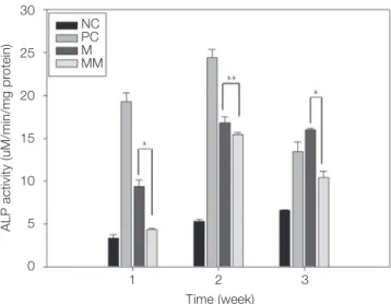
After culturing, the morphology of the MC3T3-E1 of both groups was round and it spread partially over the experimental surfaces. The cells grew along the parallel grooves of the titanium surface and proliferated and cov- ered the experimental surface. The cells were flattened and well spread, and there was little difference between M and MM.For investigating the cell differentiation, alkaline phos- phatase activity was measured. An increasing ALP expres- sion means a subsequent increasing of osteoblastic activity and bone formation. Fig. 2 shows the results of the ALP activity measurement. ALP activities showed an increase inincubation time in all the groups until the 14th day. After 21 days, ALP activities decreased or maintained stable. Only in the negative control group did the ALP activity always increase concurrently with incubation time. At the 7th and 14th day of culture, cells cultivated on culture plate with cell differentiation (positive control) showed the highest ALP activity and on culture plate without cell differentiation (negative control), the lowest ALP activity was seen. MM exhibited lower ALP activity than those of M (P<.01 at 7
and 21 days, P<.05 at 14 days). There was a significant dif- ference between groups in ALP activity and differentiation.
MM group showed relatively lower ALP activity at 7 days, but explosively increased at 14 days and relatively main- tained stable at 21 days. Compared with M, ALP activities of the MM imply that magnesium incorporation did not promote differentiation of osteoblastic cells.
Quantitative real time PCR analysis of mRNA levels for osteopontin (OPN), collagen 1 (Col1), bone sialoprotein (BSP), and osteocalcin (OCN) in cells grown on different surfaces was performed on the 7th and 14th day of culture.
Levels of osteoblastic phenotype gene expression are illus- trated in Fig. 3. Enhanced alkaline phosphatase activity on M and MM on the 7th and 14th day resulted in simultaneous increases in the expression of marker genes (BSP and osteocalcin) for osteoblastic differentiation at 7th and 14th days. Enhanced expression of diverse integrins associated in cell grown on the MM at an early incubation time result- ed in consecutive increase in BSP and OCN expressions on the 7th and 14th day. Expression levels of mRNA in BSP and OCN escalated in accordance with elapsed incubation time in all groups. OPN and Col1 mRNA expression were elevated in cells grown on both surfaces than negative con- trol in every case. While the level of expression decreased as time passed, the patterns of OPN and Col1 mRNA expression were dissimilar to that of alkaline phosphatase activity on the 7th and 14th day of culture. Although MM surface did not exhibit a higher OPN, Col1, and osteocalcin mRNA expression levels on the 14th day, MM surface dis- played relative stable mRNA expression on the 7th and 14th day. Results from replicated experiments showed similar gene expression patterns.
ALP activity (uM/min/mg protein)
30
25
20
15
10
5
0
NCPC MMM
1 2 3 Time (week)
Fig. 2. ALP activity of MC3T3-E1 cells on different Ti surfaces at 1st week, 2nd week, and 3rd week of culture.
NC-negative control (machined Ti disc w/o dif.), PC-positive control (culture plate w/dif.), M-machined surface, MM-magnesium surface. Significant difference by Tukey’s multiple comparison at * P<.01, and ** P<.05.
Table 3. Contact angle(deg)
Sample Contact angle (deg)
Machined Ti 72.9 ± 7.3
Magnesium Ti 69.4 ± 2.9
OPN expression (relative to control)BSP expression (relative to control) Col1 expression (relative to control)OCN expression (relative to control) 1.2
1.0
0.8
0.6
0.4
0.2
0.0
10
8
6
4
2
0
1.2
1.0
0.8
0.6
0.4
0.2
0.0
7 6 5 4 3 2 1 0 PCM
MM
PCM MM
PCM MM
PCM MM
7 14
7 14
7 14
7 14 Time (day)
Time (day)
Time (day)
Time (day)
Fig. 3. Real Time PCR of Osteoblast cell. Quantitative real-time PCR analysis of the levels of mRNA for bone sialoprotein (BSP), and osteocalcin (OC) in cells grown on different surfaces at 7 and 14 days of culture. PC-positive control (culture plate with differentiation); M-machined surface; MM-magnesium surface.
Alizarin Red-S stain for mineralization demonstrated that osteoblasts cultured on positive control, M and MM surfaces had higher levels of mineralization; more intense red staining which indicate calcium deposition was observed. When quantified by image analysis programs, sig- nificant (P<.05) enlargement in mineral amount was noticed in osteoblasts cultured for 21 days (Fig. 4A). But no significant difference among all the groups were observed (Fig. 4B). Therefore, we concluded that ostoeblasts increased mineralization on all surfaces without significant difference.
We examined the outcome of osteoclast maturation and differentiation ability to resorb bone during bone remodel- ing. Mouse bone marrow-derived macrophages (BMMs) cells were plated on each Ti disc. Cells were stimulated with 100 μg/mL RANKL and 40 μg/mL M-CSF. RANKL and M-CSF stimulated BMMs on M and MM surfaces and cells differentiated, suggesting that the bone resorption activity of cells made them into an active state resembling osteo-
clasts, which were produced by an actin ring structure.
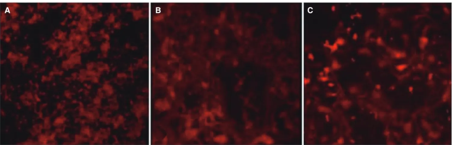
Immunofluorescence analysis was performed to find the effect on actin ring formation (Fig. 5). The majority of neg- ative control (NC) group cells without addition of RANKL and M-CSF displayed no actin rings, while M and MM group cells revealed well-formed actin rings. As expected, M and MM group cells showed many full actin rings and disrupted actin rings with more than 50% intact compared to the NC group cells. No significant variation between M and MM group were observed, but the MM group showed relatively filled actin rings.
Phenotypes of differentiated mouse bone marrow- derived macrophages (BMMs) were characterized by inves- tigating the gene expression by using RT-PCR (Fig. 6).
Markers assigned to BMMs were TRAP to characterize osteoclasts. GAPDH expression wasdecided inorder to vali- date the usage of similar amounts of RNA for RT-PCR.
Osteoclastic marker TRAP was detected. RT-PCR analysis showed the highest expression level for TRAP on MM surface.
Fig. 5. An effect on the ability of osteoclasts maturation and differentiation by actin ring formation assay. Mouse bone marrow-derived macrophages (BMMs) cells were plated on each disk; negative control (NC: machined Ti disc without differentiation)(A), Machined surface (M)(B), and Magnesium surface (MM)(C). Cells were stimulated with 100 μg/mL RANKL and 40 μg/mL M-CSF. In order to investigate the effect on actin ring formation, immunofluorescence analysis was performed. M and MM group cells showed many full actin rings and disrupted actin rings compared to NC group cells. Much fuller actin rings existed on MM compared with M.
A B C
A B
M MM
TRAP dencity (relative to control)
1.6 1.4 1.2 1.0 0.8 0.6 0.4 0.2 0.0
Fig. 6. (A) OC - RT-PCR gene expression of TRAP of machined surface (M), Mg-implanted machined surface (MM). The mRNA expression levels of the indicated genes were determined by RT-PCR. Expression of the markers was normalised to the expression of the housekeeping gene GAPDH, (B) Trap density comparison between M and MM; lowest values were set at 0.
M MM TRAP
GAPDH
A B
PT M MM
O.D. Density
0.4
0.3
0.2
0.1
0.0
Fig. 4. (A) OB-Alizarin red S Assay Alizarin red S staining for mineralized nodule formation in osteoblast cells, (B) Quantification of alizarin red S in osteoblasts. PT-positive control (culture plate with differentiation); M-machined surface; MM-magnesium surface.
PT M MM
DISCUSSION
Currently, biochemical bonding mechanisms have been observed utilizing a variety of chemical modifications of titanium surfaces; i.e. ion implantation.3,5 Plasma source ion implantation (PSII) was produced to implant ions into the Ti surface by changing the surface chemistry but not alter- ing the surface topography. Changes in surface chemistry influence osteogenesis and bone remodeling. Previous stud- ies have proven that Mg incorporated oxide surface manu- factured by an anodic oxidation process makes stronger bone integration.6,7 Because a stronger bone response was investigated, Mg ion implantations have the possibility of higher osteoconductivity. Therefore it is necessary to understand the osteoblast and osteoclast responses on the Mg-incorporated titanium implant surface.
In this study, the surface topography which may be trig- gered by bombarding changed slightly after Mg ion implan- tation. Throughout this energetic bombardment of ions, the transfer of kinetic energy from incident particle to atoms on the surface of the specimen results in some of the atoms being removed from the surface. Recent studies suggest that these textures can also have an effect on the osteoblasts.8 This change can also provide a chance for sta- ble cellular proliferation and differentiation because of good biocompatibility. Mg ion implantation has potential enhancement in the cell response at initial stage of osseoin- tegration.9,10 In this study, the surface topography and sur- face chemistry of the substrates appeared to influence the initial cell response of osteoblast and osteoclast. But the SEM assay showed no significant difference between the surface morphology. Therefore, we can speculate that the initial cellular response was not affected by the surface roughness but that the surface chemistry influenced the cel- lular response through the initial and early stages, especially on osteoclasts.
By measuring the ALP activity of MC3T3-E1 cells cul- tured on M and MM, we found that Mg incorporation did not enhance the differentiation of MC3T3-E1 cells. But on both Mg surfaces that were incorporated and machined, MC3T3-E1 cells showed increased and maintained ALP activity and expression of various osteoblast phenotype gene which induce osteoblastic differentiation. Specific osteoblast phenotype genes (OPN, Col1, OCN, and BSP) appeared during active osteoblastic differentiation.11,12 Our findings conclude that the expression of osteoblastic phe- notype genes, especially on BSP and OCN, parallels a simi- lar induction to ALP activity which is in accord with previ- ous literatures. BSP and OCN genes are responsive to osteoblastic differentiation. It is understood that magne- sium ions aid the binding of the physiologic ligand to acti- vated cation-binding A-domain.13 Therefore elevated levels of ligand-binding integrins activate integrin-mediated intra- cellular signaling pathways and can positively influence the consequent cell behavior on titanium substrates.14 Olivares- Navarrete et al.15 demonstrated that integrin a2b1 can medi- ate osteoblastic response to the microscopicsurface topog-
raphy and chemistry of titanium substrate. Promoted dif- ferentiation and adhesion of osteoblastic cells induce the expression of transcription factor gene and osteoblast marker genes such as BSP and OCN.16 The improved osteoblast differentiation with incorporated magnesium ions in the titanium oxide layer may be accredited to increase magnesium ion-mediated binding to critical integ- rins activating consequent osteoblastic differentiation.9
Increased ALP activity and gene expression demon- strate the capability to differentiate into osteoblastic cells.
Through the alizarin red S staining, mineralization was observed in every surface cell layer during osteoblast differ- entiation of the MC3T3-E1 cells. Therefore our data indi- cate that the MC3T3-E1 cells proliferated and differentiat- ed in osteoblast cell on both M and MM without significant difference.
At the end of incubation, osteoclasts were stained with rhodamin-conjugated phalloidin for actin and DAPI for nucleus. A fluorescence microscope was utilized to detect and visualize the distribution of actin rings. Osteoclasts dis- played full actin rings or disrupted actin rings with more than 50% intact.17,18 M and MM group cells revealed well- formed actin rings compared to the NC group cells which showed no actin rings. The expression levels of tartrate- resistant acid phosphatase (TRAP) mRNA were evaluated by semi-quantitive RT-PCR duringculture. As shown in Fig.
6, TRAP was also confirmed by quantitative RT-PCR. This data shows that MM has more affect in differentiation of the BMMs than M. In the BMMs, various intracellular sig- naling pathways are activated in osteoclast precursor cells and differentiating osteoclasts on MM. Previous studies showed that various intracellular signaling pathways involv- ing ERK, JNK, p38, and Akt/protein kinase B (PKB) are involved in the differentiation of osteoclasts.19,20 In particu- lar, osteoclast activity was higher in the MM group.
Such outcomes suggest that both machined and magne- sium incorporated oxide surfaces enhance osteoblastic and osteoclastic cell differentiation, but Mg-incorporated oxide surfaces are able to mature and differentiate osteoclasts bet- ter. Although further detailed studies are in demand to find if this surface could also show in vivo bone healing-promot- ing effects, the limited outcomes from this in vitro study imply that the magnesium incorporated titanium oxide layer fabricated may have a synergistic effect by promoting osteoclastic differentiation.
The results obtained in this paper propose that the use of Mg-incorporated Ti oxide implant body can lead to improvements in bone remodeling by enhancing osteoclas- tic differentiation. From analysis of the response on the magnesium surface, there is anticipationof the magnesium ion implantation for application as a potential biochemical bond. Further studies are needed to examine the precise mechanism of magnesium ion implantation and the long term effects of Mg ion implants in humans.
CONCLUSION
Both machined titanium surface (M) and Mg-incorporated Ti oxide discs (MM) have a good effect on osteoblastic and osteoclastic cell differentiation, but MM may speed the bone remodeling process by substantially activating on osteoclast differentiation. Mg ion coating may be an effec- tive approach in improving the osteoconductivity and bone remodeling of commercial titanium implants.
REFERENCES
1. Sul YT, Johansson C, Albrektsson T. Which surface proper- ties enhance bone response to implants? Comparison of oxi- dized magnesium, TiUnite, and Osseotite implant surfaces.
Int J Prosthodont 2006;19:319-28.
2. Zreiqat H, Howlett CR, Zannettino A, Evans P, Schulze- Tanzil G, Knabe C, Shakibaei M. Mechanisms of magne- sium-stimulated adhesion of osteoblastic cells to commonly used orthopaedic implants. J Biomed Mater Res 2002;62:175- 84.
3. Sul YT, Johansson P, Chang BS, Byon ES, Jeong Y. Bone tis- sue responses to Mg-incorporated oxidized implants and ma- chine-turned implantsin the rabbit femur. J Appl Biomater Biomech 2005;3:18-28.
4. Sul YT, Johansson CB, Kang Y, Jeon DG, Albrektsson T.
Bone reactions to oxidized titanium implants with electro- chemical anion sulphuric acid and phosphoric acid incorpo- ration. Clin Implant Dent Relat Res 2002;4:78-87.
5. Maitz MF, Poon RW, Liu XY, Pham MT, Chu PK. Bioactivity of titanium following sodium plasma immersion ion implan- tation and deposition. Biomaterials 2005;26:5465-73.
6. Sul YT, Johansson C, Byon E, Albrektsson T. The bone re- sponse of oxidized bioactive and non-bioactive titanium im- plants. Biomaterials 2005;26:6720-30.
7. Cho LR, Kim DG, Kim JH, Byon ES, Jeong YS, Park CJ.
Bone response of Mg ion-implanted clinical implants with the plasma source ion implantation method. Clin Oral Implants Res 2010;21:848-56.
8. Sammons RL, Lumbikanonda N, Gross M, Cantzler P.
Comparison of osteoblast spreading on microstructured den- tal implant surfaces and cell behaviour in an explant model of osseointegration. A scanning electron microscopic study.
Clin Oral Implants Res 2005;16:657-66.
9. Park JW, Kim YJ, Jang JH, Song H. Osteoblast response to magnesium ion-incorporated nanoporous titanium oxide sur- faces. Clin Oral Implants Res 2010;21:1278-87.
10. Takebe J, Itoh S, Okada J, Ishibashi K. Anodic oxidation and hydrothermal treatment of titanium results in a surface that causes increased attachment and altered cytoskeletal mor- phology of rat bone marrow stromal cells in vitro. J Biomed Mater Res 2000;51:398-407.
11. Samee N, Geoffroy V, Marty C, Schiltz C, Vieux-Rochas M, Levi G, de Vernejoul MC. Dlx5, a positive regulator of osteo- blastogenesis, is essential for osteoblast-osteoclast coupling.
Am J Pathol 2008;173:773-80.
12. Tadic T, Dodig M, Erceg I, Marijanovic I, Mina M, Kalajzic
Z, Velonis D, Kronenberg MS, Kosher RA, Ferrari D, Lichtler AC. Overexpression of Dlx5 in chicken calvarial cells accelerates osteoblastic differentiation. J Bone Miner Res 2002;17:1008-14.
13. Ajroud K, Sugimori T, Goldmann WH, Fathallah DM, Xiong JP, Arnaout MA. Binding Affinity of Metal Ions to the CD11b A-domain Is Regulated by Integrin Activation and Ligands. J Biol Chem 2004;279:25483-8.
14. Zreiqat H, Valenzuela SM, Nissan BB, Roest R, Knabe C, Radlanski RJ, Renz H, Evans PJ. The effect of surface chem- istry modification of titanium alloy on signalling pathways in human osteoblasts. Biomaterials 2005;26:7579-86.
15. Olivares-Navarrete R, Raz P, Zhao G, Chen J, Wieland M, Cochran DL, Chaudhri RA, Ornoy A, Boyan BD, Schwartz Z. Integrin alpha2beta1 plays a critical role in osteoblast re- sponse to micron-scale surface structure and surface energy of titanium substrates. Proc Natl Acad Sci USA 2008;105:
15767-72.
16. Keselowsky BG, Wang L, Schwartz Z, Garcia AJ, Boyan BD.
Integrin alpha(5) controls osteoblastic proliferation and dif- ferentiation responses to titanium substrates presenting dif- ferent roughness characteristics in a roughness independent manner. J Biomed Mater Res A 2007;80:700-10.
17. Wilson SR, Peters C, Saftig P, Brömme D. Cathepsin K activ- ity-dependent regulation of osteoclast actin ring formation and bone resorption. J Biol Chem 2009;284:2584-92.
18. Flanagan AM, Massey HM, Wilson C, Vellodi A, Horton MA, Steward CG. Macrophage colony-stimulating factor and receptor activator NF-kappaB ligand fail to rescue osteoclast- poor human malignant infantile osteopetrosis in vitro. Bone 2002;30:85-90.
19. Boyle WJ, Simonet WS, Lacey DL. Osteoclast differentiation and activation. Nature 2003;423:337-42.
20. Burgess TL, Qian Y, Kaufman S, Ring BD, Van G, Capparelli C, Kelley M, Hsu H, Boyle WJ, Dunstan CR, Hu S, Lacey DL. The ligand for osteoprotegerin (OPGL) directly acti- vates mature osteoclasts. J Cell Biol 19993;145:527-38.