Received May 30, 2019, Revised July 1, 2019, Accepted for publication July 5, 2019
Corresponding author: Gwang Seong Choi, Department of Dermatology, Inha University School of Medicine, 27 Inhang-ro, Jung-gu, Incheon 22332, Korea. Tel: 82-32-890-2238, Fax: 82-32-890-2236, E-mail: garden@
inha.ac.kr
ORCID: https://orcid.org/0000-0002-5766-0179
This is an Open Access article distributed under the terms of the Creative Commons Attribution Non-Commercial License (http://creativecommons.
org/licenses/by-nc/4.0) which permits unrestricted non-commercial use, distribution, and reproduction in any medium, provided the original work is properly cited.
Copyright © The Korean Dermatological Association and The Korean Society for Investigative Dermatology
Ann Dermatol Vol. 31, No. 5, 2019 https://doi.org/10.5021/ad.2019.31.5.538
ORIGINAL ARTICLE
Therapeutic Effect of Glucosamine on an Atopic Dermatitis Animal Model
Hee Seong Yoon, Ji Won Byun, Jeonghyun Shin, Young Hyo Kim1, Gwang Seong Choi
Department of Dermatology, Inha University School of Medicine, 1Department of Otorhinolaryngology-Head and Neck Surgery, Inha University School of Medicine, Incheon, Korea
Background: Recent studies have reported that glucosamine (GlcN) showed therapeutic effects in allergic diseases such as asthma and rhinitis, and its mechanisms include the sup- pression of T helper type 2 immune responses and the nu- clear factor-κB pathway. Objective: We aimed to inves- tigate the effect of GlcN on atopic dermatitis (AD) in an ani- mal model. Methods: Twenty-five BALB/c mice were div- ided into five groups (groups A∼E). Group A was the phos- phate-buffered saline (PBS)-treated group without AD in- duction. Group B was the PBS control group with AD in- duction. Groups C to E were the AD induction groups, which were treated with three different doses of GlcN (10 mg, 20 mg, and 40 mg, respectively). Histopathological examina- tion was performed after GlcN administration. Interleukin (IL)-4, IL-13, and IL-17 cytokine levels were measured by en- zyme-linked immunosorbent assay using skin biopsy speci- mens. Serum total immunoglobulin E (IgE) concentrations were measured before and after administration with GlcN or PBS. Results: Clinical dermatitis scores decreased with in- creasing GlcN dose (p<0.001). Concentrations of tissue IL-13 and IL-17 decreased after GlcN administration (each group: p=0.002 and p<0.001, respectively), but the con- centrations of tissue IL-4 did not show differences across
groups. Serum IgE levels tended to be lower after GlcN ad- ministration (p=0.004). Histopathological scores were not significantly different among groups B∼E (p=0.394). Con- clusion: GlcN improved AD symptoms and decreased tissue IL-13, IL-17, and serum total IgE levels in an animal model.
(Ann Dermatol 31(5) 538∼544, 2019) -Keywords-
Allergy and immunology, Anti-allergic agents, Atopic der- matitis, Glucosamine
INTRODUCTION
Atopic dermatitis (AD) is a chronic inflammatory skin dis- ease that is caused by a combination of genetic, environ- mental, and immunological factors1. Immunologically, it is characterized by the T helper type 2 (Th2) immune re- sponse. When allergens and antigens enter through the damaged skin, Langerhans cells and keratinocytes secrete cytokines and chemokines, including interleukin (IL)-12 and IL-18, and chemoattract various immune cells1. In the acute stage of AD, Th1, Th2, Th17, and Th22 immune re- sponses are all present, with the Th2 immune response mainly contributing to the pathogenesis; this leads to the increased production of IL-4, IL-5, and IL-132. Recently, it was demonstrated that the proportion of Th17 cells were increased in peripheral blood mononuclear cells of pa- tients with AD and were infiltrated more markedly in the acute lesions than in the chronic lesions of AD3. IL-17 plays a role in keratinocyte stimulation to produce vas- cular endothelial growth factor, tumor necrosis factor-α, IL-8, granulocyte-macrophage colony-stimulating factor, and chemokine (C-X-C motif) ligand 10. In contrast, in chronic AD, the production of IL-4 and IL-13 is reduced
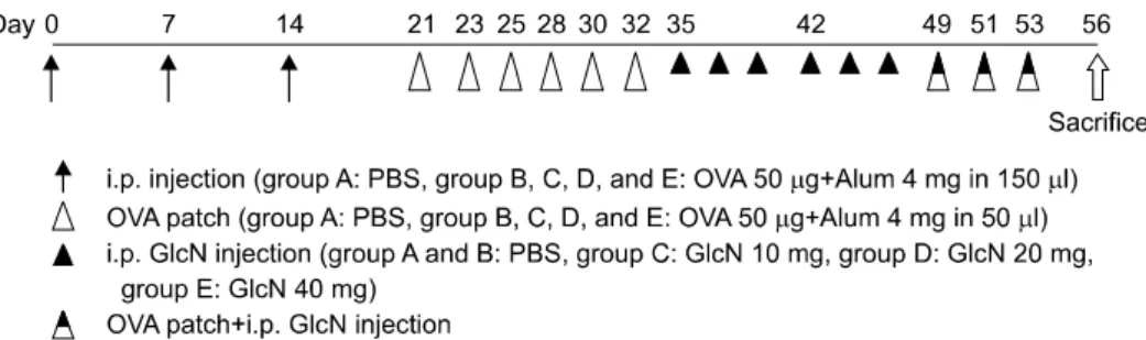
Fig. 1. Study protocol for this experiment. Normal (group A) and atopic dermatitis (AD) groups (groups B∼E). AD was induced by ovalbumin (OVA) and aluminum hydroxide gel (Alum). Group A: normal, group B: phosphate-buffered saline (PBS) control, group C: glucosamine (GlcN) 10 mg, group D: GlcN 20 mg, group E: GlcN 40 mg administration group. i.p.: intraperitoneal.
and the production of IL-5 and IL-12 is increased, suggest- ing that Th1 cells are predominant in the chronic lesions of AD2. Interferon secreted by Th1 cells causes keratinocyte apoptosis, skin remodeling, and hypoxia in Th22 cells1. Most of the patients with AD (80%∼90%) have been re- ported to have high serum immunoglobulin E (IgE) levels4. Although the role of IgE in the pathogenesis of AD has not yet been fully understood1, it has been reported that se- rum IgE levels can be used as a parameter of severity for AD4.
Treatment of AD includes moisturizers, steroids and im- munosuppressive agents, narrow-band ultraviolet B (NB- UVB) therapy, and biologics, and the treatment focuses on improving skin barrier function and controlling the im- mune system5. However, systemic medicines have various side effects, especially when used for long periods of time.
In the case of NB-UVB, it presents disadvantages such as requiring frequent hospital visits. Recently, dupilumab has been developed and used in the treatment of severe AD as it inhibits IL-4 and IL-13 signaling6. In Korea, however, du- pilumab is not easily accessible as it is expensive and is not yet covered by health insurance.
Glucosamine (2-amino-2-deoxy-D-glucose; GlcN) is an amino sugar synthesized from glucose by the hexosamine biosynthetic pathway that is present in cells of the body and has anti-inflammatory and immunomodulatory effects7. Previous studies have reported the efficacy of GlcN in the treatment of osteoarthritis and in improving the symptoms of rheumatoid arthritis8,9. A recent study demonstrated that GlcN has anti-allergic effects in allergic asthma and rhini- tis animal models7.
We expected that GlcN, which inhibits the Th2 immune response, may be effective in the treatment of AD. We con- ducted this study to investigate the therapeutic effect of GlcN by performing clinical and histopathological exami- nation, as well as measuring the concentrations of tissue IL-4, IL-13, and IL-17 and serum total IgE levels.
MATERIALS AND METHODS
AD animal model and subjects
This study was carried out in female BALB/c mice (Orient Bio Inc., Seongnam, Korea) aged 8 weeks with AD induced by ovalbumin (OVA; Sigma-Aldrich Korea, Yongin, Korea).
Twenty-five BALB/c mice were divided into five groups of five mice: control group without induction of AD (group A), AD group with null treatment by phosphate-buffered saline (PBS) (group B), and AD groups treated with 10 mg, 20 mg, and 40 mg of GlcN (Sigma-Aldrich Korea) admin- istration (groups C, D, and E, respectively). For the AD in- duction groups (groups B∼E), 1.5 ml of OVA and 3 ml of aluminum hydroxide gel (Thermo Fisher Scientific, Waltham, MA, USA) were mixed, and 150 μl of the mixture was in- traperitoneally injected into the mice three times a week for three weeks. After a week of OVA injection, mice were epicutaneously sensitized with OVA patches. The patches were made by dripping 50 μl of OVA (1 mg/ml) in a gauze (1×1 cm) which was placed on the back skin three times per week for two weeks. Group A was injected with PBS instead of OVA. This experiment was conducted in specific-pathogen-free environment and the mice received an OVA-free diet. This study was reviewed and approved by the Institutional Animal Care and Use Committee of Inha University (INHA 181120-601).
Administration of glucosamine
After AD was induced, we injected 100 μl of GlcN intra- peritoneally at concentrations of 1 mg/10 μl, 1 mg/5 μl, and 1 mg/2.5 μl into the mice of groups C, D, and E, re- spectively three times per week for three weeks. After a week of GlcN administration, OVA patches were attached three times for one week. For groups A and B, 100 μl of PBS was administered instead of GlcN (Fig. 1).
Clinical dermatitis scores
Two dermatologists examined the skin of the AD-induced
mice after GlcN administration. The skin condition was scored from 0 to 3 for erythema, dryness, excoriation, and edema: 0 (absence), 1 (mild), 2 (moderate), and 3 (severe).
Clinical dermatitis scores were defined as the sum of all scores.
Measurement of concentration of cytokine in tissue Biopsy specimens from the epidermis to the subcutaneous layer were frozen in liquid nitrogen and stored at −80oC.
Then, the specimens were added with RIPA buffer (Pierce Biotechnology, Rockford, IL, USA) supplemented with pro- tease inhibitor (Pierce Biotechnology) and were homogen- ized on ice with a micro tissue homogenizer (DWK Life Sciences, Millville, NJ, USA). The homogenized specimens were centrifuged and the supernatant was used to meas- ure IL-4, IL-13, and IL-17 levels with individual enzyme- linked immunosorbent assay (ELISA) kits (R&D Systems, Minneapolis, MN, USA) following the manufacturer’s in- structions.
Measurement of total IgE in serum
Blood samples were obtained before and after GlcN ad- ministration in all groups. Blood (50 μl) was collected from the orbital vein after AD induction and from the heart after GlcN administration. The serum was separated and IgE levels were measured using a BD OptEIA Mouse IgE ELISA kit (Pharmingen, San Diego, CA, USA) according to the manufacturer’s instructions.
Histopathological analysis
After GlcN administration, Tiletamine/Zolazepam (0.008 ml/10 g) and Xylazine (0.002 ml/10 g) were injected for general anesthesia. Then, skin biopsy was performed. Speci- mens were fixed in 10% formaldehyde and embedded in paraffin. The tissue sections were stained with hematox- ylin and eosin (H&E). Severity of inflammation in the der- mis was evaluated grossly in H&E slides by scoring on a scale from 0 to 5: 0 (none), 1 (mild), 2 (mild to moderate), 3 (moderate), 4 (moderate to severe), and 5 (severe).
Statistical analysis
Jonckheere–Terpstra test was performed to determine the likelihood of changes in clinical dermatitis scores, concen- trations of tissue cytokines, and serum total IgE levels ac- cording to the GlcN dose. Comparison of Serum IgE level between before and after GlcN or PBS administration for each groups were performed by Wilcoxon signed rank test. Fisher’s exact test was performed to compare histo- pathological scores among groups. Statistical analysis was performed using PASW Statistics ver. 18.0 (IBM Corp., Armonk, NY, USA). A value of p<0.05 was considered
statistically significant.
RESULTS
Clinical improvement of AD in animal models
We confirmed grossly that AD-like skin lesions were sim- ilarly induced by OVA among the B∼G group before GlcN administration. GlcN improved AD symptoms and reduced clinical dermatitis scores in groups B∼E (Fig. 2).
Clinical dermatitis scores decreased with increasing GlcN dose (p<0.001).
Decreased IL-13 and IL-17 levels in GlcN-treated mice, but not IL-4
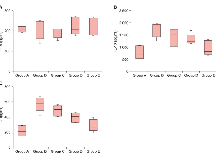
Tissue IL-13 and IL-17 levels decreased with increasing GlcN dose (each group: p=0.002 and p<0.001, respect- ively) (Fig. 3B, C). However, concentrations of IL-4 were not significantly different among groups B∼E (p=0.501) (Fig. 3A).
Low total serum IgE level in GlcN treated groups Serum total IgE levels after induction of AD were not sig- nificantly different among groups B∼E (Fig. 4A). Serum IgE levels tended to be lower with GlcN administration in comparison among group B∼E after GlcN or PBS admin- istration (p=0.004) (Fig. 4B). Serum IgE levels increased after treatment compared to before treatment in all groups, which were statistically significant only in the group A and B (both groups, p=0.043).
Improvement of dermal inflammation in histopathological examination
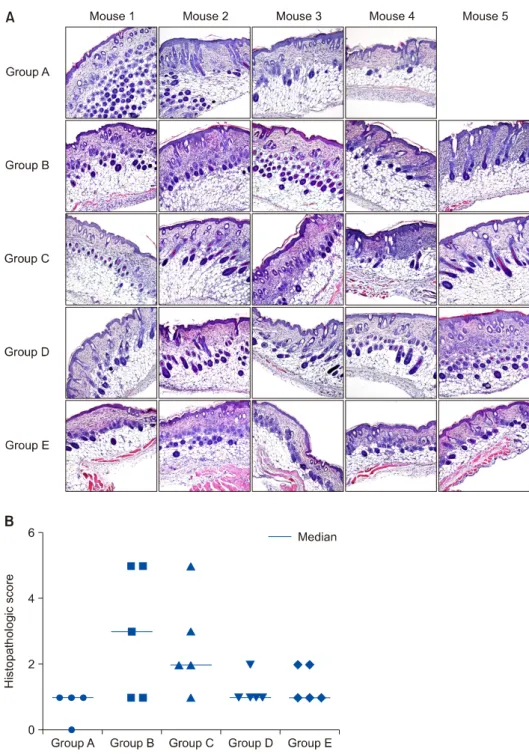
Inflammatory cell infiltration was lower in tissues of groups D and E compared to those in group B (Fig. 5A). However, the histopathological scores did not show statistically sig- nificantly differences in groups B∼E (p=0.393) (Fig. 5B).
DISCUSSION
In this study, we found that GlcN was clinically and histo- logically effective in improving AD and inflammation, as well as in decreasing the concentrations of tissue IL-13, IL-17, and serum total IgE.
GlcN reduced clinical dermatitis scores and showed a gross decrease in infiltration of inflammatory cells. Recent- ly, there have been studies on the effects of GlcN in vari- ous allergic diseases. Previously, a study using NC/Nga mice with Dermatophagoides farina (Df)-induced AD-like skin lesions also reported that GlcN treatment reduced clinical dermatitis scores10. Another study showed that crys- tallinity controlled N-acetyl glucosamine (CCG) reduced
Fig. 2. Clinical features and clinical dermatitis scores of mice in each group after administration with phosphate-buffered saline (PBS;
groups A and B) or glucosamine (GlcN; groups C, D, and E). In a total of 25 mice, a mouse in group A died after 50 days. (A) Compared with that of group B, the clinical dermatitis scores of groups D and E improved. (B) Clinical dermatitis scores tended to decrease with increasing GlcN dose (p<0.001). Group A: normal, group B: PBS control, group C: GlcN 10 mg, group D: GlcN 20 mg, group E: GlcN 40 mg.
histamine release, IL-1β production, and caspase-1/nu- clear factor-κB (NF-κB) activation in human mast cell line-1 cells, and demonstrated the anti-allergic and anti-in- flammatory effects of GlcN11. They also reported that CCG reduces systemic anaphylaxis, ear swelling, and passive cutaneous anaphylaxis in a mouse model11.
In this study, the concentrations of tissue IL-13 and IL-17 were decreased after GlcN administration, but tissue IL-4 levels did not change. Previously, an in vitro study showed that GlcN inhibits the secretion of IL-5 in primary mixed lymphocyte cultures (MLC) using splenocytes from C57BL/6 or BALB/c mice and inhibits IL-4 and IL-5 secretion in sec- ondary Th2-polarized MLC12. Another study using NC/Nga mice AD models showed that Th2 cytokines including IL-4 and IL-5 were significantly reduced by GlcN treat- ment, in contrast to our results10. We supposed that this is due to differences in experimental methods. They injected GlcN once a day and induced AD using Df body oint- ment, while we injected GlcN three times per week and
induced AD using OVA. In addition, they measured cyto- kine levels in the spleen, while we measured cytokine lev- els using skin biopsy specimens. A study using BALB/c mouse allergic asthma and rhinitis models reported that IL-4, IL-5, IL-6, IL-17, and eosinophil counts decreased in the bronchoalveolar lavage fluid after GlcN administration, and that GlcN inhibits Th2 and Th17 cytokines7.
We also found that serum IgE levels tended to be lower af- ter GlcN administration. A previous study using NC/Nga AD mouse models also reported that GlcN reduced serum IgE levels, similar to our results10. Another study using al- lergic rhinitis and asthma mice models also evaluated se- rum total IgE and OVA-specific IgE levels and reported a decrease in both IgE levels after GlcN administration7. Serum total IgE levels before and after treatment were elevated in all groups including group A. We considered it the charac- teristic of the mouse. Previously, a study using inbred BN-rats showed that total serum IgE levels increased from the age of 1 month to the age of 5 months13.
Fig. 3. Effects of glucosamine (GlcN) on cytokine (interleukin [IL]-4, IL-13, and IL-17) levels in biopsy specimens. Concentrations of tissue IL-4, IL-13, and IL-17 after GlcN administration. (A) IL-4 did not show differences in all groups. (B) IL-13 and (C) IL-17 showed decreased with increasing GlcN dose (each group: p=0.002 and p<0.001, respectively). Group A: normal, group B:
phosphate-buffered saline control, group C: GlcN 10 mg, group D: GlcN 20 mg, group E: GlcN 40 mg.
Fig. 4. Serum total immunoglobulin E (IgE) before and after administration of phosphate-buffered saline (PBS; group A and B) and glucosamine (GlcN; groups C, D, and E). (A) Serum IgE levels after induction of atopic dermatitis were not significantly different in groups B∼E. (B) Serum IgE levels tended to be lower after GlcN administration (p=0.004). Group A: normal, group B: PBS control, group C: GlcN 10 mg, group D: GlcN 20 mg, group E: GlcN 40 mg.
GlcN showed a decrease in infiltration of inflammatory cells. A previous study using NC/Nga mice with Df-in- duced AD-like skin lesions reported that GlcN reduced in-
filtration of dermal inflammatory cells, including mast cells and eosinophils10. In another study using allergic asthma and rhinitis BALB/c mouse models, GlcN showed anti-al-
Fig. 5. Histopathological findings and histopathological scores of each group after administration with pho- sphate-buffered saline (PBS; group A and B) or glucosamine (GlcN;
group C, D, and E). In a total of 25 mice, 1 mouse in group A died after 50 days. (A) The infiltration of inflammatory cells was lower in groups D and E than in group B (hematoxylin and eosin, original mag- nification ×40). (B) Histopathologi- cal scores did not show statistically significant differences among groups B∼E according to Fisher’s exact test (p=0.394). Group A: normal, group B: PBS control, group C: GlcN 10 mg, group D: GlcN 20 mg, group E: GlcN 40 mg.
lergic effects and decreased the infiltration of inflammatory cells in lung and nasal tissues7.
GlcN regulates factors involved in inflammation and the NF-κB pathway, and inhibits the Th2 immune responses14. Exogenous GlcN enters the cell through the glucose trans- porters (GLUT-1, GLUT-2, and GLUT-4) and increases intra- cellular UDP-N-acetyl-GlcN (UDP-GlcNAc)15. UDP-GlcNAc is involved in the O-GlcNAcylation of nuclear and cyto- plasmic transcription factors, as well as lipopolysaccha- ride-related inflammatory factors7,15. In addition, GlcN pre- vents IκB degradation and decreases NF-κB nuclear accu-
mulation and NF-κB reporter activity16. It is suggested that GlcN mediates its anti-inflammatory effects via this mech- anism. In addition, GlcN regulates the antigen presenta- tion of dendritic cells, and inhibits the proliferation of CD4+ T cells and the secretion of Th2 cytokines such as IL-4 and IL-512.
This study has some limitations. First, our experimental model is an animal model with AD-like skin lesions, and thus does not fully represent true AD. Therefore, further studies are needed to demonstrate the effect of GlcN in humans with AD. Second, the sample size is small. Third,
the optimal dose of GlcN is unknown. Finally, there was no positive control to compare the effect of GlcN. In fur- ther studies, it would be desirable to conduct experiments with a positive control such as cyclosporine or a systemic steroid.
In conclusion, GlcN improved AD-like skin lesions and decreased tissue IL-13 and IL-17, as well as serum total IgE levels in an animal model. These results may be a basis for the future use of GlcN as a therapeutic agent for AD.
ACKNOWLEDGMENT
This work was supported by the Inha University Research Grant.
CONFLICTS OF INTEREST
The authors have nothing to disclose.
ORCID
Hee Seong Yoon, https://orcid.org/0000-0001-8997-9697 Ji Won Byun, https://orcid.org/0000-0003-0317-6725 Jeonghyun Shin, https://orcid.org/0000-0002-4995-9533 Young Hyo Kim, https://orcid.org/0000-0002-3623-1770 Gwang Seong Choi, https://orcid.org/0000-0002-5766-0179
REFERENCES
1. Peng W, Novak N. Pathogenesis of atopic dermatitis. Clin Exp Allergy 2015;45:566-574.
2. Egawa G, Weninger W. Pathogenesis of atopic dermatitis: a short review. Cogent Biol 2015;1:1103459.
3. Koga C, Kabashima K, Shiraishi N, Kobayashi M, Tokura Y.
Possible pathogenic role of Th17 cells for atopic dermatitis.
J Invest Dermatol 2008;128:2625-2630.
4. Vaneckova J, Bukač J. The severity of atopic dermatitis and the relation to the level of total IgE, onset of atopic der- matitis and family history about atopy. Food Agric Immunol 2016;27:734-741.
5. Sidbury R, Davis DM, Cohen DE, Cordoro KM, Berger TG,
Bergman JN, et al. Guidelines of care for the management of atopic dermatitis: section 3. Management and treatment with phototherapy and systemic agents. J Am Acad Dermatol 2014;71:327-349.
6. Gooderham MJ, Hong HC, Eshtiaghi P, Papp KA. Dupilumab:
a review of its use in the treatment of atopic dermatitis. J Am Acad Dermatol 2018;78(3 Suppl 1):S28-S36.
7. Jung AY, Heo MJ, Kim YH. Glucosamine has an antiallergic effect in mice with allergic asthma and rhinitis. Int Forum Allergy Rhinol 2017;7:763-769.
8. Nakamura H, Masuko K, Yudoh K, Kato T, Kamada T, Kawahara T. Effects of glucosamine administration on patients with rheumatoid arthritis. Rheumatol Int 2007;27:213-218.
9. Reginster JY, Deroisy R, Rovati LC, Lee RL, Lejeune E, Bruyere O, et al. Long-term effects of glucosamine sulphate on osteoarthritis progression: a randomised, placebo-controlled clinical trial. Lancet 2001;357:251-256.
10. Kim CH, Cheong KA, Park CD, Lee AY. Glucosamine im- proved atopic dermatitis-like skin lesions in NC/Nga mice by inhibition of Th2 cell development. Scand J Immunol 2011;73:536-545.
11. Jin SE, Jung J, Jun J, Jeon DW, Kim HM, Jeong HJ. Anti- allergic activity of crystallinity controlled N-acetyl gluco- samine. Immunopharmacol Immunotoxicol 2012;34:991-1000.
12. Forchhammer L, Thorn M, Met O, Gad M, Weidner MS, Claesson MH. Immunobiological effects of glucosamine in vitro. Scand J Immunol 2003;58:404-411.
13. Pauwels R, Bazin H, Platteau B, van der Straeten M. The effect of age on IgE production in rats. Immunology 1979;
36:145-149.
14. Rafi MM, Yadav PN, Rossi AO. Glucosamine inhibits LPS- induced COX-2 and iNOS expression in mouse macro- phage cells (RAW 264.7) by inhibition of p38-MAP kinase and transcription factor NF-kappaB. Mol Nutr Food Res 2007;51:587-593.
15. Hwang JS, Kwon MY, Kim KH, Lee Y, Lyoo IK, Kim JE, et al.
Lipopolysaccharide (LPS)-stimulated iNOS induction is in- creased by glucosamine under normal glucose conditions but Is inhibited by glucosamine under high glucose con- ditions in macrophage cells. J Biol Chem 2017;292:1724- 1736.
16. Zahedipour F, Dalirfardouei R, Karimi G, Jamialahmadi K.
Molecular mechanisms of anticancer effects of Glucosamine.
Biomed Pharmacother 2017;95:1051-1058.