82
책임저자: 배자성, 서울시 서초구 반포4동 505
137-701, 가톨릭대학교 의과대학 외과학교실 Tel: 02-2258-6107, Fax: 02-595-2992
E-mail: [email protected]
접수일:2009년 10월 6일, 게재승인일:2009년 11월 23일
갑상선 미세유두상암에서 림프절 전이의 위험인자
가톨릭대학교 의과대학 외과학교실, 1방사선과학교실, 2병원병리학교실, 3내과학교실
이남섭ㆍ배자성ㆍ정소령
1ㆍ정찬권
2ㆍ임동준
3ㆍ박우찬ㆍ김정수ㆍ김승남
Risk Factors of Lymph Node Metastasis in Papillary Thyroid Microcarcinoma
Nam Seop Lee, M.D., Ja Seong Bae, M.D., So-Ryeong Jeong, M.D.1, Chan Kwon Jung, M.D.2, Dong Jun Lim, M.D.3, Woo Chan Park, M.D., Jeong Soo Kim, M.D., Seung Nam Kim, M.D.
Departments of Surgery, 1Radiology, 2Pathology and 3Internal Medicine, The Catholic University College of Medicine, Seoul, Korea
Purpose: Despite the overall excellent prognosis for patients with papillary thyroid microcarcinoma (PTMC), these tumors are also associated with a 5% relatively high lymph node (LN) recurrence rate and the optimal surgical extent of papillary thyroid microcarcinoma has been controversial. Cervical LN metastases (LNMs) are found in about 40∼65% of patients with PTMC. The aim of this study is to identify the factors affecting lymph node metastases (LNMs) in patients with PTMC.
Methods: We performed a retrospective study of 335 patients with PTMC who underwent total thyroidectomy or lobectomy with elective central lymph node dissection (CLND) at Kangnam St. Mary’s Hospital between Jan.
2006 and Dec. 2008. We investigated the association of LNMs and clinicopathologic factors such as sex, age, multiplicity, extrathyroidal extension, and tumor size.
Results: LNMs were present in 88 patients (26.3%). Univariate analysis showed that less than 45 years of age, male, multiplicity, a tumor size of greater than 5 mm, thyroid capsular invasion and extrathyroidal extension were predictive factors for LNMs (P<0.05). Of these, the age, male, tumor size and extrathyroidal extension were independent predictive factors for LNMs on multivariate analysis.
Conclusion: A tumor size (>5 mm), male, age (<45) and extrathyroidal extension were determined as the predictive factors for LNMs, which occurred in about one fourth of the patients with PTMC. Therefore, elective CLND should be considered in patients with PTMC who have these factors through a thorough investigation before surgery. (J Korean Surg Soc 2010;78:82-86)
Key Words: Papillary thyroid microcarcinoma, Central lymph node metastasis 중심 단어: 갑상선 미세유두상암, 중심 림프절 전이
서 론
갑상선 유두상암은 갑상선암의 가장 흔한 조직형으로 전 체 갑상선 암의 80% 이상을 차지한다. 세계보건기구(WHO)
에서는 이 중 10 mm 이하의 갑상선 유두상암을 갑상선 미 세유두상암(papillary thyroid microcarcinoma)으로 정의하는 데,(1,2) 최근 건강검진의 증가 및 초음파 진단 기술의 발달 로 이러한 갑상선 미세유두상암의 진단이 급격히 증가하고 있다.(3-6) 갑상선 미세유두상암은 암의 일반적인 특징인 전이나 침습과 같은 행동이 흔하지 않아 전반적인 예후가 아주 좋은 것으로 알려져 있다. 그러나 전체적으로 약 1%의 갑상선 미세유두상암과 관련된 사망률과 5% 정도의 림프 절 재발 그리고 약 2.5% 정도의 원격 전이율을 나타내고
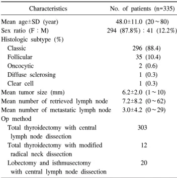
Table 1. Demographics of patients with papillary microcarcinoma Characteristics No. of patients (n=335)
Mean age±SD (year) 48.0±11.0 (20∼80)
Sex ratio (F:M) 294 (87.8%):41 (12.2%)
Histologic subtype (%)
Classic 296 (88.4)
Follicular 35 (10.4)
Oncocytic 2 (0.6)
Diffuse sclerosing 1 (0.3)
Clear cell 1 (0.3)
Mean tumor size (mm) 6.2±2.0 (1∼10) Mean number of retrieved lymph node 7.2±8.2 (0∼62) Mean number of metastatic lymph node 3.0±4.2 (0∼29) Op method
Total thyroidectomy with central 303 lymph node dissection
Total thyroidectomy with modified 12 radical neck dissection
Lobectomy and isthmusectomy 20 with central lymph node dissection
있고,(7) 이에 따른 다양한 치료 방법 및 수술 범위에 대한 의견이 제시되고 있어 재발 및 사망과 연관된 예후 인자를 찾는 것이 중요하며 여러 연구들이 시행되고 있다. 갑상선 유두상암에 있어 림프절 전이는 국소 재발이나 원격전이에 영향을 미칠 수 있는 중요한 예후인자 중의 하나로 알려져 있다.(7) 문헌 보고에 의하면 약 40∼65%까지 중앙 림프절 전이의 발견을 보고하고 있지만(6,8,9) 중앙 림프절 절제 (central lymph node dissection)의 실제적인 역할에 대해서는 아직 논란의 여지가 있으며 또한 중앙 림프절 절제술을 시 행할 경우 저칼슘혈증이나 되돌이 후두신경(recurrent lar- yngeal nerve) 손상 같은 갑상선 수술의 합병증이 증가할 수 있기 때문에 갑상선 미세유두상암의 수술에 있어 일상적인 중앙 림프절 절제술(elective central lymph node dissection) 시행이 반드시 추천되고 있지는 않다.(8,10) 본 연구는 갑상 선 미세유두상암의 적절한 수술 범위 및 치료 전략을 수립 하기 위해 일상적인 동측 중앙 림프절 절제술 시행 후 나타 난 림프절 전이 및 그와 연관된 인자를 조사하였다.
방 법
2006년 1월부터 2008년 12월까지 가톨릭대학교 의과대학 강남성모병원에서 갑상선암 수술을 받은 환자 중 갑상선 미세유두상암 환자 335명을 대상으로 하였다. 환자는 수술 전 초음파 및 세침흡인검사에서 갑상선암 의심 혹은 갑상 선암으로 진단되었고 수술은 갑상선 전절제술 및 동측 중 앙 림프절 절제술을 원칙으로 하였으며 수술 전 측경부 림 프절 전이가 의심될 경우 변형 근치 경부 곽청술(modified radical neck dissection)을 시행하였다. 또한 환자가 원하거나 갑상선 엽절제술 후 최종 조직검사 결과에서만 갑상선암으 로 판명된 경우에는 더 이상의 수술은 시행하지 않았다. 중 앙 림프절 절제술은 동측의 기관주위 림프절(paratracheal) 과 기관앞 림프절(pretracheal) 절제술을 원칙으로 하였으며, 수술 전 양측 갑상선암으로 진단 받은 경우에는 양측 중앙 림프절 절제술을 시행하였다. 모든 환자에 있어 성별, 나이, 종양의 다발성(multiplicity), 종양의 피막형성(tumor encapsu- lation), 갑상선외 침범(extrathyroidal extension) 유무, 종양의 크기, 위치, 측경부 림프절 전이 유무(lateral cervical LN meta.), 피막 침윤(capsular invasion)과 같은 임상병리학적 특 징에 대하여 후향적 연구를 시행하였다. 통계학적 분석은 SPSS version 12.0를 사용하였으며 Chi-square test을 통하여 단변량 분석을 시행하였고, 이를 바탕으로 로지스틱 회귀
분석(logistic regression analysis)을 통해 다변량 분석을 시행 하여 림프절 전이와 연관된 인자를 찾고자 하였다. P<0.05 인 경우를 통계학적으로 의미 있는 것으로 해석하였다.
결 과
총 환자는 335명이었으며 여자는 294명(87.8%) 남자는 41명(12.2%)이었다. 평균 나이는 48.0±11.0세였으며 20세에 서 80세까지 분포하였다. 조직학적 아형(subtype)은 296명 (88.4%)이 classic type이었고 follicular variant가 35명(10.4%), oncocytic variant 2명, diffuse sclerosing variant, clear cell var- iant 각각 1예였다. 갑상선암의 평균 크기는 6.2±2.0 mm이었 고 평균 제거된 림프절 개수는 7.2±8.2개, 평균 전이 림프절 개수는 3.0±4.2개였다. 수술은 303명에서 갑상선 전절제술 및 중앙 림프절 절제술을 시행하였으며 12명의 환자에서 갑상선 전절제술 및 변형 근치 경부 곽청술을 시행하였다.
또한 20명의 환자에서 갑상선 일엽절제술 및 중앙 림프절 절제술을 시행하였다(Table 1). 125명(37.3%)은 5 mm 이하, 210명은 5 mm 이상이었고 121명(36.1%)에서 갑상선외 침 범이 있었다. 종양의 다발성은 118명(35.2%)에서 있었고, 이 중 35명(10.4%)은 일측성, 83명(24.8%)은 양측성이었다.
림프절 전이가 있는 환자는 88명(26.3%)이었고, 이 중 중앙 림프절과 측경부 림프절 전이가 함께 나타난 경우는 5명, 중앙림프절 전이 없이 측경부 림프절로 도약 전이(skip
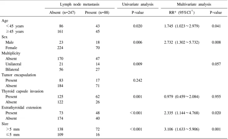
Table 2. Univariate and multivariate analysis of association between clinicopathologic features and central lymph node metastasis in patients with papillary microcarcinoma
Lymph node metastasis Univariate analysis Multivariate analysis
Absent (n=247) Present (n=88) P-value RR* (95%CI†) P-value
Age
<45 years 86 43 0.020 1.745 (1.023∼2.979) 0.041
≥45 years 161 45
Sex
Male 23 18 0.006 2.732 (1.302∼5.732) 0.008
Female 224 70
Multiplicity
Absent 170 47
Unilateral 21 14 0.009 0.057
Bilateral 56 27
Tumor encapsulation
Present 83 17 0.242
Absent 184 71
Thyroid capsule invasion
Present 125 62 0.001 0.979 (0.459∼2.084) 0.955
Absent 122 26
Extrathyroidal extension
Present 73 48 <0.001 2.335 (1.144∼4.768) 0.020
Absent 174 40
Size
>5 mm 138 72 <0.001 3.106 (1.633∼5.906) 0.001
≤5 mm 109 16
*RR = relative risk; †95%CI = 95% confidence interval.
metastasis)가 나타난 경우는 4명(1.2%)이었다. 중앙림프절 전이만 있는 환자 중 수술전 검사에서 림프절 전이가 의심 되는 환자는 없었다. 단변량 분석에서 45세 미만의 나이, 남 자, 다발성 병변, 피막 침윤, 갑상선외 침범 그리고 크기 5 mm 이상 등이 통계적으로 의미 있게 림프절 전이와 관련된 인자로 나타났다(P<0.05)(Table 2). 이를 토대로 다변량 분 석을 시행하였는데 45세 미만의 나이, 남자, 크기가 5 mm 이상 그리고 갑상선외 침범 등이 림프절 전이의 독립적인 위험인자로 나타났다(Table 2).
고 찰
갑상선 유두상암은 모든 갑상선 암의 약 80∼90%를 차지 하여 가장 흔한 형태이고 이 중 약 30% 정도는 미세유두상 암의 형태를 취하고 있다.(11) 최근 국내에서는 이러한 갑 상선 미세유두상암 진단이 급격히 증가하고 있는데,(3-5) 갑상선 미세유두상암의 치료방법 및 수술 범위에 대해서는 아직까지 논란의 여지가 남아있다. 일부에서는 사망률이
낮고 대체로 양호한 예후를 보이기 때문에 최소한의 치료 만을 해야 한다고 주장하고,(12) 또 다른 일부에서는 높은 림프절 전이율을 보고하고 원격전이가 가능하기 때문에 공 격적인 수술적 치료와 방사선 동위원소 치료, 갑상선 호르 몬 억제요법 등이 필요하다고 보고하였다.(7,8) NCCN gui- deline에서는 절단면이 침범되지 않고, 반대쪽엽이 침범되 지 않은 경우, 림프절 전이가 없는 경우, 양성질환으로 갑상 선 절제술을 시행한 후 우연히 발견된 1 cm 이하의 갑상선 유두상암의 경우 등은 일엽절제술로 충분하다고 하고 있 다. 또한 ATA와 ETA guideline에서도 고위험군이 아닌 1 cm 이하의 갑상선에 국한된 단일 종양의 경우 일엽절제술 로 충분하다고 제시하고 있다.(10,13)
갑상선 미세유두상암에서 림프절 전이는 40∼65%까지 나타나고 이들 중 약 5%에서는 림프절 전이가 갑상선 암보 다 먼저 진단된다.(14) 일반적으로 림프절 전이는 먼저 중 심 림프절, 동측 측경부 림프절로 전이되고 반대측 측경부 림프절 및 종격 상부(superior mediastinal) 림프절 순으로 나 타난다. 이러한 림프절 전이는 생존율에 영향을 미치지는
않지만 국소재발 및 원격전이의 위험을 증가시킨다.(7,15-17) 따라서 중심 림프절의 전이 여부를 파악하여 림프절 절제 범위를 결정하는 것이 국소재발을 줄이는 데 있어 중요하 다. 그러나 경부 초음파를 통한 중심 림프절 전이의 진단은 민감도가 떨어져 약 10% 정도로 보고되고 있다.(6) 본 연구 에서 림프절 전이를 보인 경우는 335명 중 88명으로 26.3%
였고 이 중 수술전 림프절 전이를 의심할 수 있는 경우는 없었으며 단지 수술전 검사에서 측경부 림프절 전이가 의 심되어 변형 근치 경부 곽청술을 시행한 경우는 12명으로 3.6%였다. 따라서 갑상선 미세유두상암에서도 비교적 높은 림프절 전이율을 보이기 때문에 수술전 검사결과와 관계없 이 선택적 중앙 림프절 절제술을 시행하는 것이 타당하다 고 생각된다.
갑상선 미세유두상암에서 중심림프절 전이를 예측할 수 있는 인자들에 관해서는 여러 연구가 보고되었다. Ozaki 등 (18)과 Sampson 등(19)은 중심림프절 전이와 피막 침윤이 관련되어 있다고 보고하였고, Kasai와 Sakamoto(20)는 5 mm 이상의 종양에서 5 mm 이하에 비해 중심림프절 전이 가 높다고 보고하였다. 본 연구에서는 종양의 크기가 5 mm 이상인 경우, 종양의 갑상선외 침범이 높은 중심 림프절 전 이와 관련 있는 인자로 나타났다.
성별과 림프절전이와의 관계에서는 기존 연구에서 상반 된 결과를 보이고 있는데 Roh 등(21)은 성별과 림프절전이 와 관계없다는 연구결과를 보였으며 Besic 등(22)은 남자에 서 더 림프절전이가 많다고 보고하였다. 본 연구에서는 남 자에서 중심 림프절 전이가 높은 빈도로 나타났다.
일반적으로 고분화 갑상선 암에서 진단 당시의 나이가 예후의 중요한 인자로 알려져 있고 젊은 환자에서 림프절 전이의 빈도가 높다.(23,24) 본 연구에서도 45세 미만의 나 이에서는 129명 중 43명(33.3%)으로 45세 이상의 206명 중 45명(21.8%)과 비교하여 통계적으로 의미 있게 높게 나타 났다(P=0.020). 그러나 NCCN guideline에서는 일엽절제술의 조건으로 나이가 15세에서 45세까지로 한정하고 있어 결과 해석 및 적용에는 주의를 해야 할 것으로 생각된다.
종양의 다발성은 여러 연구에서 중심 림프절 전이의 독 립적인 예측 인자로 인식되었지만,(7,8,25) Lee 등(26)의 연 구에서는 종양의 다발성은 중심 림프절 전이의 위험 인자 가 아니라고 하였다. 다발성 병변은 갑상선내 전이(intrathy- roidal metastasis)라고 간주되었지만 Sugg 등(27)의 분자생물 학적 연구에서 동일 환자의 다발성 병변에서 서로 다른 Ret/PTC rearrangements이 발견됨으로써 다발성 병변은 서
로 다른 종양으로 발생한다는 것이 밝혀졌다. 본 연구에서 118명(35.2%)에서 다발성 병변이 확인되었으나 통계학적인 의미가 없어 림프절 전이의 예측 인자로서는 관련성이 낮 았지만, 양측성 다발성의 빈도가 24.8%로 높게 나타나기 때 문에 갑상선 미세유두상암의 수술적 치료로 특히 수술전 검사에서 반대측 갑상선에 종괴가 있을 경우 갑상선 전절 제술을 고려해야 한다고 생각된다.
본 연구에서는 수술 후 추적기간이 길지 않아 갑상선 미 세유두상암에 있어 국소재발, 원격전이, 예후 등에 대해서 는 상관관계를 파악할 수는 없었으나 장기간의 추적관찰이 이루어진다면 갑상선 미세유두상암의 예후 및 예후 인자에 대한 보다 더 정확한 자료를 얻을 수 있을 것으로 생각되며, 이에 따른 적절한 치료계획을 수립할 수 있을 것으로 생각 된다. 하지만 45세 미만의 남자, 종양의 크기가 5 mm 이상, 갑상선외 침범 등의 소견을 보이는 경우 우선적으로 갑상 선 전절제술 및 선택적 중심 림프절 절제술을 고려해야 한 다고 생각된다.
결 론
미세 갑상선 유두암은 비교적 높은 비율의 중앙림프절 전이의 빈도를 보였으며 이와 연관된 인자로는 크기가 5 mm 이상, 남자, 45세 미만의 나이 그리고 갑상선외 침범이 있는 경우에 림프절 전이와 독립적으로 연관이 있는 것으 로 나타났다. 그러나 수술전 검사에서 림프절 전이 유무를 알기 어렵기 때문에 이를 좀 더 효과적으로 파악할 수 있는 여러 연구가 선행되어야 할 것으로 생각된다.
REFERENCES
1) Yokozawa T, Miyauchi A, Kuma K, Sugawara M. Accurate and simple method of diagnosing thyroid nodules the modified technique of ultrasound-guided fine needle aspiration biopsy.
Thyroid 1995;5:141-5.
2) Hedinger C, Williams ED, Sobin LH. The WHO histological classification of thyroid tumors: a commentary on the second edition. Cancer 1989;63:908-11.
3) Yoo YS, Kim SS, Mun SP, Kim KJ, Chang JH, Min YD, et al. Clinicopathologic findings of micropapillary carcinomas, according to tumor size. J Korean Surg Soc 2009;76:348-54.
4) Kim JH, Yang JH. Papillary microcarcinoma of the thyroid.
J Korean Surg Soc 2001;61:485-90.
5) Lee KJ, Kim HR, Kim SJ, Lee SC, Kim JG, Sung GY, et al. Analysis of the relationship between central cervical lymph
node metastasis from papillary thyroid carcinoma and the asso- ciated factors according to the tumor size. J Korean Surg Soc 2008;75:156-61.
6) Ito Y, Tomoda C, Uruno T, Takamura Y, Miya A, Kobayashi K, et al. Clinical significance of metastasis to the central com- partment from papillary microcarcinoma of the thyroid. World J Surg 2006;30:91-9.
7) Chow SM, Law SC, Chan JK, Au SK, Yau S, Lau WH.
Papillary microcarcinoma of the thyroid-Prognostic signifi- cance of lymph node metastasis and multifocality. Cancer 2003;98:31-40.
8) Wada N, Duh QY, Sugino K, Iwasaki H, Kameyama K, Mimura T, et al. Lymph node metastasis from 259 papillary thyroid microcarcinomas: frequency, pattern of occurrence and recurrence, and optimal strategy for neck dissection. Ann Surg 2003;237:399-407.
9) Shindo M, Wu JC, Park EE, Tanzella F. The importance of central compartment elective lymph node excision in the stag- ing and treatment of papillary thyroid cancer. Arch Otolaryn- gol Head Neck Surg 2006;132:650-4.
10) Cooper DS, Doherty GM, Haugen BR, Kloos RT, Lee SL, Mandel SJ, et al. Revised American Thyroid Association man- agement guidelines for patients with thyroid nodules and dif- ferentiated thyroid cancer. Thyroid 2009;19:1167-214.
11) Yamamoto Y, Maeda T, Izumi K, Otsuka H. Occult papillary carcinoma of the thyroid. A study of 408 autopsy cases. Can- cer 1990;65:1173-9.
12) Harach HR, Franssila KO, Wasenius VM. Occult papillary carcinoma of the thyroid. A “normal” finding in Finland. A systematic autopsy study. Cancer 1985;56:531-8.
13) Shaha AR, Tuttle RM, Shah JP. Papillary microcarcinoma of the thyroid. J Surg Oncol 2007;95:532-3.
14) Appetecchia M, Scarcello G, Pucci E, Procaccini A. Outcome after treatment of papillary thyroid microcarcinoma. J Exp Clin Cancer Res 2002;21:159-64.
15) Sakorafas GH, Giotakis J, Stafyla V. Papillary thyroid micro- carcinoma: a surgical perspective. Cancer Treat Rev 2005;31:
423-38.
16) Hay ID, Grant CS, van Heerden JA, Goellner JR, Ebersold
JR, Bergstralh EJ. Papillary thyroid microcarcinoma: a study of 535 cases observed in a 50-year period. Surgery 1992;112:
1139-46; discussion 46-7.
17) Harwood J, Clark OH, Dunphy JE. Significance of lymph node metastasis in differentiated thyroid cancer. Am J Surg 1978;136:107-12.
18) Ozaki O, Ito K, Kobayashi K, Suzuki A, Manabe Y. Modified neck dissection for patients with nonadvanced, differentiated carcinoma of the thyroid. World J Surg 1988;12:825-9.
19) Sampson RJ, Oka H, Key CR, Buncher CR, Iijima S.
Metastases from occult thyroid carcinoma. An autopsy study from Hiroshima and Nagasaki, Japan. Cancer 1970;25:803-11.
20) Kasai N, Sakamoto A. New subgrouping of small thyroid carcinomas. Cancer 1987;60:1767-70.
21) Roh JL, Kim JM, Park CI. Central cervical nodal metastasis from papillary thyroid microcarcinoma: pattern and factors predictive of nodal metastasis. Ann Surg Oncol 2008;15:
2482-6.
22) Besic N, Zgajnar J, Hocevar M, Petric R. Extent of thyroi- dectomy and lymphadenectomy in 254 patients with papillary thyroid microcarcinoma: a single-institution experience. Ann Surg Oncol 2009;16:920-8.
23) Cady B, Sedgwick CE, Meissner WA, Wool MS, Salzman FA, Werber J. Risk factor analysis in differentiated thyroid cancer.
Cancer 1979;43:810-20.
24) Scheumann GF, Gimm O, Wegener G, Hundeshagen H, Dralle H. Prognostic significance and surgical management of locore- gional lymph node metastases in papillary thyroid cancer.
World J Surg 1994;18:559-67; discussion 67-8.
25) Gulben K, Berberoglu U, Celen O, Mersin HH. Incidental pap- illary microcarcinoma of the thyroid--factors affecting lymph node metastasis. Langenbecks Arch Surg 2008;393:25-9.
26) Lee SH, Lee SS, Jin SM, Kim JH, Rho YS. Predictive factors for central compartment lymph node metastasis in thyroid pap- illary microcarcinoma. Laryngoscope 2008;118:659-62.
27) Sugg SL, Ezzat S, Rosen IB, Freeman JL, Asa SL. Distinct multiple RET/PTC gene rearrangements in multifocal papillary thyroid neoplasia. J Clin Endocrinol Metab 1998;83:4116-22.