Pedicle screws are widely used to provide stable fu- sion of the spine and to restore its alignment (1-5). To minimize the area of vertebral fusion, rods or plates are used for screw fixation, though hardware motion may lead to loosening and repeated movements of loose ap-
pliances can lead to bone resorption around a screw (1, 2). This is manifested as a radiolucent zone that can be easily assessed at plain radiography. Bone resorption may, however, also occur in the normal course of events.
In this retrospective study, we analyzed the radi- ographic patterns of bone resorption occurring around pedicle screws after pedicle screw plate fixation of tho- racic, lumbar and sacral spine, and assessed its frequen- cy, level, distribution, onset, and progression.
Materials and Methods
Pre-operative, post-operative, and follow-up radi-
─ 331 ─
Bone Resorption Around Pedicle Screws After Pedicle Screw Plate Fixation
1Sun-Won Park, M.D.1, 2, Joo-Hyuk Lee, M.D.1, 3, Shigeru Ehara, M.D.4, Su Ok Seong, M.D.1, Joo-Tae Park, M.D.5
Purpose: To determine the frequency, level, distribution, onset, and pattern of pro- gression of bone resorption that occurring around pedicle screws after pedicle screw plate fixation.
Materials and Methods: Bone resorption around 902 pedicle screws was analyzed in post-operative, and follow-up radiographs obtained from 156 patients who underwent pedicle screw plate fixation. To determine the resorption degree, categorized arbitrari- ly as grade 1 (less than 1 mm), grade 2 (1 mm or more, but less than 2 mm), or grade 3 (2 mm or more), the width of radiolucent zones was measured. In 39 patients in whom resorption was graded 1, 2, or 3, the pattern of progression of 78 screws was evaluated.
Results: Resorption occurred around 78 (8.6%) screws in 39 (25%) patients, 26 of whom had more than one lesion. For 99% of screws, there was evidance of resorption within 12 weeks of pedicle screw plate fixation. During follow-up, 61.5% of screws (48/78) remained stable, while 38.5% (30 screws) showed progression to higher grades.
The possibility of progression to a higher grade is less when the initial grade is lower.
Conclusion: An understanding of the radiographic patterns of bone resorption is use- ful for monitoring a patient after pedicle screw plate fixation.
Index words : Spine, radiography Spine, bone resorption Pedicle screws, instability
1Department of Radiology, Cheongju St. Mary’s Hospital
2Department of Diagnostic Radiology, Inha University College of Medicine
3Department of Diagnostic Radiology, National Cancer Center
4Department of Radiology, Iwate Medical University, School of Medicine
5Department of Orthopedic Surgery, Cheongju St. Mary’s Hospital Received November 28, 2002; Accepted February 24, 2003
Address reprint requests to : Joo-Hyuk Lee, M.D., Department of Radiology, National Cancer Center, 809 Madu 1-dong, Ilsan-gu, Koyang, Kyonggi 411-351, South Korea.
Tel. 82-31-920-1172 Fax. 82-2-920-1171 E-mail: [email protected]
ographs of 902 screws in 156 patients (55 men and 101 women aged 16-82 (mean, 49) years) who underwent pedicle screw plate fixation of the thoracic, lumbar and sacral spine were analyzed retrospectively to evaluate bone resorption around pedicle screws. The indications for pedicle screw plate fixation were spinal trauma with associated fracture, instability caused by degenerative disease, spondylolisthesis, and scoliosis correction. The system applied included the Diapason spinal system (Stryker Spine, Bordeaux, France).
Follow up radiographs of anteroposterior (AP), lateral and oblique projections were obtained weekly during the first six weeks, biweekly thereafter up to 12 weeks and then monthly until follow up, lasting 91-382 (mean, 236) days, was lost. All radiographs were retro- spectively reviewed by two radiologists (J.-H. L., S.- W. P) whose conclusions were reached by consensus (k = 0.834, index of interobserver agreement, p=0.0001). The presence or absence of a radiolucent zone at the screw-bone interface was noted, and imme- diate post-operative and follow-up radiographs were compared. If equivocal radiolucency around pedicle screws was observed on immediate post-operative radi- ographs but did not progress to overt radiolucency dur- ing follow-up, this was regarded as an absence of bone resorption. Of 902 screws in 56 patients, 78 screws in 39 showed overt radiolucent zones around screws. Where maximal width was observed, this zone was measured
at the screw- bone interface by one experienced radiolo- gist (J.-H. L.), using a caliper, and the degree of bone re- sorption was thus determined. This was graded accord- ing to the width of the radiolucent zone, as follows:
grade 1: less than 1 mm; grade 2: 1mm or more but less than 2 mm; grade 3: 2 mm or more (Figs. 1, 2). Grade 3 was considered to be abnormal (6-8). For 78 screws in 39 patients in whom bone resorption was observed, the level, distribution, onset, and pattern of progression were analyzed. A total of 254 pedicle screws had been inserted at multiple levels in these patients : at two lev- els in five, three levels in 22, four levels in nine, and five levels in three (Table 1). Lesions were described as sin- gle, bilateral at a single, or multiple (more than two) at multiple (two or more) levels. The level of bone resorp- tion was classified according to its location: cranial, cau- dal, or intermediate. When resorption was noted at proximal parts among pedicle screws, it was classified as cranial; if at distal parts, as caudal. When the radiolu- cent zone around the screw appeared on follow-up radi- ographs, it was considered to indicate the onset of bone resorption. Resorption during follow-up was classified as stable or progressive.
Results
Among 902 screws in 156 patients, bone resorption was seen in 78 (8.6%) in 39 (25%) patient. When resorp-
Fig. 2. AP radiograph of loose pedicle screw (74-year-old woman). Grade 3 bone resorption (between arrows) is seen at Fig. 1. AP radiograph of bone resorption around pedicle
screws (63-year-old man). Both grade 1 (between small arrows)
tion initially appeared, 68 (7.5%) of 902 screws were classified as grade 1, eight (0.9%) as grade 2, and two (0.2%) as grade 3. In these 39 patients in whom resorp- tion occurred, no evidance of infection was noted.
Resoption manifested as a single lesion in 13 patients (13 screws); bilateral, single-level lesions in 18 (36 screws);
and multiple lesions at multiple levels in eight patients
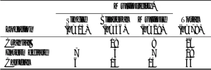
(29 screws) (Fig. 1). The location of bone resorption was cranial in 33% of the cases (26 of 78 screws), caudal in 43.6% (34 screws), and intermediate in 23.1% (18 screws) (Table 2). For 14 screws distributed among five patients, resorption was both cranial and caudal. As shown in Fig. 3, resorption first occurred within six weeks of pedicle screw plate fixation in 78.2% of cases (61 of 78 screws) and within 12 weeks of this in 98.7%
(77 of 78 screws). It first appeared at between 2 and 3 weeks (5.1%, 4 screws); most frequently, initial onset was between 3 and 4 weeks (30.8%, 24 screws).
As shown in Table 3, 61.5% (48 of 78 screws) re- mained in stable condition, while in 38.5% (30 screws), resorption pregressed. Of the 68 screws showing grade 1 resorption, 42 (61.8%) remained in stable condition, 14
─ 333 ─
A B
Fig. 4. Progression of bone resorption (A) AP radiograph of bone resorption (62-year-old man). Grade 1 bone re- sorption in L5 (between thin arrows) and grade 2 bone resorption in L4 (be- tween thick arrows) are noted. (B) AP radiograph of the same patient ob- tained 7 months later. Progression of bone resorption in L5 to grade 2 (be- tween thin arrows). The grade 2 bone resorption in L4 remained stable (be- tween thick arrows).
Fig. 3. Bar graph shows 99% of bone resorption was appeared within 12 weeks after pedicle screw plate fixation. Bone re- sorption began to appear between 2 and 3 weeks, and the peak onset was between 3 and 4 weeks.
Table 1. Level of Pedicle Screws in Patients with Bone Resorption Level No. of Patients No. of Inserted Screws
(n=39) (n=254)
2 Levels
L3, L4 01 04
L4, L5 04 06
3 Levels
T12, L1, L2 01 06
L2, L3, L4 01 06
L3, L4, L5 11 66
L4, L5, S1 09 54
4 Levels
L2, L3, L4, L5 04 32
L3, L4, L5, S1 05 40
5 Levels
L2, L3, L4, L5, S1 03 30
Table 2. Location of Bone Resorption
Multiplicity*
Single Bilateral Multiple Total Location (n=13) (n=36) (n=29) (n=78)
Cranial 18 08 26
Intermediate 7 04 07 18
Caudal 6 14 14 34
* Data are the number of screws.
Table 3. Progression of Bone Resorption during Follow-up.
Follow-up Grade*
Grade 1 Grade 2 Grade 3 Total Initial Grade (n=42) (n=18) (n=18) (n=78)
Grade 1 42 14 12 68
Grade 2 04 04 08
Grade 3 02 02
* Data are the number of screws.
Grade 1: bone resorption less than 1 mm in width Grade 2: bone resorption less than 2 mm in width
Grade 3: bone resorption more than 2 mm or more in width.
0-1 1-2 2-3 3-4 4-5 5-6 6-8 8-10 10-12 12-14 Appearance of bone resorption during follow-up(week) 30
25 20 15 10 5 0
Number of screws
(20.6%) pregressed to grade 2, and 12 (17.6%) pro- gressed to grade 3 (Fig. 4). Of the eight screws showing grade 2 resorption, the condition of four (50%) remained stable, while the other four (50%) progressed to grade 3.
Both screws showing grade 3 resorption remained sta- ble during follow-up. Overall, 18 of 902 screws (2.0%) in seven of 156 patients (4.5%) showed abnormal, grade 3 bone resorption. No screws were removed.
Discussion
Pedicle screws together with plate and rod systems have been employed to create stability in thoracolumbar spine. They are attached posteriorly to rods or plates by threaded nuts. When firmly anchored into intact bone, pedicle screws resist loads in all directions (9), but when a screw becomes loose, hardware may fail at the site of attachment to the spine. Loosening of screws that are in direct contact with bone is caused by inadequate fixa- tion, or infection (6, 10-16). Repeated movements of loose hardware produce bone resorption or erosion around instruments (2, 6, 17, 18). Bone resorption is seen on plain radiographs as a radiolucent zone, though the development of such zones is not always due to loos- ening; it may also be a normal event. Such normal re- sorption can be caused by surgical trauma, the interposi- tioning of soft tissue or blood, and micromotion (17, 18).
Analysis of sequential films can be used to distinguish physiologic change from loosening (19). The width and appearance of bone resorption are important predictors of the possibility of screw loosening.
The incidence of such loosening according to earlier reports, varies from 0.6% to 11% (8, 12-16). The fre- quency of abnormal (grade 3) bone resorption (4.5%; 7 of 156 patients) encountered during follow-up was with- in the range of previous reports. In the majority (82%, 32 of 39 patients) of our cases with bone resorption, this was less than 2 mm in width.
Bone resorption took the form of a solitary lesion in one-third of our cases and more than two lesions in two- thirds. Because of the decreased mobility of fused seg- ments, spinal motion most significantly occurs just above and below the fused segments. In 77% of our cas- es, resorption occurred cranially, caudally, or both ends, where motion was marked.
The development and progression of bone resorption seem to be rapid, appearing within 12 weeks of pedicle
grade is low. Only 7.6% of screws showing grade 1 re- sorption progressed to grade 3, while 50% of those showing grade 2 progressed to grade 3.
When abnormal bone resorption occures, differential diagnosis should include the possibility of infection. In our cases, no clinical sign of infection was noted, though screws were not removed because stability was main- tained with the help of bony fusion in the supposedly loosened 18 screws.
Two limitations of our study are the relatively short follow-up periods and lack of surgical confirmation.
Bone resorption occuring during the late postoperative period could thus not be evaluated.
In conclusion, bone resorption can occur around both pedicle screws and other similar screws or instruments applied to long bones. When spinal stability was main- tained with the help of bony fusion and without evi- dence of infection, it was not always necessary to re- move loosened screws. We believe that an understand- ing of the radiographic patterns of bone resorption aids in monitoring a patient after pedicle screw plate fixa- tion.
References
1. Slone RM, MacMillan M, Montgomery WJ, Heare M. Spinal fixa- tion. Part 2. Fixation techniques and hardware for the thoracic and lumbosacral spine. Radiographics 1993;13:521-543
2. Slone RM, MacMillan M, Montgomery WJ. Spinal fixation. Part 3.
Complications of spinal instrumentation. Radiographics 1993;13:
797-816
3. Yahiro MA. Comprehensive literature review. Pedicle screw fixa- tion devices. Spine 1994;19(20S):2274S-2278S
4. Yuan HA, Garfin SR, Dickman CA, Mardjetko SM. A historical co- hort study of pedicle screw fixation in thoracic, lumbar, and sacral spinal fusions. Spine 1994;19(20S):2279S-2296S
5. Mulholland RC. Pedicle screw fixation in the spine. J Bone Joint Surg Br 1994;76:517-519
6. Weissman BN. Imaging of joint replacement. In: Resnick D, ed.
Bone and joint imaging. 2nd ed. Philadelphia, Saunders, 1996;183- 193
7. Bergstorm B, Lidgren L, Lindberg L. Radiographic abnormalities caused by postoperative infection following total hip arthroplasty.
Clin Orthop 1974;95-102
8. Dussault RG, Goldman AB, Ghelman B. Radiologic diagnosis of loosening and infection in hip prosthesis. J Can Assoc Radiol 1977;
28:119-123
9. Krag MH. Spinal fusion: overview of options and posterior internal fixation devices. In: Frymeyer JW, Ducker TB, Hadler NM, Kotsuik JP, Weinstein JN, Whitecloud TS III, eds. The adult spine:
principles of practice. New York, Raven, 1991;1919-1945
10. Garfin SR. The useof bone screws in the vertebral pedicles.
Summation. Spine 1994;19(20S):2300S-2305S
12. Dick W. The “fixateur interne”as a versatile implant for spine surgery. Spine 1987;12:882-900
13. Horowitch A, Peek RD, Thomas JC Jr, et al. The Wiltse pedicle screw fixation system. Early clinical results. Spine 1989;14:461-467 14. Louis R. Fusion of the lumbar and sacral spine by internal fixation
with screw plates. Clin Orthop 1986;203:18-33
15. Roy-Camille R, Saillant G, Mazel C. Internal fixation of the lumbar spine with pedicle screw plating. Clin Orthop 1986;203:7-17 16. Thalgott JS, LaRocca H, Aebi M, Dwyer AP, Razza BE.
Reconstruction of the lumbar spine using AO DCP plate internal
fixation. Spine 1989;14:91-95
17. Reckling FW, Asher MA, Dillon WL. A longitudinal study of the radiolucent line at the bone-cement interface following total joint- replacement procedures. J Bone Joint Surg Am 1977;59:355-358 18. Salvati EA, Im VC, Aglietti P, Wilson PD Jr. Radiology of total hip
replacements. Clin Orthop 1976;121:74-82
19. Tapadiya D, Walker RH, Schurman DJ. Prediction of outcome of total hip arthroplasty based on initial postoperative radiographic analysis. Matched, paired comparisons of failed versus successful femoral components. Clin Orthop 1984;186:5-15
─ 335 ─
대한방사선의학회지 2003;48:331-335
척추경 나사 고정술 후 발생하는 나사 주위 골흡수1
1청주성모병원 방사선과
2인하대학교 의과대학 방사선과학교실
3국립암센터
4Department of Radiology, Iwate Medical University, School of Medicine
5청주성모병원 정형외과
박선원1,2・이주혁1,3・Shigeru Ehara4・성수옥1・박주태5
목적: 척추의 안정성을 유지시키기 위해 시행되는 척추경(spinal pedicle) 나사 고정술(screw plate fixation) 후 척 추경 나사 주위에 생기는 골흡수의 빈도, 위치, 분포, 발생 시기, 골흡수의 진행 양상을 평가하고자 한다.
대상과 방법: 척추경 나사 고정술을 시행 받은 156명 환자의 902개 척추경 나사를 대상으로 수술 직후와 추적 관
찰 기간 동안 척추경 나사 주위에 발생한 골흡수 여부를 분석하였다. 골흡수의 정도를 평가하기 위하여 방사선 투 과성 부위(radiolucent zone)의 폭을 측정하여 Grade 1(1 mm 미만), Grade 2(1 mm 이상 2 mm 미만), Grade 3(2 mm 이상)으로 등급화 하였다. 골흡수가 관찰된 39명의 78개의 척추경 고정 나사에서 골흡수의 정도, 위치, 분포, 발생 시기 및 진행 양상을 평가하였다.
결과: 척추경 나사 고정술을 시행받은 156명 902개의 척추경 중 39명(25%) 환자의 78 개(8.6%)의 척추경에서 골흡수가 관찰되었다. 39명 중 26명에서 두 곳 이상의 척추경 고정 나사 주위에서 골흡수를 보였다. 척추경 고정 나사 중 99%에서 수술 후 12주 이내에 골흡수가 관찰되었다. 추적 기간 중 62%(78 개중 48개의 척추경 고정 나 사)에서 병변의 정도는 진행하지 않았으나 38%에서는 더 높은 등급으로 골흡수의 정도가 진행하였다. 초기의 골 흡수 등급이 낮은 경우에서 높은 등급으로 골흡수가 진행하는 정도가 낮았다.
결론: 척추경 나사 고정술 후 발생하는 나사 주위의 골흡수의 방사선학적 소견에 대한 이해는 수술 후 환자의 경과 를 관찰하고 이해하는 데 도움이 될 것으로 기대한다.