www.krspine.org
Concurrent Degenerative Cervical and Lumbar Spondylolisthesis
Moon Soo Park, M.D., Ji-Hyo Hwang, M.D., Tae-Hwan Kim, M.D., Jae Keun Oh, M.D., Ho Guen Chang, M.D., Hyung Joon Kim, M.D., Kun-Tae Park, M.D., Jin Kyu Lim, M.D., K. Daniel Riew, M.D.
J Korean Soc Spine Surg 2018 Dec;25(4):154-159.
Originally published online December 31, 2018;
https://doi.org/10.4184/jkss.2018.25.4.154
Korean Society of Spine Surgery
Asan Medical Center 88, Olympic-ro 43 Gil, Songpa-gu, Seoul, 05505, Korea Tel: +82-2-483-3413 Fax: +82-2-483-3414
©Copyright 2017 Korean Society of Spine Surgery pISSN 2093-4378 eISSN 2093-4386
The online version of this article, along with updated information and services, is located on the World Wide Web at:
http://www.krspine.org/DOIx.php?id=10.4184/jkss.2018.25.4.154
This is an Open Access article distributed under the terms of the Creative Commons Attribution Non-Commercial License (http://
creativecommons.org/licenses/by-nc/4.0) which permits unrestricted non-commercial use, distribution, and reproduction in any medium, provided the original work is properly cited.
Journal of Korean Society of
Spine Surgery
Concurrent Degenerative Cervical and Lumbar Spondylolisthesis
Moon Soo Park, M.D., Ji-Hyo Hwang, M.D., Tae-Hwan Kim, M.D.
*, Jae Keun Oh, M.D., Ho Guen Chang, M.D.
†, Hyung Joon Kim, M.D., Kun-Tae Park, M.D., Jin Kyu Lim, M.D.
‡, K. Daniel Riew, M.D.
§Department of Orthopaedic Surgery, Hallym University Sacred Heart Hospital, Medical College of Hallym University, Gyeonggi-do, Republic of Korea
*Department of Orthopaedic Surgery, Gangnam Sacred Heart Hospital, Hallym University College of Medicine, Seoul, Republic of Korea
†Department of Neurosurgery, Hallym University Sacred Heart Hospital, Medical College of Hallym University, Gyeonggi-do, Republic of Korea
‡Department of Orthopaedic Surgery, Kangdong Sacred Heart Hospital, Medical College of Hallym University, Seoul, Republic of Korea
§Department of Orthopedic Surgery, Columbia University Medical Center / NY-Presbyterian Hospital, The Spine Hospital at NY- Presbyterian/Allen Hospital, New York, NY, USA
Study Design: Retrospective radiographic study.
Objective: To evaluate the characteristics of concurrent degenerative cervical and lumbar spondylolisthesis.
Summary of Literature Review: Concurrent degenerative cervical and lumbar spondylotic diseases have been reported. Given that severe spondylosis can result in spondylolisthesis, one might expect that concurrent spondylolisthesis of the cervical and lumbar spines might also be prevalent. However, the incidence of spondylolistheses in the lumbar and cervical spines might differ due to anatomical differences between the 2 areas. Nonetheless, there is minimal information in the literature concerning the incidence of concurrent cervical and lumbar spondylolisthesis.
Material and Methods: We evaluated standing cervical and lumbar lateral radiographs of 2510 patients with spondylosis. Concurrence, age group, gender, and direction of spondylolisthesis were evaluated. Lumbar spondylolisthesis was defined as at least Meyerding grade I and degenerative cervical spondylolisthesis was defined as over 2 mm of displacement on standing lateral radiographs.
Results: Lumbar spondylolisthesis was found in 125 patients (5.0%) and cervical spondylolisthesis was found in 193 patients (7.7%).
Seventeen patients had both degenerative cervical and lumbar spondylolistheses (0.7%). Lumbar spondylolisthesis is a risk factor for co- existing cervical spondylolisthesis. Lumbar spondylolisthesis was more common in females than males, independent of advancing age. In contrast, degenerative cervical spondylolisthesis was more common in older patients, independent of gender. Anterolisthesis was more common in the lumbar spine. Retrolisthesis was more common in the cervical spine.
Conclusions: There was a higher prevalence of degenerative cervical spondylolisthesis in patients with degenerative lumbar spondylolisthesis.
Key Words: Cervical vertebrae, Lumbar vertebrae, Concurrent degenerative spondylolisthesis
Received: October 14, 2018 Revised: October 19, 2018 Accepted: November 19, 2018 Published Online: December 31, 2018 Corresponding author: Moon Soo Park, M.D.
ORCID ID: Moon Soo Park: https//orcid.org/0000-0003-2833-9148 Department of Orthopaedic Surgery Hallym University Sacred Heart Hospital Medical College of Hallym University 22 Gwanpyeong-ro 170 beon-gi, Dongan-gu, Anyang-si, Gyeonggi-do, 14068, Republic of Korea TEL: +82-31-380-6000, FAX: +82-31-380-6008
E-mail: amhangpark@gmail.com
Introduction
Concomitant cervical and lumbar surgeries for concomitant cervical and lumbar degenerative diseases are not uncommon.1-3) Jacob et al1) found that in a study population of 200 patients who underwent cervical spine surgery, thirty-one percent required additional surgery in the lumbar spine. The patients who underwent both lumbar and cervical decompressive surgeries had satisfactory results after the operations.2,3)
Concurrent cervical and lumbar spondylotic diseases
Concurrent Spondylolisthesis Journal of Korean Society of Spine Surgery
www.krspine.org 155 have been reported on.4-6) Severe spondylosis can result in
degenerative spondylolisthesis. Since both the cervical and lumbar spines are prone to severe spondylosis, one might expect that concurrent spondylolisthesis of the cervical and lumbar spine might also be prevalent. On the other hand, the incidence of spondylolistheses in the lumbar and cervical spines might differ due to anatomical differences between the two areas.
There is minimal information in the literature concerning the incidence of concurrent cervical and lumbar spondylolisthesis.
The aim of this study is to identify the incidence of concurrent degenerative cervical and lumbar spondylolistheses and to evaluate the characteristics of the respective spondylolistheses.
Methods
This study was approved by the institutional review board at the institution of the corresponding author (IRB number:
2014-I105). We evaluated the standing cervical or lumbar lateral radiographs of all patients over 50 years old who had neck or back pain or radiating arm or leg pain, from February 2006 to August 2012. Patients with non-degenerative disease (trauma, infection, tumor, deformity, rheumatoid arthritis) or with a history of previous spine surgery in the lumbar and cervical spine were excluded. In addition, we excluded patients with spondylolytic spondylolisthesis in the lumbar spine. We identified 2,510 patients who had both standing cervical &
lumbar simple lateral radiographs. There were one thousand one hundred seventeen men and one thousand three hundred ninety-three women. The patients were categorized according to their age group. There were 1,156 subjects in their 50s, 674 in their 60s, 510 in their 70s, 155 in their 80s, and 15 in their 90s. Degenerative lumbar spondylolisthesis was defined as >0%
slippage (more than or equal to Meyerding grade I except 0%
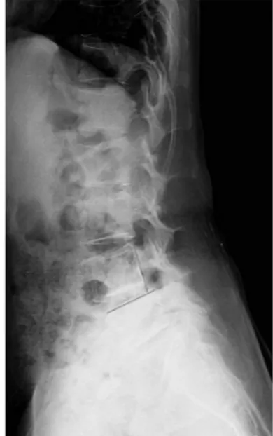
slippage) on standing lateral radiographs and degenerative cervical spondylolisthesis was defined as greater than 2 mm of displacement on standing lateral radiographs (Fig. 1).7,8)
All statistical analyses were performed with an SPSS version 24.0 for Windows (Chicago, IL, USA). It was considered significant when p was less than 0.05. The Chi-square test or Fisher’s exact test were used to analyze the differences of measuring factors on each group according to categorized variables.
Results
Out of 2,510 patients, 125 had degenerative lumbar spondylolisthesis and 193 patients had degenerative cervical spondylolisthesis (Table 1). Seventeen patients had both cervical and lumbar spondylolistheses (0.7%, 17/125 patients with lumbar spondylolisthesis (13.6%) and 17/193 patients with cervical spondylolisthesis (8.8%)). Degenerative lumbar spondylolisthesis was found to be a risk factor for co- existing degenerative cervical spondylolisthesis (lumbar spondylolisthesis odd for cervical spondylolisthesis 17/108, no lumbar spondylolisthesis odd for cervical spondylolisthesis
Fig. 1. Meyerding grading method showing grade II spondylolisthesis of L4.
Table 1. Concurrent degenerative lumbar and cervical spondylolisthesis
Cervical spondylolisthesis No cervical spondylolisthesis Total
Lumbar spondylolisthesis 17(0.7%) 108(4.3%) 125(5.0%)
No lumbar spondylolisthesis 176(7.0%) 2,209(88.0%) 2,385(95.0%)
Total 193(7.7%) 2,317(92.3%) 2,510(100%)
176/2,209, odds ratio of lumbar spondylolisthesis (17/108)/
(176/2,209)=17×2,209/108×176, odds ratio of 1.976, 95% confidence interval of 1.158 to 3.370, p=0.011, Table 1). Likewise, degenerative cervical spondylolisthesis was found to be a risk factor for co-existing degenerative lumbar spondylolisthesis (cervical spondylolisthesis odd for lumbar spondylolisthesis 17/176, no cervical spondylolisthesis odd for lumbar spondylolisthesis 108/2,209, odds ratio of cervical spondylolisthesis (17/176)/(108/2,209)=17×2,209/176×108, odds ratio of 1.976, 95% confidence interval of 1.158 to 3.370, p=0.011, Table 1).
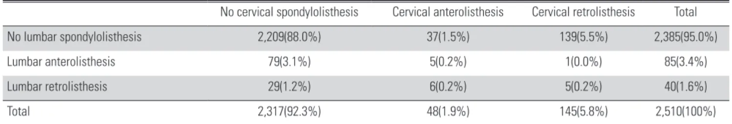
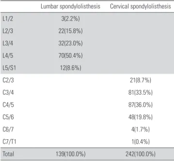
Lumbar spondylolisthesis was more common in females, with an odds ratio of 2.135 (95% confidence interval of 1.433 to 3.181, p<0.001, Table 2). In contrast, cervical spondylolisthesis had no gender differences (p=0.893, Table 2). The prevalence of degenerative lumbar spondylolisthesis was not different in the different age groups, but cervical spondylolisthesis was more common in older patients (p<0.001, Table 3). Anterolisthesis was more common than retrolisthesis in the lumbar spine (Table 4), whereas retrolisthesis was more common than anterolisthesis in the cervical spine (Table 4). The direction of cervical spondylolisthesis did not correlate with the direction of lumbar spondylolisthesis (Table 4). Degenerative lumbar spondylolisthesis was common at the disc levels of L3/4 and L4/5 and degenerative cervical spondylolisthesis was common at the disc levels of C3/4 and C4/5 (Table 5).
The disc levels of L3/4 and L4/5 in degenerative lumbar
spondylolisthesis had no correlation with those of C3/4 and C4/5 in degenerative cervical spondylolisthesis (p>0.05, Table 6). None of Meyerding grade II and grade III of degenerative lumbar spondylolisthesis was found in our study population.
We found only Meyerding grade I (slippage 1~24%) of degenerative lumbar spondylolisthesis in the study population.
It might be due to the study design that we had excluded the patients with spondylolytic spondylolisthesis in the lumbar spine.
Discussion
To our knowledge, there has been no study documenting the co-existence of degenerative cervical and lumbar spondylolistheses. The purpose of this study is to identify the incidence and characteristics of concurrent degenerative cervical and degenerative lumbar spondylolisthesis.
Master et al4) evaluated degenerative spondylosis of the cervical and lumbar spines, looking at osteophytes of the endplate and facet joints in an anatomic study of 234 cadaveric specimens. They found that lumbar spondylosis was associated with concurrent cervical spondylosis. Similar to their results, the present study showed that the presence of degenerative lumbar spondylolisthesis is a risk factor for
Table 2. Degenerative spondylolisthesis according to gender Lumbar
spondylolisthesis Cervical
spondylolisthesis Subgroup population
Female 90(6.5%) 108(7.8%) 1,393(100.0%)
Male 35(3.1%) 85(7.6%) 1,117(100.0%)
Total 125(5.0%) 193(7.7%) 2,510(100.0%)
Table 3. Degenerative spondylolisthesis according to age Lumbar
spondylolisthesis
Cervical spondylolisthesis
Subgroup population
50s 45(3.9%) 57(4.9%) 1,156(100.0%)
60s 42(6.2%) 37(5.5%) 674(100.0%)
70s 25(4.9%) 64(12.5%) 510(100.0%)
80s 12(7.7%) 30(19.4%) 155(100.0%)
90s 1(6.7%) 5(33.3%) 15(100.0%)
Total 125(5.0%) 193(7.7%) 2,510(100.0%)
Table 4. The direction of degenerative spondylolisthesis according to cervical and lumbar spine
No cervical spondylolisthesis Cervical anterolisthesis Cervical retrolisthesis Total
No lumbar spondylolisthesis 2,209(88.0%) 37(1.5%) 139(5.5%) 2,385(95.0%)
Lumbar anterolisthesis 79(3.1%) 5(0.2%) 1(0.0%) 85(3.4%)
Lumbar retrolisthesis 29(1.2%) 6(0.2%) 5(0.2%) 40(1.6%)
Total 2,317(92.3%) 48(1.9%) 145(5.8%) 2,510(100%)
Concurrent Spondylolisthesis Journal of Korean Society of Spine Surgery
www.krspine.org 157 co-existing degenerative cervical spondylolisthesis. While
degenerative spondylosis in both the cervical and lumbar spines are associated with advancing age,4-6) we found that only cervical and not lumbar spondylolisthesis is associated with older age. This suggests that, while spondylosis increases with advancing age, lumbar spondylolisthesis does not.
There are a few possible explanations for this. First, it is well known that degenerative lumbar spondylolisthesis is more common in patients in their forties. It may be that lumbar spondylolisthesis is already present at a relatively young age and doesn’t appreciably increase with increasing age. Second, spondylosis is not an all-or-none phenomenon so one would expect degenerative changes to worsen with advancing age.
In contrast, degenerative listhesis is either present or not.
Therefore, if degenerative listhesis has already occurred at vulnerable levels at a relatively young age, further aging may not affect its prevalence. Degenerative lumbar listhesis has also
been reported to be more common in female patients.9,10) We confirmed that there was a higher incidence of degenerative lumbar spondylolisthesis in females, independent of their age.
The patients with degenerative spondylolisthesis had the less lumbar lordosis, greater pelvic incidence, and positive sagittal balance than the normal volunteers.11) The positive sagittal balance was common in the cervical spine of elderly subjects.12) In the elderly patients, the cervical spondylolisthesis was common in the current study. It is a possible explanation that degenerative lumbar spondylolisthesis was found to be a risk factor for co-existing degenerative cervical spondylolisthesis.
Anterolisthesis was more common in the lumbar spine.
Retrolisthesis was more common in the cervical spine.
Anterolisthesis can only occur if the facets remodel to allow the listhesis. On the other hand, retrolisthesis can occur without facet remodeling, as the disc height decreases. Also, biomechanical forces are likely to play a critical role in the direction of listhesis. This suggests that the biomechanical forces that affect the lumbar listhesis levels, most commonly, L4-5 and L5-S1, tend to cause anterolisthesis. On the other hand, in the cervical spine, the forces acting upon the cervical levels tend to produce retrolisthesis.
Our study has several limitations. First, all patients in this study were symptomatic and were older than 50 years. Thus, there might be a selection bias, and the result may not be applicable to the general population. Second, as a retrospective study, most of the variables were not controlled. Our plan is to perform a prospective study evaluating the same parameters to determine which factors affect the prevalence of concurrent degenerative cervical and lumbar spondylolistheses. Third, we did not evaluate the clinical symptoms and signs associated with spondylolisthesis. Radiologic spondylolisthesis does not always have clinical symptoms and signs. Finally, we included the patients who had neck or back pain or radiating arm or leg pain instead of normal volunteers. Therefore, there is a possible selection bias to make a conclusion that there was a higher incidence of degenerative cervical spondylolisthesis in elderly patients because more cervical symptoms have been reported only in elderly patients. The strengths of the study include the fact that this is the first paper that examined the incidence of concurrent degenerative cervical and lumbar spondylolisthesis.
In addition, we examined a large number of subjects (2,510).
Table 5. Degenerative spondylolisthesis according to disc levels Lumbar spondylolisthesis Cervical spondylolisthesis
L1/2 3(2.2%)
L2/3 22(15.8%)
L3/4 32(23.0%)
L4/5 70(50.4%)
L5/S1 12(8.6%)
C2/3 21(8.7%)
C3/4 81(33.5%)
C4/5 87(36.0%)
C5/6 48(19.8%)
C6/7 4(1.7%)
C7/T1 1(0.4%)
Total 139(100.0%) 242(100.0%)
When there is spondylolisthesis in the multiple disc levels, the disc level with most slippage was chosen for spondylolisthesis.
Table 6. The correlation of the presence of degenerative spondylolisthe- sis between the disc levels of C3/4, C4/5, and L3/4, L4/5.
C3/4 C4/5
L3/4 0.903 0.233
L4/5 0.836 0.509
Conclusion
To our knowledge, this is the largest series to examine the prevalence of concurrent degenerative spondylolistheses of the cervical and lumbar spines. We found a higher prevalence of degenerative cervical spondylolisthesis in patients with degenerative lumbar spondylolisthesis. We had better watch out for the cervical spine of the old patients with degenerative lumbar spondylolisthesis. Unlike degenerative lumbar spondylolisthesis, which never rises much over a prevalence of 7%, regardless of age, cervical spondylolisthesis becomes more prevalent with age and is common in the elderly.
REFERENCES
1. Jacobs B, Ghelman B, Marchisello P. Coexistence of cervical and lumbar disc disease. Spine (Phila Pa 1976).
1990 Dec;15(12):1261-4. DOI: 10.1097/00007632- 199012000-00006.
2. Teng P, Papatheodorou C. Combined Cervical and Lumbar Spondylosis. Arch Neurol. 1964 Mar;10(3):298-307. DOI:
10.1001/archneur.1964.00460150068007.
3. Choudhury AR, Taylor JC. The cervicolumbar syndrome.
Ann R Coll Surg Engl. 1980 May;62(3):200-2.
4. Master DL, Eubanks JD, Ahn NU. Prevalence of concurrent lumbar and cervical arthrosis: an anatomic study of cadaveric specimens. Spine (Phila Pa 1976). 2009 Apr;34(8):E272-5.
DOI: 10.1097/BRS.0b013e318195d10b.
5. van Saase JL, van Romunde LK, Cats A, et al. Epidemiology of osteoarthritis: Zoetermeer survey. Comparison of radio- logical osteoarthritis in a Dutch population with that in 10 other populations. Ann Rheum Dis. 1989 Apr;48(4):271- 80. DOI: 10.1136/ard.48.4.271.
6. Lawrence JS. Disc degeneration. Its frequency and relation- ship to symptoms. Ann Rheum Dis. 1969 Mar;28(2):121- 38. DOI: 10.1136/ard.28.2.121.
7. Kopacz KJ, Connolly PJ. The prevalence of cervical spon- dylolisthesis. Orthopedics. 1999 Jul;22(7):677-9.
8. Lee C, Woodring JH, Rogers LF, et al. The radiographic distinction of degenerative slippage (spondylolisthesis and retrolisthesis) from traumatic slippage of the cervical spine.
Skeletal Radiol. 1986;15(6):439-43. DOI: 10.1007/
bf00355101.
9. Herkowitz HN. Spine update. Degenerative lumbar spon- dylolisthesis. Spine (Phila Pa 1976). 1995 May;20(9):1084- 90. DOI: 10.1097/00007632-199505000-00018.
10. Bolesta MJ, Bohlman HH. Degenerative spondylolis- thesis. Instructional course lectures. Instr Course Lect.
1989;38:157-65.
11. Barrey C, Jund J, Noseda O, et al. Sagittal balance of the pelvis-spine complex and lumbar degenerative diseases.
A comparative study about 85 cases. Eur Spine J. 2007 Sep;16(9):1459-67. DOI: 10.1007/s00586-006-0294-6.
12. Park MS, Moon SH, Lee HM, et al. The effect of age on cer- vical sagittal alignment: normative data on 100 asymptomatic subjects. Spine (Phila Pa 1976). 2013 Apr;38(8):E458-63.
DOI: 10.1097/BRS.0b013e31828802c2.
© Copyright 2018 Korean Society of Spine Surgery
Journal of Korean Society of Spine Surgery. www.krspine.org. pISSN 2093-4378 eISSN 2093-4386
This is an Open Access article distributed under the terms of the Creative Commons Attribution Non-Commercial License (http://creativecommons.org/licenses/by-nc/4.0/) which permits unrestricted non-commercial use, distribution, and reproduction in any medium, provided the original work is properly cited.
159
J Korean Soc Spine Surg. 2018 Dec;25(4):154-159. https://doi.org/10.4184/jkss.2018.25.4.159
Original Article
경추 및 요추 퇴행성 척추전위증의 병발성 유병률
박문수 • 황지효* • 김태환 • 오재근† • 장호근 • 김형준 • 박건태 • 임진규‡ • K. Daniel Riew§
한림대학교 한림대학교성심병원 정형외과학교실, *한림대학교 강남성심병원 정형외과학교실, †한림대학교 한림대학교성심병원 신경외과학교실
‡한림대학교 강동성심병원 정형외과학교실
연구 계획: 후향적 방사선 연구
목적: 경추와 요추에 동시에 발생한 퇴행성 척추전위증을 알아보고자 한다.
선행 연구논문의 요약: 경추와 요추에 동시에 발생한 퇴행성 척추질환에 대한 여러 보고가 있었다. 퇴행성 척추전위증은 퇴행성 변화에 의하여 시발되므 로 경추와 요추에 척추전위증이 병발할 것으로 추정된다. 반면에, 요추와 경추의 해부학적 구조가 서로 다르므로 두 질환의 진행이 동일하지 않을 것으로 추정할 수도 있다. 그러나, 경추와 요추에 동시에 발생한 퇴행성 척추전위증에 대한 보고는 적었다.
대상 및 방법: 요추 및 경추 부위에 기립위 단순방사선 검사를 둘다 시행한 퇴행성 척추 질환 환자 2,510명을 대상으로 하였다. 병발여부, 나이, 성별, 전 위증의 방향에 대하여 조사하였다. 퇴행성 요추전위증은 기립위 단순방사선영상에서 Meyerding 방법을 사용하여 grade 1 이상인 경우 진단하였으며 퇴 행성 경추전위증은 기립위 단순방사선영상에서 2 mm 이상의 전위가 보이는 경우 진단하였다.
결과: 퇴행성 요추전위증은 125명에서 관찰되었으며(5.0%) 퇴행성 경추전위증은 193명에서 관찰되었다(7.7%). 요추전위증과 경추전위증은 17명에서 같이 관찰되었다(0.7%). 요추전위증이 있는 환자가 없는 환자에 비하여 경추전위증이 더 많이 관찰되었다. 요추전위증은 남자보다 여자에서 더 흔하였으 나, 모든 연령군에서 비슷하게 발생하였다. 경추전위증은 고령의 연령군에서 더 많이 발생하였으나, 남녀의 발생 비율이 비슷하였다. 요추전위증에서는 전방으로 많이 발생하였고, 경추전위증에서는 후방으로 많이 발생하였다.
결론: 퇴행성 요추전위증이 있는 경우가 없는 경우에 비하여 퇴행성 경추전위증이 더 많이 발생하였다.
색인 단어: 경추, 요추, 병발성 퇴행성 척추 전위증 약칭 제목: 동시에 발생한 척추전위증
접수일: 2018년 10월 14일 수정일: 2018년 10월 19일 게재확정일: 2018년 11월 19일 교신저자: 박문수
경기도 안양시 동안구 관평로 170번길 22번지 한림대학교부속 한림대학교성심병원 정형외과학교실
TEL: 031-380-6000 FAX: 031-380-6008 E-mail: amhangpark@gmail.com