INTRODUCTION
An scalp arteriovenous malformation (AVM) is an abnor- mal fistulous connection between feeding arteries and drain- ing veins without an intervening capillary bed within the sub- cutaneous layer. The draining veins are typically grossly dilated and may show variceal dilation that is not life threat- ening, but can cause cosmetic deformity (1). Hemophilia A, transmitted as an X-linked recessive trait, is a common heri- table bleeding disorder caused by mutations in the gene that codes for factor VIII (2). Here, we describe an unusual case of scalp AVM in a patient with severe hemophilia A whom we treated with direct puncture embolization using coils and n- butyl-cyanoacrylate (NBCA), followed by surgical removal.
CASE REPORT
A 22-year-old man presented with a pulsatile mass on his
right parietal scalp at birth and had increased in size during the preceding year. The patient had no previous history of trauma or head injury. Moreover, he was previously diag- nosed with hemophilia A and hemophilic arthropathy, and had undergone surgery for hemophilic arthropathy of the left knee and left elbow joints 7 years prior to presentation. His plasma factor VIII was less than 1%, indicating severe hemo- philia A (normal range, 50-80%). His prothrombin time was 12.2 seconds (control, 12.5-14.7) and his activated partial thromboplastin time was 59 seconds (control, 29-43). Upon examination, we observed no ulceration of the scalp skin or active bleeding.
Enhanced computed tomography demonstrated prominent vascular enhancement with an aneurysmal sac on the right parietal scalp (Fig. 1). Digital subtraction angiography (DSA) was performed to evaluate the scalp mass and to plan treat- ment. To ensure effective hemostasis for intra-arterial catheter- ization, a bolus dose of coagulation factor VIII (4500 units)
J Korean Soc Radiol 2011;65(3):229-233
Received May 7, 2011; Accepted July 19, 2011 Corresponding author: Eui Jong Kim, MD Department of Radiology, Kyung Hee University Hospital, Kyung Hee University School of Medicine, 1 Hoegi-dong, Dongdaemun-gu, Seoul 130-702, Korea.
Tel. 82-2-958-8621, 8622, 9461 Fax. 82-2-968-0787 E-mail: [email protected]
Copyrights © 2011 The Korean Society of Radiology
We present a case of scalp arteriovenous malformation (AVM) in a patient with severe hemophilia A. The 22-year-old man presented with a pulsatile right parietal scalp mass. Digital subtraction angiography revealed an AVM in the right parietal scalp, supplied by superficial temporal and occipital arteries that drained into multiple ve- nous structures. We successfully performed direct puncture embolization followed by surgical resection of the scalp AVM in conjunction with supplemental infusion of co- agulation factor VIII before, during and after the embolization and the operation.
Index terms Scalp AVM Hemophilia A
Endovascular Treatment Direct Puncture Embolization Coagulation Factor VIII
Direct Puncture Embolization of Scalp Arteriovenous Malformation in a Patient with Severe Hemophilia A: A Case Report
1혈우병 환자에서 생긴 두피내 동정맥기형의 직접 천자 색전술: 증례 보고1
Kyung Mi Lee, MD
1, Eui Jong Kim, MD
1, Bong Jin Park, MD
2, Keon Ha Kim, MD
3Departments of 1Radiology, 2Neurosurgery, Kyung Hee University Hospital, Kyung Hee University, Graduate School of Medicine, Seoul, Korea
3Department of Radiology, Samsung Medical Center, Sungkyunkwan University School of Medicine, Seoul, Korea
(Fig. 2). There were no bleeding complications at the punc- ture. Complete removal of the scalp AVM was planned be- cause severe hemophilia A can lead to serious and life-threat- ening bleeding, even during minor trauma.
Eight days after the initial angiography, preoperative percu- taneous embolization of the scalp AVM was performed im- mediately after intravenous injection of coagulation factor VIII.
Initially, the right common femoral artery was punctured and a 6 Fr sheath was inserted into the right femoral artery, fol- lowed by a 5 Fr Berenstein catheter inserted into the right ex- ternal cerebral artery (ECA) for control angiography. The di- lated aneurysmal sac near the fistulous or arteriovenous connection point was punctured by an 18G angiocath (Fig. 3A).
Pushable detachable coils (Nester 14 mm/12 cm × 7, 14/10 × 2, 14/8 × 1, 14/6 × 2, 14/4 × 1 and Tornado coils 4 mm/2 cm × 2) were inserted into the aneurysmal sac to obliterate the arterio venous (AV) shunting and nidus (Fig. 3B). Coil embolization of the scalp AVM was attempted before NBCA injection be- cause the nidus and distal venous structural sac were too large to treat with liquid material such as NBCA, and additionally, there were too many feeding arteries and draining veins of the AVM. DSA of the right ECA showed decreases in flow and shunting, but still contained some shunting and dilated [Greenmono, monoclonal anti-FVIII antibodies (mAb) sup-
plied by Hyland Division, Baxter Healthcare Corp., Deerfield, IL, USA] was administered before angiography. DSA showed that the superficial temporal and occipital arteries supplying the nidus were dilated, along with the multiple dilated early drained venous structures and a dilated aneurysmal pouch
Fig. 1. Contrast enhanced CT reveals prominent vessels with an aneu- rysmal sac on the subcutaneous layer of the right parietal scalp.
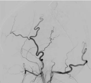
Fig. 2. Lateral projection images from right (A) and left (B) ECA angiography show opacification of an AVM of the right parietal scalp. The lesion was supplied by dilated superficial temporal and occipital arteries and were drained into multiple early dilated venous structures.
Note.-AVM = arteriovenous malformation, ECA = external cerebral artery
A B
ministerd into the draining veins and feeders. There was no significant bleeding during or after these procedures.
Two weeks after percutaneous embolization, enucleation of the scalp AVM was performed because the AVM had de- creased in size after coiling with glue injection, but still re- tained mass formation and cosmetic problems. A bolus dose venous structures. A new nidal point puncture was attempted
and two sessions of 25% NBCA injection with lipiodol were performed under manual compression of the draining veins (Fig. 4). Right ECA angiography after percutaneous emboli- zation showed complete obliteration of the AVM nidus and arteriovenous shunts (Fig. 5). A small amount of glue was ad-
Fig. 4. Lateral radiograph obtained after another direct puncture of the nidus was obtained with manual compression during injection NBCA with lipiodol.
Note.-NBCA = n-butyl-cyanoacrylate
Fig. 5. Lateral projection images from the right ECA angiography after embolization show effective devascularization of the lesion.
Note.-ECA = external cerebral artery
Fig. 3. A. Lateral projection from the direct puncture angiography shows filling of the lesion with contrast material and the venous drainage.
B. Lateral projection after insertion of detachable coils (Nester and Tornado coils) shows multiple coils filling the aneurysmal sac and vascular structures.
A B
Because scalp AVM in patients with severe hemophilia A is relatively rare, there are no standard treatment options. Al- though embolization using metallic coils or liquid material has achieved successful results in selected cases, it usually re- sults in palliation rather than cure (6). If surgical resection alone is used for AVM in hemophilia patients, patients often suffer from recurrent AVM or bleeding during the operation, as well as potential cosmetic morbidity (6, 7). Therefore, pre- operative embolization with surgical resection has been the main treatment option for scalp AVM with hemophilia.
In cases of AVM with hemophilia, preoperative transarteri- al embolization of the multiple feeding arteries may be inef- fective or technically difficult because of the bleeding tenden- cy. Without superselection of the nidus and supplying vessels, transarterial embolization often results in the occlusion of proximal arteries to the AVM and the development of promi- nent collateral vessels. To prevent this, the AV connection must be occluded by deep penetration of small particles or a liquid embolic agent such as NBCA by means of direct puncture of the nidus or venous pouch (5). Metallic coil embolization can prevent the spread of the liquid agent and decrease the size of AVM when performed before liquid embolic agent emboliza- tion (5). Scalp AVMs are less likely to communicate with the deep venous system because of their superficial locations. For these reasons, we performed manual compressions of venous drainage during glue injection to reduce undesirable washout into the distal venous outflow. To avoid exposure of the oper- ator’s hand to radiation or temporary occlusion of the venous outflow, a ring-shaped compression device can be used. But in case of an AVM with complex venous drainage, complete occlusion of the venous outflow with a simple device is diffi- cult (5, 8).
Scalp AVMs rarely induce serious hemorrhage, but minor trauma may result in serious complication such as life-threat- ening bleeding in hemophilic patients. Modern therapy, which incorporates differentiated coagulation factor replacement, makes it possible to perform major surgery on hemophilic patients with virtually no complications. Benndorf et al. (9) reported an unusual case of a mandibular AVM in a patient with severe hemophilia A and recommended endovascular treatment in mandibular AVMs, in which replacement thera- py with coagulation factors enabled safe endovascular treat- of 4,500 units of coagulation factor VIII was administered be-
fore surgery. During the operation, 4,500 units were adminis- tered daily by continuous intravenous infusion. The emboli- zed AVM was safely removed by surgery without any morbidity.
After the operation, the patient received coagulation factor VIII (1,500 units, tid). We maintained FVIII levels > 80% for 4 days. To assist with the administration of coagulation factor VIII, laboratory tests (aPTT, PT, FVIII level, platelet count) were performed on a daily basis. The patient was discharged after 4 days.
DISCUSSION
Scalp AVM is a relatively rare type of vascular lesion and usually gradually increases in size from birth. In this report, the patient complained of a recent increase in the size of the mass, which resulted in cosmetic problems. Rapid increases in size have been reported to occur at puberty, during preg- nancy and during menstruation (1). Other symptoms and signs of scalp AVM include pain, headaches, swelling of soft tissue, changes in the skin and bruit. It can even cause scalp necrosis and bleeding (3). High output cardiac failure can oc- cur with large fistulae (1).
Treatment of scalp AVM is generally considered when complications arise, including bleeding, ulceration, high out- put cardiac failure, and cosmetic disfiguration. Treatment op- tions for scalp AVM include surgical treatment, endovascular treatment, or a combination of both (3, 4). In the past, surgi- cal excision or ligation of feeding arteries was the treatment of choice for scalp AVM. However, with the advent of endovas- cular treatment techniques and new embolic agents, emboli- zation has become the preferred treatment for these lesions.
There are three endovascular treatments for scalp AVM: tran- sarterial, transvenous and direct puncture embolization (5).
The risk of necrosis of the overlying skin may be increased due to the embolization of bilateral superficial temporal arter- ies when the lesion is in or near the midline. In such situa- tions, direct puncture embolization with NBCA, alcohol, or metallic coils is preferred. In direct puncture embolization, the venous structure just distal to the AV connection is targeted.
We report a patient with severe hemophilia A, whose plas- ma coagulation factor was less than 1% of the normal value.
Otolaryngol-Head Neck Surg 2005;48:1136-1142
5. Han MH, Seong SO, Kim HD, Chang KH, Yeon KM, Han MC.
Craniofacial arteriovenous malformation: preoperative embolization with direct puncture and injection of n-bu- tyl cyanoacrylate. Radiology 1999;211:661-666
6. Jeong HS, Baek CH, Son YI, Kim TW, Lee BB, Byun HS. Treat- ment for extracranial arteriovenous malformations of the head and neck. Acta Otolaryngol 2006;126:295-300 7. Widlus DM, Murray RR, White RI Jr, Osterman FA Jr, Sch-
reiber ER, Satre RW, et al. Congenital arteriovenous mal- formations: tailored embolotherapy. Radiology 1988;169:
511-516
8. Ryu CW, Whang SM, Suh DC, Kim SM, Jang YJ, Kim HJ, et al. Percutaneous direct puncture glue embolization of high-flow craniofacial arteriovenous lesions: a new circu- lar ring compression device with a beveled edge. AJNR Am J Neuroradiol 2007;28:528-530
9. Benndorf G, Kim DM, Menneking H, Klein M. Endovascular management of a mandibular arteriovenous malformation in a patient with severe hemophilia a. AJNR Am J Neurora- diol 2004;25:614-617
ment in hemophiliac patients. In previous studies, including our case, hemophilia and hemostasis were achieved by either intermittent bolus injection or continuous intravenous infu- sion of coagulation factor.
In conclusion, replacement therapy that includes normal levels of coagulation factor enables the safe and effective en- dovascular treatment of scalp AVM by direct puncture embo- lization in hemophiliac patients.
REFERENCES
1. Fisher-Jeffes ND, Domingo Z, Madden M, de Villiers JC.
Arteriovenous malformations of the scalp. Neurosurgery 1995;36:656-660; discussion 660
2. Bowen DJ. Haemophilia A and haemophilia B: molecular insights. Mol Pathol 2002;55:1-18
3. Nagasaka S, Fukushima T, Goto K, Ohjimi H, Iwabuchi S, Maehara F. Treatment of scalp arteriovenous malforma- tion. Neurosurgery 1996;38:671-677; discussion 677 4. Jeong HS, Choi JY, Lee HJ, Kim TW, Kim MB, So YK, et al.
Diagnosis and Treatment of Extracranial Arteriovenous Malformations in the Head and Neck Region. Korean J
혈우병 환자에서 생긴 두피내 동정맥기형의 직접 천자 색전술:
증례 보고1
이경미
1· 김의종
1· 박봉진
2· 김건하
3본 증례 보고는 혈우병 환자에서 생긴 두피내 동정맥기형에 관한 내용이다. 22세 남자가 오른쪽 두정엽 두피내에 위치하 는 박동성의 종괴를 주소로 내원하였다. 디지털 감산 혈관조영술에서 오른쪽 두정엽 두피내에 동정맥기형이 관찰되었으 며, 종괴는 측두엽 및 후두엽 동맥 및 여러개의 정맥으로 이루어져 있었다. 두피내 동정맥기형에 대해 직접 천자 색전술과 함께 수술적 제거를 시행하였으며, 색전술과 수술 전후로 혈액응고인자 8을 투여함으로써 성공적으로 동정맥기형을 제 거할 수 있었다.
경희대학교 의과대학 경희의료원 1영상의학과학교실, 2신경외과학교실, 3성균관대학교 의과대학 삼성서울병원 영상의학과학교실