ABSTRACT
Background: Fifty to sixty percent of patients with anorectal malformation (ARM) have at least one associated anomaly (AA). We determined the incidence of AA with the subtypes of ARM classified in accordance with the Krickenbeck classification and analyzed differences in the incidence rates of major and minor AAs according to organ system.
Methods: From January 1999 to May 2017, we retrospectively analyzed congenital anomalies in patients who underwent an anoplasty for ARM at our institution. The AAs were divided into nine organ systems. To analyze the difference in the incidence of AAs, we calculated odds ratios (ORs) using cases of perineal fistula as the base group.
Results: Of the 460 patients, 256 (55.7%) were male, 299 (65%) had at least one anomaly, and 274 (59.6%) had major AAs. According to organ system, AAs were most common in the genitourinary (28%), cardiovascular (25%), and spinal/vertebral systems (22.6%). Major AA was most common in the cardiovascular (23%) and spinal/vertebral and genitourinary systems (19.3%). According to ARM subtype, AAs were common in the order of cloaca (93.9%), rectovaginal fistula (85.7%), and recto-bladder neck fistula (85%). For the incidence of AAs, cloaca (OR, 15.7) and recto-bladder neck fistula (OR, 5.74) showed significantly higher ORs. In the analysis of major AAs, the cloaca (OR, 19.77) showed the highest OR, followed by no fistula (OR, 4.78) and recto-bladder neck fistula (OR, 3.83).
Conclusion: A considerable number of patients with ARM had AAs. Our data are useful for predicting AAs in patients with ARM.
Keywords: Anorectal Malformation; Imperforate Anus; Associated Anomaly; Organ System, Major Congenital Anomaly
INTRODUCTION
Anorectal malformations (ARMs) are relatively common congenital anomalies in pediatric surgery, with an incidence of 1 in 4,000 to 5,000 live births, and slightly more common in males than in females.1,2 Moreover, 50%–60% of patients with ARM are known to have at least one other congenital malformation.3 Associated anomalies (AAs) were reported to have more AAs in the higher ARM type when Wingspread classification was applied.4 The most commonly associated organ systems are the genitourinary, spinal, and cardiovascular
Original Article
Received: Nov 2, 2019 Accepted: Feb 7, 2020 Address for Correspondence:
Hyun-Young Kim, MD, PhD Department of Pediatric Surgery, Seoul National University Children's Hospital, Seoul National University College of Medicine, 101 Daehak-ro, Jongno-gu, Seoul 03080, Korea.
E-mail: spkhy02@snu.ac.kr
© 2020 The Korean Academy of Medical Sciences.
This is an Open Access article distributed under the terms of the Creative Commons Attribution Non-Commercial License (https://
creativecommons.org/licenses/by-nc/4.0/) which permits unrestricted non-commercial use, distribution, and reproduction in any medium, provided the original work is properly cited.
ORCID iDs Chaeyoun Oh
https://orcid.org/0000-0001-9734-0869 Joong Kee Youn
https://orcid.org/0000-0002-6345-5745 Ji-Won Han
https://orcid.org/0000-0001-6435-9627 Hee-Beom Yang
https://orcid.org/0000-0002-5343-0448 Hyun-Young Kim
https://orcid.org/0000-0003-0106-9969 Sung-Eun Jung
https://orcid.org/0000-0003-3155-2900 Disclosure
The authors have no potential conflicts of interest to disclose.
Chaeyoun Oh ,1 Joong Kee Youn ,2 Ji-Won Han ,2 Hee-Beom Yang ,2 Hyun-Young Kim ,2 and Sung-Eun Jung 2
1Division of Pediatric Surgery, Department of Surgery, Korea University College of Medicine, Seoul, Korea
2 Department of Pediatric Surgery, Seoul National University Children's Hospital, Seoul National University College of Medicine, Seoul, Korea
Analysis of Associated Anomalies in Anorectal Malformation: Major and Minor Anomalies
Surgery
Author Contributions
Conceptualization: Oh C, Kim HY. Data curation: Oh C. Formal analysis: Oh C, Youn JK.
Investigation: Oh C, Yang HB. Methodology:
Oh C, Han JW. Writing - original draft: Oh C.
Writing - review & editing: Kim HY, Jung SE.
systems, and many studies have evaluated AAs in patients with ARM.4-8 However, some studies are limited by their small sample sizes.6,7 Other studies used a classification of ARM that most current physicians are not familiar with.4,8 In addition, in some studies, the protocolized screening examination for AA was not performed in all the patients.8
Associated congenital anomalies can lead to overall mortality and contribute to serious morbidity. Among these congenital anomalies, the major ones are generally defined as structural abnormalities of surgical, medical, functional, or cosmetic importance. Many studies have assessed AAs in patients with ARM, but to our knowledge, no studies have yet classified AAs as major and minor congenital anomalies.
This study analyzed the incidence of congenital anomalies associated with the subtypes of ARM classified in accordance with the Krickenbeck classification in a relatively large number of patients enrolled at a single center and the differences in the incidence rates of major and minor AAs according to organ system among patients with ARM.
METHODS
Study design
From January 1999 to May 2017, we retrospectively analyzed congenital anomalies in patients who underwent an anorectoplasty for ARM at Seoul National University Children's Hospital. ARM subtypes are classified in accordance with the Krickenbeck classification.9 AAs were divided into specific categories by nine organ systems as follows: chromosomal, cardiovascular, gastrointestinal, spinal or vertebral, genitourinary, musculoskeletal, craniofacial, respiratory, and brain anomalies. We have identified minor congenital anomalies associated with congenital malformations by using the European surveillance of congenital anomalies (EUROCAT Guide 1.4 definition).10 Major AAs were defined as all malformations other than minor congenital anomalies. Chromosomal anomaly was not classified into major or minor.
Data collection
The data of the patients' AAs were based on the result of the protocolized examination in Seoul National University Children's Hospital. Since March 1998, our institute has applied a protocol for urinalysis, infantography, echocardiography, and abdominal ultrasonography, including a detailed physical examination for all patients with ARM. However, at this time, spinal ultrasonography or magnetic resonance imaging (MRI) was not included in our protocol. Since January 2002, spinal examination has been included in the protocols for all patients with ARM in our institution. Spinal ultrasonography was performed in patients aged < 3 months, and spinal MRI was performed in patients aged > 3 months. As with most other institutions, detailed screening examination of chromosomal, musculoskeletal, craniofacial, respiratory, and brain is not routinely performed in our institution. Voiding cystourethrography (VCUG) was performed in all subtypes except perineal or vestibular fistula. VCUG was not performed routinely in patients with perineal or vestibular fistula but was performed in patients with abnormal urinalysis or suspected reflux nephropathy.
In this study, patent ductus arteriosus (PDA) was defined when the condition persisted after 1 month of life or when surgery for PDA was performed before the first month of life. In addition, atrial septal defect was defined as a heart defect in which blood continues to flow
between the atria even after 6 months of age. All AAs according to organ system in patients with ARM at our institution are described in Supplementary Table 1.
Statistical analysis
Correlations between variables were evaluated using the χ2 test. To analyze the difference in incidence of AAs, we calculated odds ratios (ORs) using cases of perineal fistula as the base group. We performed logistic regression to calculate the OR and 95% confidence intervals.
Statistical differences were considered significant at P values of < 0.05. Statistical analysis was performed using the SPSS version 20 software (SPSS Inc., Chicago, IL, USA).
Ethics statement
This study was approved by the Institutional Review Board (IRB) of Seoul National University Hospital (IRB file No. 1807-076-958). Because this study was a retrospective study, informed consent was not provided.
RESULTS
Of the 460 patients, 256 (55.7%) were male, with a mean gestational age of 38.34 ± 2.27 weeks and a mean birth weight of 2.99 ± 0.59 kg. The most common ARM subtype was perineal fistula (157, 34.1%), which was found in 102 male patients. The second most common subtype was the vestibular fistula, which was found in 92 patients (20%). Of the total patients, 299 (65%) had at least one AA. No statistical significance was found in the presence of AAs between boys and girls, although AAs were slightly more likely to occur in boys than in girls (67.2% in boys and 62.2% in girls, P = 0.281) (Table 1).
According to organ system, AAs were most common in genitourinary anomalies (28%), followed by cardiovascular (25%) and spinal/vertebral anomalies (22.6%). However, major AAs were most common in cardiovascular anomalies (23%), followed by spinal/vertebral (20.6%) and genitourinary anomalies (19.3%) (Fig. 1). The ARM subtype analysis revealed that 93.9% of cloaca had an AA, followed by rectovaginal (85.7%) and recto-bladder neck fistula (85%). In the perineal fistula, 49.7% of the patients had AAs and 57.6% of vestibular Table 1. Characteristics of the patients
Details Values
Male 256 (55.7)
Gestational age, wk 38.34 ± 2.27
Birth weight, kg 2.99 ± 0.59
Type
Perineal fistula 157 (male 102, female 55) (34.1) Recto-bulbous urethral fistula 70 (15.2)
Recto-prostatic urethral fistula 53 (11.5)
Recto-bladder neck fistula 20 (4.4)
Vestibular fistula 92 (20)
Cloacal defect 33 (7.2)
Recto-vaginal fistula 7 (1.5)
Without fistula 19 (male 8, female 11) (4.1)
Rectal stenosis 5 (male 3, female 2) (1.1)
H fistula 4 (male 1, female 3) (0.9)
With associated anomalies 299 (male 172, female 127) (65)
VACTERL association 76 (16.5)
Follow-up duration, yr 6.64 ± 4.9
Data are presented as mean ± standard deviation or number (%).
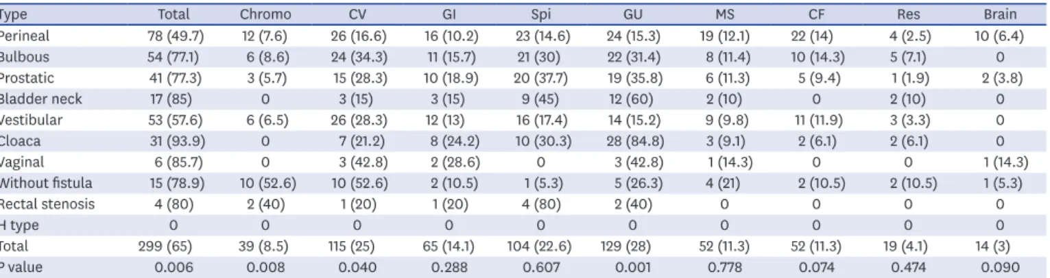
fistula had AAs. In H type, none of the patients had AAs. Some statistically significant difference was observed in the incidence of AAs among the subtypes. Chromosomal (P = 0.008) and cardiovascular anomalies (P = 0.04) showed the highest incidence in the group without fistulas, while genitourinary anomaly (P = 0.001) was the most frequent in the group with cloaca (Table 2).
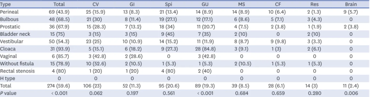
In the analysis of major AAs, the difference in incidence between the subtypes was similar to the total AA incidence. Cloaca (93.9%) had the highest incidence, followed by rectovaginal fistula (85.7%) and rectal stenosis (80%). Major cardiovascular anomaly showed the highest incidence in the without fistulas, but the difference between subtypes was not statistically significant (P = 0.062). However, the incidence of the subtypes in the major genitourinary anomalies was statistically significant (P < 0.001) (Table 3).
In the comparison of the ORs between the ARM subtypes using the cases of perineal fistula as the base group, cloaca (OR, 15.7) and recto-bladder neck fistula (OR, 5.74) showed the highest OR in the total AAs. Chromosomal anomaly (OR, 13.43) and cardiovascular anomaly (OR, 5.6) showed the highest OR in the without fistula. The category of gastrointestinal
0 30
20
centPer 10
All Major
Chromo CV GI Spi GU MS CF Res Bra
8.5 25.023.0
14.1 11.3
22.620.6 28.0
19.3
11.3 8.5
11.3
6.1 4.13.0 3.02.4
Fig. 1. Incidence of associated anomalies in anorectal malformation according to organ system.
Chromo = chromosomal, CV = cardiovascular, GI = gastrointestinal, Spi = spinal/vertebral, GU = genitourinary, MS
= musculoskeletal, CF = craniofacial, Res = respiratory, Bra = brain.
Table 2. Incidence of anomalies associated with the anorectal malformation subtypes
Type Total Chromo CV GI Spi GU MS CF Res Brain
Perineal 78 (49.7) 12 (7.6) 26 (16.6) 16 (10.2) 23 (14.6) 24 (15.3) 19 (12.1) 22 (14) 4 (2.5) 10 (6.4)
Bulbous 54 (77.1) 6 (8.6) 24 (34.3) 11 (15.7) 21 (30) 22 (31.4) 8 (11.4) 10 (14.3) 5 (7.1) 0
Prostatic 41 (77.3) 3 (5.7) 15 (28.3) 10 (18.9) 20 (37.7) 19 (35.8) 6 (11.3) 5 (9.4) 1 (1.9) 2 (3.8)
Bladder neck 17 (85) 0 3 (15) 3 (15) 9 (45) 12 (60) 2 (10) 0 2 (10) 0
Vestibular 53 (57.6) 6 (6.5) 26 (28.3) 12 (13) 16 (17.4) 14 (15.2) 9 (9.8) 11 (11.9) 3 (3.3) 0
Cloaca 31 (93.9) 0 7 (21.2) 8 (24.2) 10 (30.3) 28 (84.8) 3 (9.1) 2 (6.1) 2 (6.1) 0
Vaginal 6 (85.7) 0 3 (42.8) 2 (28.6) 0 3 (42.8) 1 (14.3) 0 0 1 (14.3)
Without fistula 15 (78.9) 10 (52.6) 10 (52.6) 2 (10.5) 1 (5.3) 5 (26.3) 4 (21) 2 (10.5) 2 (10.5) 1 (5.3)
Rectal stenosis 4 (80) 2 (40) 1 (20) 1 (20) 4 (80) 2 (40) 0 0 0 0
H type 0 0 0 0 0 0 0 0 0 0
Total 299 (65) 39 (8.5) 115 (25) 65 (14.1) 104 (22.6) 129 (28) 52 (11.3) 52 (11.3) 19 (4.1) 14 (3)
P value 0.006 0.008 0.040 0.288 0.607 0.001 0.778 0.074 0.474 0.090
Data are presented as number (%).
Chromo = chromosomal, CV = cardiovascular, GI = gastrointestinal, Spi = spinal/vertebra, GU = genitourinary, MS = musculoskeletal, CF = craniofacial, Res = respiratory, Perineal = perineal fistula, Bulbous = recto-bulbous urethral fistula, Prostatic = recto-prostatic urethral fistula, Bladder neck = recto-bladder neck fistula, Vestibular = vestibular fistula, Vaginal = recto-vaginal fistula.
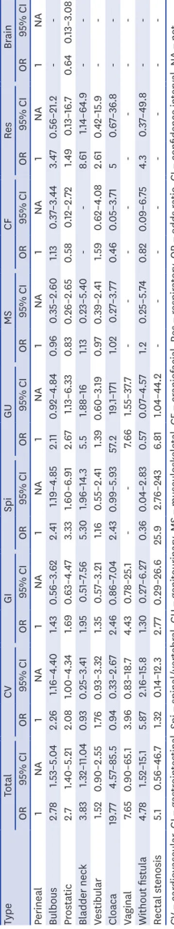
anomaly showed a significant OR only in cloaca (OR, 2.82). Spinal/vertebral anomaly showed the highest OR in rectal stenosis (OR, 23.3), followed by recto-bladder neck fistula (OR, 4.77) and recto-prostatic urethral fistula (OR, 3.53). Genitourinary anomaly showed the highest OR in cloaca (OR, 30.8), followed by recto-bladder neck fistula (OR, 8.25) (Table 4).
In the analysis of total major AAs, cloaca (OR, 19.77) showed the highest OR, followed by no fistula (OR, 4.78) and recto-bladder neck fistula (OR, 3.83). Cardiovascular major AA showed the highest OR in the without fistula (OR, 5.87), and gastrointestinal major AA showed no statistical significance for each subtype. Spinal/vertebral and genitourinary anomalies showed the highest OR in rectal stenosis and cloaca, respectively (Table 5).
DISCUSSION
Our study was conducted with a relatively large number of patients with ARM in a single center and facilitated communication with other physicians and researchers by applying the Krickenbeck classification, which is currently the most frequently used ARM classification system (Fig. 2). Moreover, our protocolized screening examination for AAs has been applied since 1999, which is a strength of this study. However, the spinal screening examination in our center has been applied strictly to all patients with ARM since January 2002. In our institution, newborns with sacral dimple or signs of occult or apert spinal dysraphism, such as incontinence, loss of sensation, and extremity weakness or paralysis, were examined with spinal ultrasonography or MRI because spinal anomaly might be present. In our study, 89 patients underwent anorectoplasty between 1999 and 2001, and 51 of them underwent spinal examination, but 38 (8.3% in our study population) did not. Spinal cord anomaly was found in 9 of the 51 patients. The ARM subtypes of the 38 patients who had not undergone spinal screening examination were perineal fistula in 21, recto-urethral fistula in 6, no fistula in 3, vestibular fistula in 6, recto-vaginal fistula in 1, and H-type in 1. However, these did not belong to either the cloaca or recto-bladder neck fistula subtype. Therefore, excluding these 38 patients would have affected the actual incidence and proportion of ARM subtypes in this study. It would also have affected the actual incidence of AAs other than spinal anomaly.
Therefore, we conducted a study including these 38 patients. The incidence of spinal anomaly in our study may be underestimated owing to these 38 patients.
Table 3. Incidence of major anomalies associated with the anorectal malformation subtypes
Type Total CV GI Spi GU MS CF Res Brain
Perineal 69 (43.9) 25 (15.9) 13 (8.3) 21 (13.4) 14 (8.9) 14 (8.9) 10 (6.4) 2 (1.3) 9 (5.7)
Bulbous 48 (68.5) 21 (30) 8 (11.4) 19 (27.1) 12 (17.1) 6 (8.6) 5 (7.1) 3 (4.3) 0
Prostatic 36 (67.9) 15 (28.3) 7 (13.2) 18 (34) 11 (20.7) 4 (7.5) 2 (3.8) 1 (1.9) 2 (3.8)
Bladder neck 15 (75) 3 (15) 3 (15) 9 (45) 7 (35) 2 (10) 0 2 (10) 0
Vestibular 50 (54.3) 23 (25) 10 (10.9) 14 (15.2) 11 (11.9) 8 (8.7) 9 (9.8) 3 (3.3) 0
Cloaca 31 (93.9) 5 (15.1) 6 (18.2) 9 (27.3) 28 (84.8) 3 (9.1) 1 (3) 2 (6.1) 0
Vaginal 6 (85.7) 3 (42.8) 2 (28.6) 0 3 (42.8) 0 0 0 0
Without fistula 15 (78.9) 10 (52.6) 2 (10.5) 1 (5.3) 1 (5.3) 2 (10.5) 1 (5.3) 1 (5.3) 0
Rectal stenosis 4 (80) 1 (20) 1 (20) 4 (80) 2 (40) 0 0 0 0
H type 0 0 0 0 0 0 0 0 0
Total 274 (59.6) 106 (23) 52 (11.3) 95 (20.6) 89 (19.3) 39 (8.5) 28 (6.1) 14 (3) 11 (2.4)
P value < 0.001 0.062 0.197 0.561 < 0.001 0.684 0.659 0.280 0.006
Data are presented as number (%).
CV = cardiovascular, GI = gastrointestinal, Spi = spinal/vertebral, GU = genitourinary, MS = musculoskeletal, CF = craniofacial, Res = respiratory, Perineal = perineal fistula, Bulbous = recto-bulbous urethral fistula, Prostatic = recto-prostatic urethral fistula, Bladder neck = recto-bladder neck fistula, Vestibular = vestibular fistula, Vaginal = recto-vaginal fistula.
Table 5. Comparison of major associated anomalies between the anorectal malformation subtypes with perineal fistula cases as the base group TypeTotalCVGISpiGUMSCFResBrain OR95% CIOR95% CIOR95% CIOR95% CIOR95% CIOR95% CIOR95% CIOR95% CIOR95% CI Perineal1NA1NA1NA1NA1NA1NA1NA1NA1NA Bulbous2.781.53–5.042.261.16–4.401.430.56–3.622.411.19–4.852.110.92–4.840.960.35–2.601.130.37–3.443.470.56–21.2-- Prostatic2.71.40–5.212.081.00–4.341.690.63–4.473.331.60–6.912.671.13–6.330.830.26–2.650.580.12–2.721.490.13–16.70.640.13–3.08 Bladder neck3.831.32–11.040.930.25–3.411.950.51–7.565.301.96–14.35.51.88–161.130.23–5.40--8.611.14–64.9-- Vestibular1.520.90–2.551.760.93–3.321.350.57–3.211.160.55–2.411.390.60–3.190.970.39–2.411.590.62–4.082.610.42–15.9-- Cloaca19.774.57–85.50.940.33–2.672.460.86–7.042.430.99–5.9357.219.1–1711.020.27–3.770.460.05–3.7150.67–36.8-- Vaginal7.650.90–65.13.960.83–18.74.430.78–25.1--7.661.55–37.7-------- Without fistula4.781.52–15.15.872.16–15.81.300.27–6.270.360.04–2.830.570.07–4.571.20.25–5.740.820.09–6.754.30.37–49.8-- Rectal stenosis5.10.56–46.71.320.14–12.32.770.29–26.625.92.76–2436.811.04–44.2-------- CV = cardiovascular, GI = gastrointestinal, Spi = spinal/vertebral, GU = genitourinary, MS = musculoskeletal, CF = craniofacial, Res = respiratory, OR = odds ratio, CI = confidence interval, NA = not applicable, Perineal = perineal fistula, Bulbous = recto-bulbous urethral fistula, Prostatic = recto-prostatic urethral fistula, Bladder neck = recto-bladder neck fistula, Vestibular = vestibular fistula, Vaginal = recto-vaginal fistula.
Table 4. Comparison of associated anomalies between anorectal malformation subtypes with perineal fistula cases as the base group TypeTotalChromoCVGISpiGUMSCFResBrain OR95% CIOR95% CIOR95% CIOR95% CIOR95% CIOR95% CIOR95% CIOR95% CIOR95% CIOR95% CI Perineal1NA1NA1NA1NA1NA1NA1NA1NA1NA1NA Bulbous3.421.80–6.481.130.40–3.152.631.37–5.021.640.72–3.752.51.27–4.912.521.29–4.900.940.38–2.251.020.45–2.292.940.76–11.3-- Prostatic3.461.69–7.070.720.19–2.671.990.95–4.132.050.87–4.853.531.73–7.183.071.51–6.250.930.34–2.460.640.23–1.780.740.08–6.730.580.12–2.72 Bladder neck5.741.62–20.4--0.890.24–3.251.550.41–5.894.771.77–12.78.253.05–22.30.810.17–3.75--4.250.72–24.8-- Vestibular1.380.82–2.310.840.30–2.321.981.06–3.681.320.59–2.931.230.61–2.460.990.48–2.020.790.34–1.820.830.38–1.811.290.28–5.89-- Cloaca15.73.63–67.8--1.360.53–3.452.821.09–7.292.531.06–6.0130.810.8–87.60.730.20–2.610.400.09–1.772.470.43–14.1-- Vaginal6.080.71–51.6--3.780.79–17.93.520.63–19.7--4.120.86–19.61.210.13–10.6----2.450.27–22.3 Without fistula3.81.20–11.913.434.57–39.35.62.07–15.11.040.22–4.900.320.04–2.541.960.64–5.961.940.58–6.440.720.15–3.344.50.76–26.40.820.09–6.75 Rectal stenosis4.050.44–37.18.061.22–52.91.260.13–11.72.20.23–20.923.32.49–2173.670.58–23.1-------- Chromo = chromosomal, CV = cardiovascular, GI = gastrointestinal, Spi = spinal/vertebral, GU = genitourinary, MS = musculoskeletal, CF = craniofacial, Res = respiratory, OR = odds ratio, CI = confidence interval, NA = not applicable, Perineal = perineal fistula, Bulbous = recto-bulbous urethral fistula, Prostatic = recto-prostatic urethral fistula, Bladder neck = recto-bladder neck fistula, Vestibular = vestibular fistula, Vaginal = recto-vaginal fistula.
In general, major congenital anomalies define structural anomalies such as significant medical, social, or cosmetic consequences for the affected individual, and usually require medical or surgical intervention. Conversely, minor congenital anomalies are structural changes that pose no significant health problem in the neonatal period and tend to have limited social or cosmetic consequences for the affected individual.11 A large number of congenital anomalies are difficult to classify by only two stages, major and minor. Congenital anomalies such as anencephaly and gastroschisis, which are lethal or require surgical correction, may be easily classified as major congenital anomalies. However, congenital malformations such as atrial septal defect or intestinal malrotation are sometimes fatal but may not cause any life-threatening symptoms. Therefore, major and minor congenital anomalies are difficult to distinguish. Here, minor congenital anomalies of congenital malformations were identified using the EUROCAT Guide 1.4 definition,1 and major AAs were defined as all malformations other than minor congenital anomalies in this study.
The incidence of major AA according to organ system did not differ significantly from the Chromo
All Major
CV GI Spi GU MS CF Res Bra 0
15 20
5 10
Percent
Perineal fistula (n = 157)
Chromo
All Major
CV GI Spi GU MS CF Res Bra 0
30 40
10 20
Percent
Recto-bulbous urethral fistula (n = 70)
Chromo
All Major
CV GI Spi GU MS CF Res Bra 0
30 40
10 20
Percent
Recto-prostatic urethral fistula (n = 53)
Chromo
All Major
CV GI Spi GU MS CF Res Bra 0
60 80
20 40
Percent
Recto-bladder neck fistula (n = 20)
Chromo
All Major
CV GI Spi GU MS CF Res Bra 0
20 30
centPer 10
Vestibular fistula (n = 92)
Chromo
All Major
CV GI Spi GU MS CF Res Bra 0
80 100
20 60 centPer 40
Cloaca (n = 33)
Chromo
All Major
CV GI Spi GU MS CF Res Bra 0
40 50
10 30 centPer 20
Recto-vaginal fistula (n = 7)
Chromo
All Major
CV GI Spi GU MS CF Res Bra 0
40 60
centPer 20
Without fistula (n = 19)
Chromo
All Major
CV GI Spi GU MS CF Res Bra 0
80 100
40 20 60
Percent
Rectal stenosis (n = 5)
Fig. 2. Incidence of associated anomalies according to anorectal malformation subtype.
Chromo = chromosomal, CV = cardiovascular, GI = gastrointestinal, Spi = spinal/vertebra, GU = genitourinary, MS = musculoskeletal, CF = craniofacial, Res = respiratory, Bra = brain.
total AA incidence, including minor cases, perhaps because a small number of congenital malformations were classified as minor anomalies. Indexes dividing major and minor congenital anomalies for each organ system more clearly would have made our study more meaningful. We described the major AAs in patients with ARM by applying the concept of major and minor congenital malformations. Our data will be of great significance in reporting major congenital anomalies that should be considered more carefully in the AAs of ARM.
The time when the screening examination of AAs in patients with ARM was first systematically performed is unclear. To understand ARM-related congenital anomalies, the VATER/VACTERL association must be understood. The VATER association, which was first described in 1973, is a spectrum of AAs that refers to patients with coexistence of at least three of the anomalies of the vertebrae, ARMs, esophageal atresia/tracheoesophageal fistula, and radial and renal anomalies.12 Shortly thereafter, cardiac and limb malformations were added to the anomalous feature of the VATER association, which was called the VACTERL association.13 Therefore, patients with suspected VACTERL association should undergo diagnostic tests (radiography, echocardiography, and abdominal ultrasonography including the kidney, etc.) based on careful physical examination. The incidence of VACTERL association in patients with ARM is known to be 15.4%–17.8%.14,15 Thus, for decades, patients with ARM would have been given diagnostic tests for the VACTERL association. In our study, 76 patients (16.5%) were eligible for VACTERL association.
Here, patients with spinal cord or vertebral anomalies, including sacral anomalies, accounted for 22.6% of all patients with ARM. The tethered cord refers to the state in which the conus medullaris is located below the L2 vertebra and is associated with the anomalies of spinal dysraphism. Levitt et al.16 reported that 24% of patients with ARM had a tethered cord. In this study, 69 patients (15%) presented with a tethered cord when evaluated on the basis of the above-mentioned diagnostic criteria of tethered cord. The prevalence of tethered cord, in fact, reported differed depending on the definition of tethered cord and the diagnostic tool used. In a recent report of pediatric surgeons in 24 European centers (members of the ARM- Net Consortium), the overall prevalence of tethered cord in patients with ARM was 46%
in the respondents who reported a prevalence of < 15%, 29% in respondents who reported 15% to 30%, and 4% in the respondents who reported 30% to 45%.17 Several reports that diagnosed spinal anomaly by performing MRI on all patients with ARM reported that the prevalence of tethered cord or spinal anomaly ranged from 35% to 60%.18-21 Other reports indicated that the prevalence of spinal anomaly varies according to ARM subtype. In a study of 416 patients with ARM in 2004, the incidence of spinal cord anomaly was found to be 4.4% in the low type and 10% in the high type, showing a significant difference (P = 0.04).8 Other studies also reported that the prevalence of spinal anomalies was higher in the high ARM type than in the low ARM type (31.2% vs. 50%18 and 11.4% vs. 47.3%21). On the other hand, in some studies, the prevalence of tethered spinal cord was higher in the low ARM type than in the high ARM type (50% vs. 26.8%19 and 53.3% vs. 38.5%20). In our study, tethered cord was found in 10.8% of the low ARM type cases and 17.3% of the high ARM type cases, without statistical significance (P = 0.077). We therefore believe that screen examination for spinal/vertebral anomaly should be performed on all ARM patients.
The association between the genitourinary system and ARM has long been known.22 In 1971, a survey by the members of the surgical section of the American Academy of Pediatrics reported that 26% of 1,166 patients with ARM had urological malformations.23 Renal agenesis, renal dysplasia, and vesicoureteral reflux (VUR) are the most common anomalies that can affect the
morbidity or mortality of patients with genitourinary anomaly.24 Abdominal ultrasonography can easily detect renal abnormalities, but VUR cannot be easily diagnosed without VCUG.
Therefore, it is argued that VCUG should be applied to all patients with ARM.25,26 Previous studies showed that the incidence of VUR in patients with ARM was varied, ranging from 2.3% to 47%.5,24,27-30 One recent study reported that VCUR was performed in 133 patients with ARM, of whom 41 (31%) had VUR.31 Of the 41 patients with VUR in this study, 56% had a VUR of grade ≥ 3. This study also showed that VUR is not related to the location of the fistula.
In our study, VUR was confirmed in 46 patients (10%) and 30 patients (65.2%) with grade ≥ 3 VUR. The incidence of VUR according to ARM subtype was 3.2% in perineal fistula (5), 11.4%
in recto-bulbous urethral fistula (8), 17% in recto-prostatic urethral fistula (9), 20% in recto- bladder neck fistula (4), 5.4% in vestibular fistula (5), 30.3% in cloaca (10), 15.8% in no fistula (3), and 40% in rectal stenosis (2). In our study, fistula location and VUR incidence were statistically significant when cross-tabulation analysis was performed (P = 0.006). However, in our study, the VUR incidence in perineal and vestibular fistulas may be underestimated because VCUG was performed only in some patients with perineal and vestibular fistulas. In many cases, reflux nephropathy can be prevented by medical therapy, and low-grade VUR tends to improve over time.26,29 Although the incidence of VUR is not high among patients with ARM, considering that a considerable number of VURs were grade ≥ 3, we believe that VCUG should be performed as an initial screening test in all patients with ARM.
Many studies have evaluated musculoskeletal, craniofacial, respiratory, or brain anomalies in patients with ARM who have AAs. However, no consensus has been reached on the screening test for these organ systems, such as limb radiography or brain imaging, probably because of the relatively low incidence as compared with those of heart, spinal, genitourinary, and gastrointestinal anomalies, and anomaly of these organ systems do not significantly affect the morbidity or corrective procedure of ARM. Recently, a study of 506 patients with ARM reported that only 15.2% of patients underwent limb radiography or ultrasonography as a screening test for limb anomalies.32 In our study, radiography or ultrasonography was used for screening limb anomalies in only 16.3% of the patients, most of whom were patients with limb anomalies that could be identified on visual examination.
The retrospective design is one limitation of the present study. Also, the protocol for evaluation of AAs is not consistent over study period. These are the weak points of our study.
If a prospective study complements these points, it would be a more powerful study. As mentioned in the methods, to analyze the difference in incidence of AAs, we calculated ORs using cases of perineal fistula as the base group. The reason why we used the perineal fistula patients as the base group was that there were lot of patients and incidence of AAs was the lowest in this group. If there was an analysis of congenital anomaly in healthy children born during the study period, and if it used as a base group, it would be a better study.
In conclusion, of the patients, 65% had at least one anomaly and 59.6% had major AAs.
The most common AAs were genitourinary, cardiovascular, and spinal/vertebral anomalies, and the major AAs were cardiovascular, spinal/vertebral, and genitourinary anomalies, in that order. The subtype with the highest AA incidence was cloaca (93.9%). By organ system, cardiovascular anomaly was most frequent in the without fistula, and gastrointestinal anomaly was most frequent in recto-vaginal fistula. Moreover, spinal/vertebral anomaly was more frequent in rectal stenosis and recto-bladder neck, and genitourinary anomaly was most frequent in cloaca. These data will be effective for predicting AAs in patients with ARM.
Altogether, all patients with ARM should undergo a thorough systematic evaluation.
SUPPLEMENTARY MATERIAL
Supplementary Table 1
Associated anomalies of anorectal malformation patients according to organ system Click here to view
REFERENCES
1. Santulli TV. The treatment of imperforate anus and associated fistulas. Surg Gynecol Obstet 1952;95(5):601-14.
PUBMED
2. Trusler GA, Wilkinson RH. Imperforate anus: a review of 147 cases. Can J Surg 1962;5:269-77.
PUBMED
3. Smith ED, Saeki M. Associated anomalies. Birth Defects Orig Artic Ser 1988;24(4):501-49.
PUBMED
4. Endo M, Hayashi A, Ishihara M, Maie M, Nagasaki A, Nishi T, et al. Analysis of 1,992 patients with anorectal malformations over the past two decades in Japan. Steering Committee of Japanese Study Group of Anorectal Anomalies. J Pediatr Surg 1999;34(3):435-41.
PUBMED | CROSSREF
5. Stoll C, Alembik Y, Dott B, Roth MP. Associated malformations in patients with anorectal anomalies. Eur J Med Genet 2007;50(4):281-90.
PUBMED | CROSSREF
6. Nah SA, Ong CC, Lakshmi NK, Yap TL, Jacobsen AS, Low Y. Anomalies associated with anorectal malformations according to the Krickenbeck anatomic classification. J Pediatr Surg 2012;47(12):2273-8.
PUBMED | CROSSREF
7. Bălănescu RN, Topor L, Moga A. Anomalies associated with anorectal malformations. Chirurgia (Bucur) 2013;108(1):38-42.
PUBMED
8. Ratan SK, Rattan KN, Pandey RM, Mittal A, Magu S, Sodhi PK. Associated congenital anomalies in patients with anorectal malformations--a need for developing a uniform practical approach. J Pediatr Surg 2004;39(11):1706-11.
PUBMED | CROSSREF
9. Holschneider A, Hutson J, Peña A, Beket E, Chatterjee S, Coran A, et al. Preliminary report on the International Conference for the Development of Standards for the Treatment of Anorectal Malformations. J Pediatr Surg 2005;40(10):1521-6.
PUBMED | CROSSREF
10. European Surveillance of Congenital Anomalies. EUROCAT guide 1.4 and reference documents. http://
www.eurocat-network.eu/content/EUROCAT-Guide-1.4-Full-Guide.pdf. Updated 2018. Accessed December 26, 2018.
11. WHO/CDC/ICBDSR. Birth Defects Surveillance: a Manual for Programme Managers. Geneva: World Health Organization; 2014.
12. Quan L, Smith DW. The VATER association. Vertebral defects, anal atresia, T-E fistula with esophageal atresia, radial and renal dysplasia: a spectrum of associated defects. J Pediatr 1973;82(1):104-7.
PUBMED | CROSSREF
13. Temtamy SA, Miller JD. Extending the scope of the VATER association: definition of the VATER syndrome.
J Pediatr 1974;85(3):345-9.
PUBMED | CROSSREF
14. Cuschieri A; EUROCAT Working Group. Anorectal anomalies associated with or as part of other anomalies. Am J Med Genet 2002;110(2):122-30.
PUBMED | CROSSREF
15. Totonelli G, Catania VD, Morini F, Fusaro F, Mosiello G, Iacobelli BD, et al. VACTERL association in anorectal malformation: effect on the outcome. Pediatr Surg Int 2015;31(9):805-8.
PUBMED | CROSSREF
16. Levitt MA, Patel M, Rodriguez G, Gaylin DS, Pena A. The tethered spinal cord in patients with anorectal malformations. J Pediatr Surg 1997;32(3):462-8.
PUBMED | CROSSREF
17. Fanjul M, Samuk I, Bagolan P, Leva E, Sloots C, Giné C, et al. Tethered cord in patients affected by anorectal malformations: a survey from the ARM-Net Consortium. Pediatr Surg Int 2017;33(8):849-54.
PUBMED | CROSSREF
18. Heij HA, Nievelstein RA, de Zwart I, Verbeeten BW, Valk J, Vos A. Abnormal anatomy of the lumbosacral region imaged by magnetic resonance in children with anorectal malformations. Arch Dis Child 1996;74(5):441-4.
PUBMED | CROSSREF
19. Golonka NR, Haga LJ, Keating RP, Eichelberger MR, Gilbert JC, Hartman GE, et al. Routine MRI evaluation of low imperforate anus reveals unexpected high incidence of tethered spinal cord. J Pediatr Surg 2002;37(7):966-9.
PUBMED | CROSSREF
20. Uchida K, Inoue M, Matsubara T, Otake K, Koike Y, Okugawa Y, et al. Evaluation and treatment for spinal cord tethering in patients with anorectal malformations. Eur J Pediatr Surg 2007;17(6):408-11.
PUBMED | CROSSREF
21. Totonelli G, Morini F, Catania VD, Schingo PM, Mosiello G, Palma P, et al. Anorectal malformations associated spinal cord anomalies. Pediatr Surg Int 2016;32(8):729-35.
PUBMED | CROSSREF
22. Parrott TS. Urologic implications of anorectal malformations. Urol Clin North Am 1985;12(1):13-21.
PUBMED
23. Santulli TV, Schullinger JN, Kiesewetter WB, Bill AH Jr. Imperforate anus: a survey from the members of the Surgical Secton of the American Academy of Pediatrics. J Pediatr Surg 1971;6(4):484-7.
PUBMED | CROSSREF
24. Rich MA, Brock WA, Peña A. Spectrum of genitourinary malformations in patients with imperforate anus. Pediatr Surg Int 1988;3(2-3):110-3.
CROSSREF
25. Parrott TS, Woodard JR. Importance of cystourethrography in neonates with imperforate anus. Urology 1979;13(6):607-9.
PUBMED | CROSSREF
26. Shaul DB, Harrison EA. Classification of anorectal malformations--initial approach, diagnostic tests, and colostomy. Semin Pediatr Surg 1997;6(4):187-95.
PUBMED
27. Misra D, Mushtaq I, Drake DP, Kiely EM, Spitz L. Associated urologic anomalies in low imperforate anus are capable of causing significant morbidity: a 15-year experience. Urology 1996;48(2):281-3.
PUBMED | CROSSREF
28. Hoekstra WJ, Scholtmeijer RJ, Molenaar JC, Schreeve RH, Schroeder FH. Urogenital tract abnormalities associated with congenital anorectal anomalies. J Urol 1983;130(5):962-3.
PUBMED | CROSSREF
29. Rickwood AM, Spitz L. Primary vesicoureteric reflux in neonates with imperforate anus. Arch Dis Child 1980;55(2):149-50.
PUBMED | CROSSREF
30. Narasimharao KL, Prasad GR, Mukhopadhyay B, Katariya S, Mitra SK, Pathak IC. Vesicoureteric reflux in neonates with anorectal anomalies. Br J Urol 1983;55(3):268-70.
PUBMED | CROSSREF
31. Sanchez S, Ricca R, Joyner B, Waldhausen JH. Vesicoureteral reflux and febrile urinary tract infections in anorectal malformations: a retrospective review. J Pediatr Surg 2014;49(1):91-4.
PUBMED | CROSSREF
32. Minneci PC, Kabre RS, Mak GZ, Halleran DR, Cooper JN, Afrazi A, et al. Screening practices and associated anomalies in infants with anorectal malformations: results from the Midwest Pediatric Surgery Consortium. J Pediatr Surg 2018;53(6):1163-7.
PUBMED | CROSSREF