RIVIEW ARTICLE
비만과 소화기암과 관련된 인자
김동준
인제대학교 의과대학 일산백병원 내과학교실
Obesity and Gastrointestinal Cancer-related Factor
Dong-Jun Kim
Department of Internal Medicine, Ilsan Paik Hospital, Inje University College of Medicine, Seoul, Korea
Despite a higher incidence and less favorable outcome of malignant tumors in obese patients, much less recognized is the link between obesity and cancer. The mechanism of the association of obesity with carcinogenesis remains incompletely understood. Postulated mechanisms include insulin resistance, insulin-like growth factor signaling, chronic inflammation, im- munomodulation, hyperglycemia-induced oxidative stress, and changes of intestinal microbiome. Insulin resistance leads to direct mitogenic and antiapoptotic signaling by insulin and the insulin-like growth factor axis. Obesity can be considered to be a state of chronic low-grade inflammation. In obesity, numerous proinflammatory cytokines are released from adipose tissue which may involve in carcinogenesis. Hyperglycemia in susceptible cells results in the overproduction of superoxide and this process is the key to initiating all damaging pathways related to diabetes. This hyperglycemia-induced oxidative stress could be one possible link among obesity, diabetes, and cancer development. The role of obesity-related changes in the intestinal microbiome in gastrointestinal carcinogenesis deserves further attention. (Korean J Gastroenterol 2012;59:8-15)
Key Words: Obesity; Gastrointestinal neoplasms; Insulin resistance; Inflammation
CC This is an open access article distributed under the terms of the Creative Commons Attribution Non-Commercial License (http://creativecommons.org/licenses/
by-nc/3.0) which permits unrestricted non-commercial use, distribution, and reproduction in any medium, provided the original work is properly cited.
교신저자: 김동준, 411-706, 고양시 일산서구 대화동 2240, 인제대학교 의과대학 일산백병원 내과
Correspondence to: Dong-Jun Kim, Department of Internal Medicine, Ilsan Paik Hospital, Inje University College of Medicine, 2240 Daehwa-dong, Ilsanseo-gu, Goyang 411-706, Korea. Tel: +82-31-910-7205, Fax: +82-31-913-5095, E-mail: [email protected]
Financial support: None. Conflict of interest: None.
서 론
비만과 암은 각각 전 세계적으로 그 발생이 늘고 있으며 인류 건강에 큰 위협이 되는 질환으로 그 동안 두 질환 간의 연관성에 대한 많은 연구들이 있었으며 비만이 여러 종류의 암 발생을 증가시키는 것이 반복적으로 확인되고 있다. 국제 암연구소(International Agency for Research on Cancer)는 2002년 비만과 관련된 암들을 발표하였는데 비만과 연관성이 확실한 암으로 대장/직장암, 폐경 후 유방암, 자궁내막암, 신 장암, 식도 선암을 열거하였으며 비만에 따라 그 발생이 증가 하는 것으로 생각하나 아직 그 증거가 확정적이지 않은 암으 로 난소암, 전립선암, 췌장암, 담낭암, 흑색종, 자궁경부암을 열거하였다.1 2007년 세계암연구재단(World Cancer Re- search Fund)은 대장/직장암, 폐경 후 유방암, 자궁내막암,
신장암, 식도선암과 함께 췌장암을 비만과의 연관성이 확실한 암으로 분류하였고 담낭암을 비만과 연관성이 있을 수 있는 암으로 분류하였다.2 1966년에서 2007년까지 발표되었던 282,137예의 암 발생을 포함한 221개의 data sets을 포함하 는 연구결과들을 메타 분석하여 이들 암 발생과 체질량지수와 의 관계가 조사되었는데 남자에서는 식도선암, 갑상선암, 대 장암, 신장암의 발생이 비만에 따라 증가하였으며 여자에서는 자궁내막암, 담낭암, 식도선암, 신장암의 발생이 증가하는 것 으로 관찰되었다.3
비만과 암 발생에 대한 우리나라 연구로는 국민건강보험 자료를 통한 약 10년 추적 검사 후의 암 발생에 대한 조사들 이 있는데, 흡연과 음주량으로 보정 후 체질량지수 ≥30 kg/m2에서 전체 암 발생이 증가하는 것을 보여주었으며 남자 에서 위암, 대장암, 간암, 담낭암이, 여자에서 간암, 췌장암,
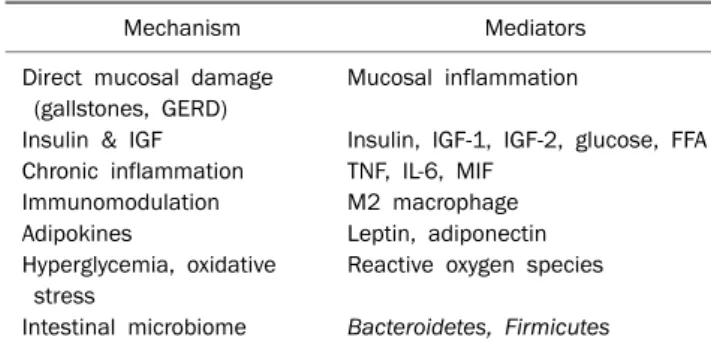
Table 1. Mechanisms Linking Obesity and Gastrointestinal Cancer Development
Mechanism Mediators
Direct mucosal damage Mucosal inflammation (gallstones, GERD)
Insulin & IGF Insulin, IGF-1, IGF-2, glucose, FFA Chronic inflammation TNF, IL-6, MIF
Immunomodulation M2 macrophage
Adipokines Leptin, adiponectin
Hyperglycemia, oxidative Reactive oxygen species stress
Intestinal microbiome Bacteroidetes, Firmicutes
GERD, gastroesophageal reflux; IGF, insulin-like growth factor; FFA, free fatty acid; TNF, tumor necrosis factor; IL, interleukin; MIF, machrophage migration inhibitory factor.
유방암이 증가함을 보여주었다.4-6
이렇듯 그간의 여러 연구들이 여러 다른 종류의 암과 더불 어 소화기암도 비만에서 증가한다는 것을 보여주고 있다. 이 에 저자는 비만에서 소화기암 발생의 증가를 설명할 수 있는 작용 기전들에 대한 문헌들을 고찰하고자 한다.
본 론
에너지 조절에 관여하는 인체 시스템이 비만과 암 발생에 관여할 가능성이 제기되고 있으며 인슐린 저항성, 인슐린유사 성장인자(insulin-like growth factor, IGF), 만성 염증(chronic inflammation), 면역 반응(immunomodulation), 아디포카인, 고혈당으로 인한 산화스트레스, 장내 세균 구성의 변화 등이 비만과 암 발생의 연결 인자로 생각되고 있다. 비만이 암 발생 률을 증가시키는 원인으로 위에서 언급한 일반적인 원인과 함 께 위치 특이적인(site-specific) 원인을 생각할 수 있다. 위치 특이적인 원인의 예로, 비만이 담석증을 잘 발생시키므로 이 에 따라 담낭암 발생이 증가할 가능성, 비만에 따라 역류성 식도염이 증가하여 식도선암 발생이 증가할 가능성, 비만에 따 라 비알코올성 지방간염(nonalcoholic steatohepatitis) 발생이 증가하면서 간암 발생이 증가할 가능성을 들 수 있다.7-9 이와 같이 비만이 소화기암 발생에 관여하는 여러 기전들이 제시되 고 있으나, 한 가지 가설로 설명하기는 어렵고 여러 인자들은 서로 긴밀하게 연결되고 복합적으로 작용하여 비만에서 소화 기 암 발생을 증가시킨다고 생각된다. 그러나 이해를 돕기 위 하여 그 인자들을 구분하여 연구 결과들을 정리하였다(Table 1).
1. 인슐린 및 인슐린유사성장인자
비만에서 암 발생을 증가시키는 중요한 기전으로 인슐린 저항성이 생각되고 있으며 인슐린 저항성은 인슐린 및 인슐린
유사성장인자 신호 전달체계의 변화를 통해 유사분열(mito- genesis)을 증가시키고 항자연사멸사(antiapoptosis)를 억제 시켜 암 발생을 유발할 것으로 생각되고 있다.10
Physician’s Health study에서는 혈중 C-peptide 상위 사 분위의 남자가 하위 사분위에 비해 체질량지수와 운동으로 보 정 후 대장/직장암의 상대적 위험도가 2.7배였으며 여러 대사 증후군 인자들을 추가 보정한 후에는 그 상대적 위험도가 오 히려 3.4배로 증가하였다.11 Nurse’s Health study에서는 혈 중 C-peptide 상위 사분위의 여자가 하위 사분위에 비해 체질 량지수와 신체활동량으로 보정 후 대장암의 상대적 위험도가 1.76배였다.12 그 간의 전향적 연구들을 메타 분석한 결과 혈 중 C-peptide/insulin 농도가 높은 경우 대장/직장암이 잘 발 생하는 것이 관찰되었는데(남자, 상대적 위험도 2.34 [95%
CI, 1.48-3.72], highest vs. lowest category; 여자, 상대적 위험도 1.28 [95% CI, 0.86-1.92]), 남자에서 여자보다 그 연 관성이 저명하였다.13 혈중 C-peptide/insulin 농도가 높은 경 우 췌장암이 약 2-4배 정도 증가한다고 보고되고 있다.13,14 한 연구는 쥐에서 10시간 동안 정상혈당 클램프(euglyce- mic clamp)를 시행하였을 때 혈중 인슐린 농도 의존적으로 대장/직장암 상피세포 증식이 일어남을 관찰하였다.15 흥미로 운 것은 고혈당이나 지방 주입을 통해서는 추가적인 세포증식 을 관찰할 수 없었다는 것인데 이는 고혈당, 고중성지방혈증 에 비해 고인슐린혈증이 암 발생에 관여하는 주된 인자임을 시사한다.
말단비대증에서 대장/직장암의 발생이 증가하고 제2형 당 뇨병과 대사증후군에서 대장/직장암과 췌장암이 증가하는 것 은 인슐린 및 IGF signaling이 비만과 연관된 암 발생에 관여 한다는 것을 시사한다.16-18 비만으로 야기된 인슐린 저항성이 암 발생을 일으키는 가능성 있는 기전들 중의 첫번째는 인슐 린 저항성으로 인한 IGF 농도의 증가이다. 비만과 제2형 당뇨 병 환자에서 관찰되는 고인슐린혈증은 간에서 IGF 결합 단백 질(IGF binding protein, IGFBP) 1,2의 합성을 감소시키고, 이는 생활성(bioactive) IGF-1, IGF-2의 혈중 농도를 증가시 킨다. 증가된 IGF-1, IGF-2는 각기 수용체와 결합한 후 세포 내 신호 전달과정을 통해 대장세포의 증식을 자극하게 된다.19 자극된 IGF-1, IGF-2 수용체는 extracelluar-signal-regulated kinase와 phosphatidylinositol 3 kinase 신호 전달 과정을 통하여 세포의 증식을 일으키게 되는데,20 최근 mammalian target of rapamycin (mTOR)과 S6 kinase가 주목을 받고 있 다.21
최근에 시행된 11개 연구의 메타분석은 제2형 당뇨병에서 널리 쓰이는 메트포르민이 다른 당뇨병 치료 약제에 비하여 암 발생 위험을 31% 감소시킨다고(summary relative risk=
0.69, [95% CI 0.61-0.79]) 보고하였다.22 당뇨병이 있으면서
유방암을 진단받았던 2,529명을 후향적으로 조사한 연구에 서, 당뇨병 치료제로 메트포르민을 투여했던 사람들의 수술 전 선행 항암치료(neoadjuvant chemotherapy)에 대한 병리 학적 완전관해율(pathologic complete response rate)이 24%였고, 메트포르민 이외의 당뇨병 약을 투여받았던 사람들 은 8.0%였다.23 이는 비록 후향적 연구이기는 하지만 메트포 르민의 항암효과를 나타내는 결과라고 생각한다. 메트포르민 의 항암기전은 혈당을 감소시켜 혈중 인슐린을 낮추는 것 이 외에도 LKB1 매개 activated protein kinase (AMPK) 의존 에너지 스트레스 반응을 유발하여 암세포의 생존에 영향을 미
치고,24-26 라파마이신(rapamycin) 신호 전달과정의 phos-
phoinositide 3-kinase/Akt/mammalian target을 저해하여 암세포의 성장을 저해하는 것으로 설명되고 있다.27 그간의 연 구 결과들을 종합하면 메트포르민은 암 발생을 억제하는 효과 가 있는 것으로 생각되며 종양학 분야에서는 메트포르민을 화 학치료의 보조치료(adjuvant therapy)로 추가하는 유용성에 대한 연구가 진행 중이다.28
인슐린 저항성으로 인한 고인슐린혈증 자체가 암 발생을 증가시킬 가능성도 제기되고 있다. 인슐린은 유사분열촉진제 (mitogen)로 작용하여 암 발생에 관여하는 것으로 생각된 다.29 인슐린 수용체와 인슐린유사성장인자 수용체는 50% 이 상의 구조적 유사성을 보이고30 인슐린과 인슐린유사성장인 자도 40-50%의 구조적 유사성을 보인다.31 이런 구조적 유사 성 때문에 인슐린과 인슐린유사성장인자는 결합력이 상대적 으로 미약하지만 각기 서로의 수용체에 결합하여 작용할 수 있다.32 따라서 인슐린은 인슐린 수용체에 결합하여 대사작용 을 일으키는 것이 그 주된 작용이지만 고농도의 인슐린은 인 슐린유사성장인자 수용체에 결합하여 세포 성장과 분화에 관 여할 가능성이 있다. 또 유방암 세포,33 전립선암 세포34에서 인슐린 수용체가 과발현된 것이 관찰되었으며 이는 비만이나 제2형 당뇨병과 같이 혈중 인슐린 농도가 올라간 경우 고농도 의 인슐린에 암세포가 반응할 가능성을 제기한다. 인슐린 수 용체에는 isoform A (IR-A, fetal form)와 isoform B (IR-B, adult form)가 있는데 IR-B는 인슐린 작용의 주 작용기관인 근육과 간에 많이 존재하고 IR-A는 우리 몸 전체에 존재한다.
IR-B는 인슐린 이외에도 강력한 fetal and cancer growth factor인 IGF-2에 높은 친화력을 보이므로 세포의 유사분열 촉진효과(mitogenic effect)를 보인다.35,36 이는 어떤 조직의 IR-A가 과발현된다면 이로 인해 암 발생이 유발될 가능성을 시사한다. 체내의 인슐린 수용체와 인슐린유사성장인자 수용 체는 구조적 유사성을 보이므로 정상적으로 hybrid IR/IGF-IR receptor를 형성할 수 있다. 인슐린 수용체는 IR-A와 IR-B가 있으므로 hybrid receptor도 hybrid-RA와 hybrid-RB가 생기 게 되는데 hybrid-RB는 인슐린유사성장인자 수용체와 같이
작용하여 인슐린에는 친화력을 보이지 않고 오직 IGF-1과만 결합하게 되지만 hybrid-RA는 인슐린과도 결합하여 그 작용 을 나타낸다. 따라서 어떤 조직에 hybrid-RA가 과발현되어 있 고 비만으로 인해 고인슐린혈증에 장기적으로 노출된다면 암 발생이 증가할 가능성이 생길 수 있다.37 이런 기전은 비만 환자에서 암의 예후가 좋지 않은 것을 설명할 수 있는데 고인 슐린혈증은 간, 유방, 대장암의 성장 속도와 분화도에 영향을 미친다는 보고들이 있으며38-40 대부분의 암에는 덜 분화된 세 포형을 나타내는 IR-A가 과발현되는 것으로 알려져 있다.41 마지막으로 인슐린 저항성이 소화관의 점막세포에 포도당, 중성지방, 유리지방산 등의 영양분을 더 많이 공급함으로써 종양 성장을 촉진할 가능성도 생각할 수 있다.
대장암 세포에서 IGF-1 수용체가 관찰되었다는 실험실 연 구가 있었으며42 동물실험에서 IGF-1 수용체를 불활성화시켜 IGF-1 농도를 낮춤으로써 종양 증식속도를 낮출 수 있음이 관찰되었다.43,44 한 연구는 식도 편평세포암의 침윤 깊이, 병 리학적 병기, 나쁜 예후와 혈중 IGF-1 농도가 연관성을 보인 다고 보고하였다.45 다섯 코호트 연구들을 종합하여 최근 발표 된 메타 분석은 IGF-1 농도로 대장/직장암 발생을 예측할 수 있음을 보고하였다.46 대장/직장암에 반해 대장/직장선종과 IGF-1 농도에 대한 연구들은 일치된 결과를 보여주지 않고 있 는데 한 연구는 혈중 인슐린 농도와 혈중 IGF-1/IGFBP3 ra- tio가 대장/직장선종 발생과 비례함을 보고하였으나,47 한 연 구는 오직 고위험 선종의 경우에서만 비례관계가 관찰되었으 며48 또 다른 연구에서는 연관성을 관찰할 수 없었다.49 대장/
직장암 이외에 췌장암에서 IGFBP-1 농도가 감소되었다는 보 고가 있었으며50 정상 췌장세포에 비하여 췌장암 세포에서 IGF-1이 과발현되어 있다는 보고도 있었다.51
2. 만성염증과 면역반응
비만 환자에서 CRP, interleukin-6 (IL-6) 등의 염증 표지 자들이 증가되어 있는 것으로 알 수 있듯이 비만은 만성적인, 경도의 염증이 지속되는 상태로 생각할 수 있다.52
지방세포에 의한 아디포카인 생성은 지방조직에서의 면역 세포 구성에 의해 지대한 영향을 받는다. 비만한 사람에서는 대식세포가 증가하며 비만 정도와 대식세포 수는 비례한다고 알려져 있다.53 말초 단핵구는 monocyte chemoattractant protein 1과 TNF (tumor necrosis factor)-α에 의해 활성 대 식세포(activated macrophage)로 분화하게 되는데54 이 활성 대식세포는 지방세포 기능에 관여하여 인슐린 저항성을 악화 시키게 된다.55,56 지방세포와 대식세포를 함께 배양하면 지방 세포에 의한 아디포카인과 염증성 사이토카인 분비가 증가하 는 것이 관찰된다.57 정상적인 면역반응은 종양 형성을 억제하 지만 어떤 상황에서는 반대로 종양 형성에 면역반응이 기여할
수 있다. 고전적 활성화(classically activated) 또는 M1 대식 세포는 종양 형성을 억제하지만, 대체적 활성화(alternatively activated) 또는 M2 대식세포는 항원 제시(antigen-present- ing) 능력이 떨어지며 혈관 형성, 조직 재형성(tissue remod- eling), 창상 치유(wound healing) 등에 관여하여 종양 형성 과 진행에 기여한다.58 한 연구에서는 종양세포가 대식세포를 유인하는 사이토카인을 분비하여 대식세포를 M2 분화시키는 것이 관찰되었다.59
IL-6, TNF, macrophage migration inhibitory factor 등과 같은 지방세포에서 분비되는 많은 사이토카인과 성장인자들 이 소화기암 발생에 관여한다고 생각되고 있는데60 증가된 사 이토카인들이 내분비 방식(endocrine manner)을 통하여 종 양 형성에 기여하는지, 혹은 전신적 만성염증이 국소 부위의 염증을 일으켜 사이토카인들이 분비되고 분비된 사이토카인 들이 주변분비 방식(paracrine manner)을 통하여 종양 형성 에 관여하는지는 아직 밝혀져 있지 않다. 최근 Poullis 등61은 국소적 장 염증의 표지자인 fecal calprotectin이 혈중 CRP로 보정 후에도 비만 정도와 비례함을 관찰하였다. 비알콜성지방 간 또한 국소적 염증반응이 종양을 유발하는 또 다른 예라고 할 수 있다.62 최근 한 연구는 식사요법으로 인한 10%의 체중 감소가 대장/직장 점막에서 염증유발 유전자의 발현을 25-50% 억제시킴을 보고하였다.63
만성염증 반응은 nuclear factor κB (NFκB) 의존적으로 인슐린 수용체를 통한 신호 전달과정을 방해함으로써 인슐린 저항성을 악화시킬 수 있다.64 염증성 사이토카인들은 NFκB pathway를 통하여 활성산소(reactive oxygen species, ROS)의 생성을 증가시키고 증가된 사이토카인과 ROS는 인 슐린 저항성을 악화시키며 이에 따라 혈중 포도당, 유리지방 산, 인슐린 농도가 증가하게 되어 다시 염증반응을 유발하는 악순환에 빠지게 한다.65 지방세포에서 분비된 사이토카인에 의해 활성화된 NFκB pathway와 signal transducer and activation of transcription 3 pathway는 세포 증식, 혈관 생성 등을 매개하는 유전자들을 발현시키게 된다.66 여러 성장 인자들 중 vascular endothelial growth factor는 대장/직장 암 치료에 있어 그 중요성이 대두되고 있다.67 그 간의 연구들 은 지방세포에서 분비되는 사이토카인들과 성장인자들이 내 장지방과는 연관성을 보이나 피하지방과는 연관성이 없음을 보고하고 있다.68-70
여덟 개의 환자-대조군 연구들에 대한 메타 분석 결과 혈중 CRP가 대장/직장암에 대한 독립적인 위험인자였는데(상대적 위험도 1.1 [95% CI 1.0-1.3]), 이는 여자보다는 남자에서, 직 장암보다는 대장암에서 저명하였다.71 그러나 한 환자-대조군 연구는 혈중 CRP와 대장/직장선종과의 연관성을 관찰할 수 없었다고 보고하였으며72 다른 단면 연구는 대장/직장선종이
혈중 IL-6, TNF와는 연관성을 보였으나 혈중 CRP와는 연관 성이 없었다고 보고하였다.73 아직까지 염증 표지자와 위장관 암과 간암 발생에 대한 전향적 연구는 없다.
3. 아디포카인(adipokines)
지방세포는 아디포카인으로 총칭되는 호르몬을 생산하는 내분비 기관으로 알려져 있다. 지방세포에서 분비되는 렙틴 (leptin), 아디포넥틴 등의 아디포카인과 TNF-α, IL-6, IL-8, IL-10 등의 사이토카인은 세포증식과 혈관생성 등을 촉진함 으로써 암 발생에 관여한다.74 아디포카인 농도와 소화기암 발 생이 연관되어 있다는 임상연구 결과들이 있고 몇몇 동물실험 들도 비슷한 연관성을 보이나 아디포카인이 암 발생에 관여하 는 정확한 기전에 대해서는 아직 알려진 바가 많지 않다.
1) 렙틴
혈중 렙틴 농도는 비만에서 증가한다.75 실험실 연구들은 렙틴이 대장/직장암에서 과발현되어 있으며,76 장 상피세포의 증식을 유발하고,77 대장직장 암세포의 자연사멸사를 감소시 키는 것을 보고하고 있으며78 생쥐에서 azoxymethane 유발 대장/직장종양의 성장을 촉진하는 것을 보고하고 있다.79 또 한 렙틴은 식도선암 세포에서 mitogenesis를 촉진하고 자연 사멸사를 감소시킨다고 보고되었다.80 한 대장내시경 연구에 서 혈중 렙틴 농도 상위 사분위에서 하위 사분위에 비해 체질 량지수로 보정 후에도 대장직장 선종이 관찰될 확률이 3.3배 로 보고되었다.81 비록 두 개의 추적 연구들이 연관성을 보여 주기는 하였지만,82,83 대장직장암과 혈중 렙틴과의 연관성은 아직 불명확하다.
2) 아디포넥틴
아디포넥틴은 인슐린 감수성을 개선시키며 체질량지수와 역상관관계를 보인다.84 아디포넥틴이 AMPK pathway와 mTOR signaling을 통하여 대장/직장암 세포의 성장을 억제 한다는 보고가 있다.85 몇몇 연구들은 혈중 아디포넥틴 농도가 대장/직장선종의 유무나 개수와 역상관관계가 있음을 보고하
였으며,86-88 체질량지수로 보정 후에도 조기 대장/직장암과 역
상관관계가 있음을 보고하였다.89 두 개의 환자대조군 연구는 혈중 아디포넥틴 농도와 위암이 역상관관계가 있음을 보고하 였다.90,91
4. 고혈당, 산화 스트레스
체중의 증가는 혈당을 높이고 비만 환자는 당뇨병을 동반 할 확률이 높아서, 암 발생에 관해서 비만과 혈당 증가의 효과 는 혼재되어 나타난다. 비만에 따른 혈당 증가로 인한 산화 스트레스 증가가 비만에서 암 발생을 증가시키는 기전 중의 하나로 생각되고 있다.
당뇨병 환자에서 mitochondrial electron-transport chain
에 의해 superoxide가 과다 생산되는 것이 당뇨병성 합병증 을 일으키는 주요 기전으로 생각되고 있다.92 췌장 선암세포인 Capan-1에서 고혈당은 aldose reductase의 활성화를 통해 polyol pathway를 자극함이 관찰되었으며, Busik 등93은 제1 형 당뇨병에서 고혈당으로 유발된 polyol pathway의 활성화 에 의해 췌장 분비가 감소한다고 하였다. 또 다른 연구에서는 혈관 상피세포에서 mitochondrial ROS를 정상화시킴에 따 라 고혈당으로 유발되는 소르비톨 축적과 NFκB pathway 활 성화를 막을 수 있음이 관찰되었으며 electron transport chain complex II 저해제, uncoupler of oxidative phos- phorylation, uncoupling protein-1, manganese super- oxide dismutase (MnSOD)를 투여함으로써 ROS 수준을 낮 출 수 있음을 관찰하였다.94 이런 연구 결과들은 고혈당이 직 접적으로 산화 스트레스에 관여함을 시사한다. MnSOD 활성 도는 정상 췌장세포에 비해 췌장암 세포에서 감소되어 있으며 췌장암의 배가시간(doubling time)과 연관성을 보이고, 더욱 이 adenovirus 유전자 주입(gene transfection)을 통해 MnSOD 활성도를 증가시켜 췌장암 세포주인 MIA PaCa-2의 성장을 늦출 수 있음이 관찰되었다.95
5. 장내 세균 구성의 변화
정상 체중을 가진 사람과 비만 환자의 장내 세균 구성이 다르며 이런 차이가 비만을 유발할 수 있다는 보고들이 있
다.96-98 마른 사람에 비해 비만에서 Bacteroides의 양이 작고
식사요법으로 인한 체중 감소가 Bacteroidetes를 증가시키고 Firmicutes를 감소시킨다는 보고가 있다.96 또한 비만대사 수 술(bariatric surgery) 후 장내 세균 구성이 변화하는 것도 관 찰되었다.99 한 환자-대조군 연구에서는 대장/직장선종이 있 는 경우 Bacteroidetes가 적은 것이 관찰되었다.100
최근 장내 세균이 대장/직장암 발생에 관여할 수 있다는 연구 결과들이 보고되고 있는데, azoxymethane 유발 암 발 생 쥐 모델에서 통상적으로 집락화된 자손(conventionally colonized littermates)에 비해 무균(germ-free) 쥐에서 발생 한 종양의 숫자가 더 적고 그 크기가 더 작았다는 보고가 있 다.101 또한 대장/직장암 환자에서 점막내 세균 수가 높다는 보고들이 있었다.102,103 이들 결과는 비만환자에서 장내 세균 의 변화가 비만에 관여하고 소화기암 발생에 관여할 가능성을 시사한다.
결 론
그 간 여러 역학 연구들이 비만이 소화기암 발생을 증가시 킨다는 것을 반복적으로 보여주고 있다. 비만이 암 발생을 증 가시키는 기전에 대해서는 여러 가설이 제시되고 있으나, 비
만과 소화기암 발생이 여러 복합적인 인자들의 상호작용으로 일어나므로 그 정확한 기전을 명확히 밝히기란 쉽지 않으며 한 가지 원인으로 설명할 수는 없을 것이라고 생각한다. 그러 나 폭발적으로 증가한 국내 비만 인구와 식생활의 서구화로 인한 소화기암의 증가를 생각하면 두 질병 간의 연결고리는 반드시 밝혀야 할 연구과제이다. 비만과 소화기암 발생의 연 결고리에 대해 많은 사실이 확인된다면 이런 연구결과는 향후 비만한 소화기암 환자에서의 약물치료 목표를 정하는 데 많은 도움을 줄 것이다. 비만이 대중 계몽과 홍보를 통한 생활습관 의 개선으로 어느 정도 해결할 수 있는 질환임을 생각하면 학계의 노력과 더불어 정부 정책기관의 적극적인 관심이 필요 하다고 생각한다.
REFERENCES
1. IARC handbooks of cancer prevention: weight control and phys- ical activity. Vol 6. Lyon: IARC Press, 2002.
2. WCRF. World Cancer Research Fund. Food, nutrition, physical activity, and the prevention of cancer: a global perspective. 2nd ed. Washington: American Institute for Cancer Research, 2007.
3. Renehan AG, Tyson M, Egger M, Heller RF, Zwahlen M.
Body-mass index and incidence of cancer: a systematic review and meta-analysis of prospective observational studies. Lancet 2008;371:569-578.
4. Jee SH, Yun JE, Park EJ, et al. Body mass index and cancer risk in Korean men and women. Int J Cancer 2008;123:1892-1896.
5. Oh SW, Yoon YS, Shin SA. Effects of excess weight on cancer in- cidences depending on cancer sites and histologic findings among men: Korea National Health Insurance Corporation Study. J Clin Oncol 2005;23:4742-4754.
6. Song YM, Sung J, Ha M. Obesity and risk of cancer in post- menopausal Korean women. J Clin Oncol 2008;26:3395-3402.
7. Kant P, Hull MA. Excess body weight and obesity--the link with gastrointestinal and hepatobiliary cancer. Nat Rev Gastroenter- ol Hepatol 2011;8:224-238.
8. Donohoe CL, Pidgeon GP, Lysaght J, Reynolds JV. Obesity and gastrointestinal cancer. Br J Surg 2010;97:628-642.
9. Giovannucci E, Michaud D. The role of obesity and related meta- bolic disturbances in cancers of the colon, prostate, and pancreas. Gastroenterology 2007;132:2208-2225.
10. Renehan AG, Frystyk J, Flyvbjerg A. Obesity and cancer risk: the role of the insulin-IGF axis. Trends Endocrinol Metab 2006;17:
328-336.
11. Ma J, Giovannucci E, Pollak M, et al. A prospective study of plas- ma C-peptide and colorectal cancer risk in men. J Natl Cancer Inst 2004;96:546-553.
12. Wei EK, Ma J, Pollak MN, et al. A prospective study of C-peptide, insulin-like growth factor-I, insulin-like growth factor binding pro- tein-1, and the risk of colorectal cancer in women. Cancer Epidemiol Biomarkers Prev 2005;14:850-855.
13. Pisani P. Hyper-insulinaemia and cancer, meta-analyses of epi- demiological studies. Arch Physiol Biochem 2008;114:63-70.
14. Michaud DS, Wolpin B, Giovannucci E, et al. Prediagnostic plas- ma C-peptide and pancreatic cancer risk in men and women.
Cancer Epidemiol Biomarkers Prev 2007;16:2101-2109.
15. Tran TT, Naigamwalla D, Oprescu AI, et al. Hyperinsulinemia, but not other factors associated with insulin resistance, acutely en- hances colorectal epithelial proliferation in vivo. Endocrinology 2006;147:1830-1837.
16. Huxley R, Ansary-Moghaddam A, Berrington de González A, Barzi F, Woodward M. Type-II diabetes and pancreatic cancer: a meta-analysis of 36 studies. Br J Cancer 2005;92:2076-2083.
17. Larsson SC, Giovannucci E, Wolk A. Diabetes and colorectal can- cer incidence in the cohort of Swedish men. Diabetes Care 2005;28:1805-1807.
18. Clayton PE, Banerjee I, Murray PG, Renehan AG. Growth hor- mone, the insulin-like growth factor axis, insulin and cancer risk.
Nat Rev Endocrinol 2011;7:11-24.
19. Giovannucci E. Insulin, insulin-like growth factors and colon can- cer: a review of the evidence. J Nutr 2001;131(11 Suppl):3109S- 3120S.
20. Sears R, Nuckolls F, Haura E, Taya Y, Tamai K, Nevins JR. Multiple Ras-dependent phosphorylation pathways regulate Myc protein stability. Genes Dev 2000;14:2501-2514.
21. Hursting SD, Lashinger LM, Wheatley KW, et al. Reducing the weight of cancer: mechanistic targets for breaking the obe- sity-carcinogenesis link. Best Pract Res Clin Endocrinol Metab 2008;22:659-669.
22. Decensi A, Puntoni M, Goodwin P, et al. Metformin and cancer risk in diabetic patients: a systematic review and meta-analysis.
Cancer Prev Res (Phila) 2010;3:1451-1461.
23. Jiralerspong S, Palla SL, Giordano SH, et al. Metformin and pathologic complete responses to neoadjuvant chemotherapy in diabetic patients with breast cancer. J Clin Oncol 2009;27:
3297-3302.
24. Cazzaniga M, Bonanni B, Guerrieri-Gonzaga A, Decensi A. Is it time to test metformin in breast cancer clinical trials? Cancer Epidemiol Biomarkers Prev 2009;18:701-705.
25. Goodwin PJ, Ligibel JA, Stambolic V. Metformin in breast cancer:
time for action. J Clin Oncol 2009;27:3271-3273.
26. Zakikhani M, Dowling R, Fantus IG, Sonenberg N, Pollak M.
Metformin is an AMP kinase-dependent growth inhibitor for breast cancer cells. Cancer Res 2006;66:10269-10273.
27. Zhou G, Myers R, Li Y, et al. Role of AMP-activated protein kinase in mechanism of metformin action. J Clin Invest 2001;108:
1167-1174.
28. Pollak M. Metformin and other biguanides in oncology: advanc- ing the research agenda. Cancer Prev Res (Phila) 2010;3:1060- 1065.
29. Argilés JM, López-Soriano FJ. Insulin and cancer (Review). Int J Oncol 2001;18:683-687.
30. Mynarcik DC, Williams PF, Schaffer L, Yu GQ, Whittaker J.
Identification of common ligand binding determinants of the in- sulin and insulin-like growth factor 1 receptors. Insights into mechanisms of ligand binding. J Biol Chem 1997;272:18650- 18655.
31. Slieker LJ, Brooke GS, DiMarchi RD, et al. Modifications in the B10 and B26-30 regions of the B chain of human insulin alter
affinity for the human IGF-1 receptor more than for the insulin receptor. Diabetologia 1997;40 Suppl 2:S54-61.
32. Werner H, Weinstein D, Bentov I. Similarities and differences be- tween insulin and IGF-1: structures, receptors, and signalling pathways. Arch Physiol Biochem 2008;114:17-22.
33. Frasca F, Pandini G, Sciacca L, et al. The role of insulin receptors and IGF-I receptors in cancer and other diseases. Arch Physiol Biochem 2008;114:23-37.
34. Cox ME, Gleave ME, Zakikhani M, et al. Insulin receptor ex- pression by human prostate cancers. Prostate 2009;69:33-40.
35. Frasca F, Pandini G, Scalia P, et al. Insulin receptor isoform A, a newly recognized, high-affinity insulin-like growth factor II re- ceptor in fetal and cancer cells. Mol Cell Biol 1999;19:3278- 3288.
36. Pandini G, Medico E, Conte E, Sciacca L, Vigneri R, Belfiore A.
Differential gene expression induced by insulin and insulin-like growth factor-II through the insulin receptor isoform A. J Biol Chem 2003;278:42178-42189.
37. Pandini G, Frasca F, Mineo R, Sciacca L, Vigneri R, Belfiore A.
Insulin/insulin-like growth factor I hybrid receptors have differ- ent biological characteristics depending on the insulin receptor isoform involved. J Biol Chem 2002;277:39684-39695.
38. Saito K, Inoue S, Saito T, et al. Augmentation effect of post- prandial hyperinsulinaemia on growth of human hepatocellular carcinoma. Gut 2002;51:100-104.
39. Goodwin PJ, Ennis M, Pritchard KI, et al. Fasting insulin and out- come in early-stage breast cancer: results of a prospective co- hort study. J Clin Oncol 2002;20:42-51.
40. Hsing AW, Chua S Jr, Gao YT, et al. Prostate cancer risk and serum levels of insulin and leptin: a population-based study. J Natl Cancer Inst 2001;93:783-789.
41. Sciacca L, Costantino A, Pandini G, et al. Insulin receptor activa- tion by IGF-II in breast cancers: evidence for a new auto- crine/paracrine mechanism. Oncogene 1999;18:2471-2479.
42. Adachi Y, Lee CT, Coffee K, et al. Effects of genetic blockade of the insulin-like growth factor receptor in human colon cancer cell lines. Gastroenterology 2002;123:1191-1204.
43. Jones LI, Clemmons DR. Insulin-like growth-factors and their binding proteins: biological actions. Endocrine Rev 1995;16:
3-34.
44. Khandwala HM, McCutcheon IE, Flyvbjerg A, Friend KE. The ef- fects of insulin-like growth factors on tumorigenesis and neo- plastic growth. Endocr Rev 2000;21:215-244.
45. Imsumran A, Adachi Y, Yamamoto H, et al. Insulin-like growth fac- tor-I receptor as a marker for prognosis and a therapeutic target in human esophageal squamous cell carcinoma. Carcinogen- esis 2007;28:947-956.
46. Renehan AG, Zwahlen M, Minder C, O'Dwyer ST, Shalet SM, Egger M. Insulin-like growth factor (IGF)-1, IGF binding protein-3, and cancer risk: systematic review and meta-regression analysis. Lancet 2004;363:1346-1353.
47. Schoen RE, Weissfeld JL, Kuller LH, et al. Insulin-like growth fac tor-I and insulin are associated with the presence and advance- ment of adenomatous polyps. Gastroenterology 2005;129:
464-475.
48. Renehan AG, Painter JE, Atkin WS, Potten CS, Shalet SM,
O'Dwyer ST. High-risk colorectal adenomas and serum in- sulin-like growth factors. Br J Surg 2001;88:107-113.
49. Flood A, Mai V, Pfeiffer R, et al. Serum concentrations of in- sulin-like growth factor and insulin-like growth factor binding protein 3 and recurrent colorectal adenomas. Cancer Epidemiol Biomarkers Prev 2008;17:1493-1498.
50. Wolpin BM, Michaud DS, Giovannucci EL, et al. Circulating in- sulin-like growth factor binding protein-1 and the risk of pancre- atic cancer. Cancer Res 2007;67:7923-7928.
51. Bergmann U, Funatomi H, Yokoyama M, Beger HG, Korc M.
Insulin-like growth factor I overexpression in human pancreatic cancer: evidence for autocrine and paracrine roles. Cancer Res 1995;55:2007-2011.
52. Park HS, Park JY, Yu R. Relationship of obesity and visceral adiposity with serum concentrations of CRP, TNF-alpha and IL-6.
Diabetes Res Clin Pract 2005;69:29-35.
53. Neels JG, Olefsky JM. Inflamed fat: what starts the fire? J Clin Invest 2006;116:33-35.
54. Curat CA, Miranville A, Sengenès C, et al. From blood monocytes to adipose tissue-resident macrophages: induction of diaped- esis by human mature adipocytes. Diabetes 2004;53:1285- 1292.
55. Sartipy P, Loskutoff DJ. Monocyte chemoattractant protein 1 in obesity and insulin resistance. Proc Natl Acad Sci U S A 2003;
100:7265-7270.
56. Wellen KE, Hotamisligil GS. Obesity-induced inflammatory changes in adipose tissue. J Clin Invest 2003;112:1785-1788.
57. Bassols J, Ortega FJ, Moreno-Navarrete JM, Peral B, Ricart W, Fernández-Real JM. Study of the proinflammatory role of human differentiated omental adipocytes. J Cell Biochem 2009;107:
1107-1117.
58. Mantovani A, Sozzani S, Locati M, Allavena P, Sica A. Macro- phage polarization: tumor-associated macrophages as a para- digm for polarized M2 mononuclear phagocytes. Trends Immunol 2002;23:549-555.
59. Pollard JW. Tumour-educated macrophages promote tumour progression and metastasis. Nat Rev Cancer 2004;4:71-78.
60. Balkwill F, Charles KA, Mantovani A. Smoldering and polarized inflammation in the initiation and promotion of malignant disease. Cancer Cell 2005;7:211-217.
61. Poullis A, Foster R, Shetty A, Fagerhol MK, Mendall MA. Bowel in- flammation as measured by fecal calprotectin: a link between lifestyle factors and colorectal cancer risk. Cancer Epidemiol Biomarkers Prev 2004;13:279-284.
62. Starley BQ, Calcagno CJ, Harrison SA. Nonalcoholic fatty liver disease and hepatocellular carcinoma: a weighty connection.
Hepatology 2010;51:1820-1832.
63. Pendyala S, Neff LM, Suárez-Fariñas M, Holt PR. Diet-induced weight loss reduces colorectal inflammation: implications for colorectal carcinogenesis. Am J Clin Nutr 2011;93:234-242.
64. Hotamisligil GS. Inflammation and metabolic disorders. Nature 2006;444:860-867.
65. Sonnenberg GE, Krakower GR, Kissebah AH. A novel pathway to the manifestations of metabolic syndrome. Obes Res 2004;
12:180-186.
66. Aggarwal BB, Vijayalekshmi RV, Sung B. Targeting inflammatory
pathways for prevention and therapy of cancer: short-term friend, long-term foe. Clin Cancer Res 2009;15:425-430.
67. Kerbel R, Folkman J. Clinical translation of angiogenesis inhibitors. Nat Rev Cancer 2002;2:727-739.
68. Miyazawa-Hoshimoto S, Takahashi K, Bujo H, Hashimoto N, Saito Y. Elevated serum vascular endothelial growth factor is as- sociated with visceral fat accumulation in human obese subjects. Diabetologia 2003;46:1483-1488.
69. Hausman GJ, Richardson RL. Adipose tissue angiogenesis. J Anim Sci 2004;82:925-934.
70. Silha JV, Krsek M, Sucharda P, Murphy LJ. Angiogenic factors are elevated in overweight and obese individuals. Int J Obes (Lond) 2005;29:1308-1314.
71. Tsilidis KK, Branchini C, Guallar E, Helzlsouer KJ, Erlinger TP, Platz EA. C-reactive protein and colorectal cancer risk: a system- atic review of prospective studies. Int J Cancer 2008;123:
1133-1140.
72. Tsilidis KK, Erlinger TP, Rifai N, et al. C-reactive protein and color- ectal adenoma in the CLUE II cohort. Cancer Causes Control 2008;19:559-567.
73. Kim S, Keku TO, Martin C, et al. Circulating levels of inflammatory cytokines and risk of colorectal adenomas. Cancer Res 2008;
68:323-328.
74. Tilg H, Moschen AR. Adipocytokines: mediators linking adipose tissue, inflammation and immunity. Nat Rev Immunol 2006;
6:772-783.
75. Considine RV, Sinha MK, Heiman ML, et al. Serum immunor- eactive-leptin concentrations in normal-weight and obese humans. N Engl J Med 1996;334:292-295.
76. Aloulou N, Bastuji-Garin S, Le Gouvello S, et al. Involvement of the leptin receptor in the immune response in intestinal cancer.
Cancer Res 2008;68:9413-9422.
77. Liu Z, Uesaka T, Watanabe H, Kato N. High fat diet enhances co- lonic cell proliferation and carcinogenesis in rats by elevating se- rum leptin. Int J Oncol 2001;19:1009-1014.
78. Rouet-Benzineb P, Aparicio T, Guilmeau S, et al. Leptin counter- acts sodium butyrate-induced apoptosis in human colon cancer HT-29 cells via NF-kappaB signaling. J Biol Chem 2004;279:
16495-16502.
79. Hirose Y, Hata K, Kuno T, et al. Enhancement of development of azoxymethane-induced colonic premalignant lesions in C57BL/KsJ-db/db mice. Carcinogenesis 2004;25:821-825.
80. Ogunwobi O, Mutungi G, Beales IL. Leptin stimulates pro- liferation and inhibits apoptosis in Barrett's esophageal ad- enocarcinoma cells by cyclooxygenase-2-dependent, prosta- glandin-E2-mediated transactivation of the epidermal growth factor receptor and c-Jun NH2-terminal kinase activation.
Endocrinology 2006;147:4505-4516.
81. Chia VM, Newcomb PA, Lampe JW, et al. Leptin concentrations, leptin receptor polymorphisms, and colorectal adenoma risk.
Cancer Epidemiol Biomarkers Prev 2007;16:2697-2703.
82. Stattin P, Palmqvist R, Söderberg S, et al. Plasma leptin and col- orectal cancer risk: a prospective study in Northern Sweden.
Oncol Rep 2003;10:2015-2021.
83. Stattin P, Lukanova A, Biessy C, et al. Obesity and colon cancer:
does leptin provide a link? Int J Cancer 2004;109:149-152.
84. Arita Y, Kihara S, Ouchi N, et al. Paradoxical decrease of an adi- pose-specific protein, adiponectin, in obesity. Biochem Biophys Res Commun 1999;257:79-83.
85. Sugiyama M, Takahashi H, Hosono K, et al. Adiponectin inhibits colorectal cancer cell growth through the AMPK/mTOR pathway.
Int J Oncol 2009;34:339-344.
86. Wei EK, Giovannucci E, Fuchs CS, Willett WC, Mantzoros CS. Low plasma adiponectin levels and risk of colorectal cancer in men:
a prospective study. J Natl Cancer Inst 2005;97:1688-1694.
87. Nakajima TE, Yamada Y, Hamano T, et al. Adipocytokines as new promising markers of colorectal tumors: adiponectin for color- ectal adenoma, and resistin and visfatin for colorectal cancer.
Cancer Sci 2010;101:1286-1291.
88. Yamaji T, Iwasaki M, Sasazuki S, Tsugane S. Interaction be- tween adiponectin and leptin influences the risk of colorectal adenoma. Cancer Res 2010;70:5430-5437.
89. Otake S, Takeda H, Fujishima S, et al. Decreased levels of plasma adiponectin associated with increased risk of color- ectal cancer. World J Gastroenterol 2010;16:1252-1257.
90. Ishikawa M, Kitayama J, Kazama S, Hiramatsu T, Hatano K, Nagawa H. Plasma adiponectin and gastric cancer. Clin Cancer Res 2005;11:466-472.
91. Otani K, Kitayama J, Kamei T, et al. Adiponectin receptors are downregulated in human gastric cancer. J Gastroenterol 2010;45:918-927.
92. Brownlee M. Biochemistry and molecular cell biology of dia- betic complications. Nature 2001;414:813-820.
93. Busik JV, Hootman SR, Greenidge CA, Henry DN. Glucose- specific regulation of aldose reductase in capan-1 human pancreatic duct cells in vitro. J Clin Invest 1997;100:1685- 1692.
94. Nishikawa T, Edelstein D, Du XL, et al. Normalizing mitochon-
drial superoxide production blocks three pathways of hy- perglycaemic damage. Nature 2000;404:787-790.
95. Cullen JJ, Weydert C, Hinkhouse MM, et al. The role of man- ganese superoxide dismutase in the growth of pancreatic adenocarcinoma. Cancer Res 2003;63:1297-1303.
96. Ley RE, Turnbaugh PJ, Klein S, Gordon JI. Microbial ecology:
human gut microbes associated with obesity. Nature 2006;
444:1022-1023.
97. Bäckhed F, Ding H, Wang T, et al. The gut microbiota as an envi- ronmental factor that regulates fat storage. Proc Natl Acad Sci U S A 2004;101:15718-15723.
98. Bäckhed F, Manchester JK, Semenkovich CF, Gordon JI.
Mechanisms underlying the resistance to diet-induced obesity in germ-free mice. Proc Natl Acad Sci U S A 2007;104:979-984.
99. Furet JP, Kong LC, Tap J, et al. Differential adaptation of human gut microbiota to bariatric surgery-induced weight loss: links with metabolic and low-grade inflammation markers. Diabetes 2010;59:3049-3057.
100. Shen XJ, Rawls JF, Randall T, et al. Molecular characterization of mucosal adherent bacteria and associations with colorectal adenomas. Gut Microbes 2010;1:138-147.
101. Vannucci L, Stepankova R, Kozakova H, Fiserova A, Rossmann P, Tlaskalova-Hogenova H. Colorectal carcinogenesis in germ- free and conventionally reared rats: different intestinal envi- ronments affect the systemic immunity. Int J Oncol 2008;32:
609-617.
102. Martin HM, Campbell BJ, Hart CA, et al. Enhanced Escherichia coli adherence and invasion in Crohn's disease and colon cancer. Gastroenterology 2004;127:80-93.
103. Swidsinski A, Khilkin M, Kerjaschki D, et al. Association be- tween intraepithelial Escherichia coli and colorectal cancer.
Gastroenterology 1998;115:281-286.