INTRODUCTION
Reproductive aging is associated with a decline in fertility, which begins in a moderate and steady fashion starting in the third to fourth decade but then accelerates rapidly after age 35 (1). A gradual diminution in the pool of ovarian follicles seems to underlie this decline (2). This age-related decrease in the follicle number and fertility is marked by an increase in follicular-phase serum follicle-stimulating hormone (FSH) levels. This usually starts in women approaching 40 yr of age (3) and is followed several years later by an increase in luteiniz- ing hormone (LH) (4, 5). In addition, changes in estradiol (E2) levels have been described in women with advanced reproduc- tive age (5).
Various endocrinological markers have been used to assess the ovarian reserve, and the accurate and reliable determina- tions of serum ovarian aging marker levels are essential for safe and successful treatment (6). Previous studies have sug- gested that anti-Mullerian hormone (AMH) and inhibin B play important roles as ovarian aging markers (7). AMH is
secreted by granulosa cells of ovarian follicles and appears to regulate the early follicular development (8). According to a previous study, serum AMH levels decrease with increasing age (9). On the other hand, inhibin B is produced by granu- losa cells which characteristically suppress the synthesis and secretion of FSH (10). In addition to its role as a FSH mod- ulator, inhibin B appears to regulate the follicular develop- ment and promote follicular growth (11).
Other putative intraovarian regulators of follicle growth include the insulin-like growth factor (IGF) family of pro- teins. The IGF system is one of several growth factor systems that probably serve adjunctive roles in ovarian follicle devel- opment (12). Moreover, IGFs may play an important role in the human preovulatory process, and IGF-binding proteins (IGFBPs) may be valuable biochemical markers in the eval- uation of oocytes maturity (13). IGF-I in blood either circu- lates freely or is bound to binding proteins, mainly to IGFBP- 3. Furthermore, serum IGFBP-3 levels have been reported to be positively regulated by IGF-I (14). A small number of studies have explored the positive relationships between fol-
104
Sun Young Shin*, Jung Ryeol Lee� Gyung Woon Noh*, Hyun Joo Kim* Won Jun Kang*,�, Seok Hyun Kim� June-Key Chung*,�
Department of Nuclear Medicine*, Tumor Immunity Medical Research Center�; Department of Obstetrics and Gynecology�, College of Medicine, Seoul National University, Seoul, Korea
Address for correspondence June-Key Chung, M.D.
Department of Nuclear Medicine, College of Medicine, Seoul National University, 28 Yeongeon-dong, Jongno-gu, Seoul 110-744, Korea
Tel : +82.2-2072-3376, Fax : +82.2-745-7690 E-mail : [email protected] DOI: 10.3346/jkms.2008.23.1.104
Analysis of Serum Levels of Anti-Mullerian Hormone, Inhibin B, Insulin- Like Growth Factor-I, Insulin-Like Growth Factor Binding Protein-3, and Follicle-Stimulating Hormone with Respect to Age and
Menopausal Status
This study was undertaken to investigate age-dependent and postmenopausal ch- anges in the serum levels of anti-Mullerian hormone (AMH), inhibin B, insulin-like growth factor (IGF)-I, IGF-binding protein-3 (IGFBP-3), and follicle-stimulating hor- mone (FSH), and to determine which of these markers best reflects the aging pro- cess in women. A total of 144 women aged 20-59 yr were enrolled in this cross-sec- tional study. Blood samples were obtained on cycle day 3 of regularly menstruating women (n=111), or at random in postmenopausal women (n=33). Data were ana- lyzed with respect to premenopausal women age groups and compared in pre- and postmenopausal women. Area under the receiver operating characteristic curve (ROCAUC) analyses were performed to assess the ability of each marker to discrim- inate between the pre- and postmenopausal status. Serum levels of AMH, IGF-I, and IGFBP-3 decreased and serum levels of FSH increased significantly with age in premenopausal women. Serum luteinizing hormone (LH) was higher and inhibin B was lower in women in their 20-30’s than in 40’s. Serum levels of AMH and IGF-I showed a consistent decrease with all age groups. ROCAUCanalysis showed that the diagnostic accuracy of AMH for menopausal status was similar to those of FSH, LH, and inhibin B, and was better than that of IGF-I. In conclusion, the serum AMH level appears to be the best marker of the aging process in premenopausal women.
Key Words : Reproductive Aging; Anti-Mullerian Hormone; Inhibin B; Insulin-Like Growth Factor I; Insulin- Like Growth Factor Binding Protein 3; FSH
Received : 21 March 2007 Accepted : 22 June 2007
. .
. .
. .
. .
licular fluid IGF-I and ovarian reserve and between follicu- lar fluid IGF-I and serum IGF-I (5). Although serum IGF-I levels have been reported to be downregulated in elderly women and to influence ovarian function (16), the serum IGF-I level is not widely used as an ovarian aging marker and its usefulness has not been clearly documented.
The present study was focused on the usefulness of several ovarian age markers in one population sample with respect to age and menopausal status. Even though the decreases of these hormones with age have been reported (7, 9, 16), the data published to date are not comprehensive enough to reach a consensus regarding reference ranges. Moreover, no study has yet been conducted in an Asian population on these hor- mones. The purposes of this study were to investigate age- dependent and postmenopausal changes in the serum levels of AMH, inhibin B, IGF-I, IGFBP-3, and FSH and to iden- tify which of these markers better reflects the aging process in Korean women. Furthermore, this study provides diagnos- tic reference ranges of novel age markers, such as AMH, inhib- in B, and IGF-I.
MATERIALS AND METHODS Subjects
A total of 144 women participated in this study. We recruit- ed healthy, ovulatory women aged 20-29 yr (n=48), 30-39 yr (n=33), and 40-49 yr (n=30) and postmenopausal women aged 50-59 yr (n=33). Premenopausal women were required to have regular menstrual cycles (24-35 days), and postmeno- pausal women were required to have been in physiologic me- nopause for at least 1 yr. No participant had any evidence of endocrine disorders (normal prolactin and thyroid-stimulat- ing hormone levels, and no evidence of polycystic ovarian syn- drome), and all participants were required to have a body mass index (BMI) of 19-26 kg/m2, both ovaries present, and an absence of medical or reproductive disorders (including any history of infertility), and no participant was taking hormone medications.
Hormone assays
Blood samples were obtained by venipuncture on cycle day 3 from regularly menstruating women or at random from postmenopausal women to measure the serum levels of AMH, inhibin B, IGF-I, IGFBP-3, FSH, LH, and E2. Serum was separated from blood samples and stored at -20℃until as- sayed. Samples from a given subject were analyzed for each hormone in the same assay to avoid inter-assay variation.
Serum levels of LH and FSH were measured by immuno- radiometric assay (IRMA) using commercial kits (Biosource, Nivelles, Belgium). The detection limits of the assay were 0.2 mIU/mL for LH and 0.1 mIU/mL for FSH. Intra- and
inter-assay coefficients of variation (CV) were 3.2% and 6.7%
for LH, and 3.3% and 7.1% for FSH, respectively. Samples were analyzed for E2using radioimmunoassay (RIA) using commercial kits (Biosource, Nivelles, Belgium). The detec- tion limit of this assay was 10 pg/mL, and intra- and inter- assay CVs were 4.9% and 5.2%, respectively. Serum levels of IGF-I and IGFBP-3 were determined by IRMA using reagents supplied by Diagnostic Systems Laboratories (DSL Inc., Webster, U.S.A.). Its detection limits were 2.06 ng/mL for IGF-I and 0.5 ng/mL for IGFBP-3, and its intra- and inter-assay CVs were 5.1% and 9.1% for IGF-I, and 5.2%
and 7.1% for IGFBP-3, respectively.
Serum levels of AMH and Inhibin B were determined using enzyme-linked immunosorbent assays (ELISA) using commercial kits from Diagnostic Systems Laboratories (DSL Inc., Webster, U.S.A.). The detection limits of this assay were 0.017 ng/mL for AMH and 7 pg/mL for Inhibin B, and its intra- and inter-assay CVs were 4.6% and 8.0% for AMH and 5.6% and 7.6% for inhibin B, respectively.
Statistical analysis
Data were analyzed by age group in premenopausal women.
Differences between age groups were tested by one-way anal- ysis of variance (ANOVA), followed by Turkey’s multiple com- parison test. Comparisons between pre- and postmenopausal women were made using Mann-Whitney U test. The abilities of hormone levels to discriminate between pre- and postme- nopause women were investigated using receiver operating characteristic (ROC) analysis. The diagnostic values of the various hormone markers were evaluated by comparing areas under the curves (ROCAUC) (23). Statistical analyses were per- formed using SPSS 12.0 for Windows (SPSS Inc., Chicago, IL, U.S.A.), and p values of <0.05 were considered statisti- cally significant. We drew fitting plots of hormone levels versus age. Mean data values with error bar are presented in the fitting plots. The x-axis was divided into intervals of 5 yr and each curve was fitted using linear, Gaussian, Lorentzian, and sigmoid functions using Mathematica 5.0 (Wolfram Research Inc., Champaign, IL, U.S.A.)
RESULTS
The characteristics of the premenopausal subjects and their serum hormone levels on cycle day 3 are shown in Table 1.
Serum levels of AMH, IGF-I, and IGFBP-3 decreased and those of FSH increased significantly with age. Serum levels of E2, LH, and inhibin B showed no significant differences.
However, serum LH was higher and inhibin B was lower in women in their 20-30’s than in 40’s. AMH and IGF-I levels consistently decreased with age, and showed significant changes across all ages, whereas IGFBP-3 and FSH levels showed a significant difference only between women in their 30’s and
40’s.
Median ages and serum hormone levels of premenopausal and postmenopausal women are presented in Table 2. Data for AMH and inhibin B were not normally distributed in the postmenopausal women, mainly because the majority of val- ues were below the detection limit. Hence the data were pre- sented as medians and ranges, and a nonparametric method was used for comparison between two groups. Premenopausal and postmenopausal women had median ages of 31 (range 20-49) yr and 56 (range 50-59) yr, respectively. Serum levels of LH, FSH, E2, IGF-I, AMH, and inhibin B showed signif- icant differences between pre- and postmenopausal women (p<0.001). However, serum IGFBP-3 levels did not change significantly after menopause. As expected, LH and FSH lev- els in postmenopausal women were significantly higher and those of E2, IGF-I, AMH and inhibin B levels were signifi- cantly lower than in premenopausal women.
AMH was undetectable in 20 of 33 postmenopausal wo- men, whereas it was only undetectable in 2 of 111 premeno- pausal women. Inhibin B was undetectable in all postmeno- pausal women and in 10 of 111 premenopausal women;
moreover, these 10 women were older than 40 yr old.
Serum levels of AMH and IGF-I showed linear decreases from the earlier age (Fig. 1A, B), whereas FSH and LH levels
showed sigmoid increases and showed greatest increase after menopause (Fig. 1C, D). Serum E2levels were fitted using sigmoid and Lorentzian functions (Fig. 1E) and inhibin-B levels showed a sigmoid decrease (Fig. 1F), and both of these hormones decreased after menopause. Serum IGFBP-3 levels were fitted using Gaussian function with a minimum value at the boundary between pre- and postmenopause (Fig. 1G).
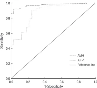
Table 3 shows the results of ROC curve analysis for serum markers. This analysis showed that the diagnostic accuracy of AMH (ROCAUC=0.943) for menopausal status was similar to those of FSH (ROCAUC=0.998), LH (ROCAUC=0.996), and inhi- bin B (ROCAUC=0.945), and was better than IGF-I (ROCAUC= 0.875). Cutoff levels providing desired sensitivities and speci- ficities can be deduced from ROC curves. When the point on a curve closest to the upper left corner of the box correspond- ing to 100% sensitivity and 100% specificity (0% false posi- tivity) was selected, it resulted in cutoff levels for AMH of
<0.46 ng/mL and for inhibin B of <0.4 pg/mL in terms of identifying postmenopausal subjects. The sensitivity and speci- ficity corresponding to these cutoffs were 92% and 97% for AMH, and 91% and 100% for inhibin B, respectively, and the cutoff level of IGF-I was <231.5 ng/mL with a sensitiv- ity and specificity of 90% and 76%, respectively. The sensi- tivity and specificity of AMH were similar to those of FSH,
Age groups 20-29 (n=48) 30-39 (n=33) 40-49 (n=30) p value*
Age (yr) 25.2±2.8 34.2±3.1 44.0±2.7 <0.001
LH (mIU/mL) 3.4±2.4 3.6±1.4 3.2±1.6 0.653
FSH (mIU/mL)� 4.3±1.4a 5.9±2.4a 8.9±5.8b <0.001
Estradiol (pg/mL) 51.3±19.4 50.1±16.7 60.7±30.2 0.119
IGF-I (ng/mL)� 463.8±111.4a 347.3±105.7b 263.7±70.8c <0.001
IGFBP-3 (ng/mL)� 4,495.1±6,38.9a 4,348.2±703.9a 3,965.9±631.0b 0.003
AMH (ng/mL)� 4.8±3.0a 2.8±2.2b 1.2±1.2c <0.001
Inhibin B (pg/mL) 52.2±39.1 52.0±37.9 36.9±35.1 0.175
Table 1. Characteristics of study subjects and serum marker levels in premenopausal women by age
LH, luteinizing hormone; FSH, follicle-stimulating hormone; IGF-I, insulin-like growth factor-I; IGFBP-3, IGF-binding protein-3; AMH, anti-Mullerian hormone.
Values are means±S.D..
*By ANOVA; �Same letters indicate non-significant differences between groups as determined by Turkey’s multiple comparison test.
LH, luteinizing hormone; FSH, follicle-stimulating hormone; IGF-I, insulin- like growth factor-I; IGFBP-3, IGF-binding protein-3; AMH, anti-Mullerian hormone; MP, menopause; ND, non-detectable.
Values are medians (range).
Pre-MP (n=111) Post-MP (n=33) p value
Age (yr) 31 (20-49) 56 (50-59) <0.001
LH (mIU/mL) 3.1 (0.8-14.1) 15.8 (6.8-55.9) <0.001 FSH (mIU/mL) 5.0 (1.6-29.2) 51.9 (11.3-116) <0.001 Estradiol (pg/mL) 49 (17-140) 20 (12-36) <0.001 IGF-I (ng/mL) 363 (87-767) 179 (35-388) <0.001 IGFBP-3 (ng/mL) 4,254 (2,796-6,071) 4,070 (2,539-6,301) 0.141 AMH (ng/mL) 2.54 (ND-12.2) ND (ND-0.80) <0.001 Inhibin B (pg/mL) 44.2 (ND-189) All ND <0.001 Table 2. Age and marker levels according to the menopausal status
LH, luteinizing hormone; FSH, follicle-stimulating hormone; IGF-I, insulin- like growth factor-I; IGFBP-3, IGF-binding protein-3; AMH, anti-Mullerian hormone.
ROCAUC, area under the receiver operating characteristic curve.
Cut-off % Sen- sitivity
% Spe- cificity
p value ROCAUC
LH 0.996 >8.7 mIU/mL 98.2 97.0 <0.001 FSH 0.998 >22.3 mIU/mL 99.1 97.0 <0.001 Estradiol 0.954 <34.5 pg/mL 83.8 97.0 <0.001 IGF-I 0.875 <231.5 ng/mL 90.1 75.8 <0.001 IGFBP-3 0.584 <4168.0 ng/mL 59.5 54.5 0.141 AMH 0.973 <0.5 ng/mL 91.9 97.0 <0.001 Inhibin B 0.955 <0.4 pg/mL 91.0 100 <0.001 Table 3. Receiver operating characteristic curve analysis results for serum markers in terms of discriminating menopausal status
. .
. . . .
LH, and inhibin B, and were better than those of IGF-I.
DISCUSSION
Several studies have investigated changes in markers of
ovarian follicular reserve. Serum FSH levels increase in old reproductive age women, a fact that has been well document- ed and recognized for many years by many investigators (5).
In women approaching 40 yr of age, serum FSH levels usu- ally begin to rise, which reflects a reduction in the number of early antral follicles present that can be recruited to ovu-
5 4 3 2 1 0 -1
Fig. 1. Fitting plots of hormone serum levels according to age. (A) AMH, y=8.438-0.156x; (B) IGF-I, y=667-8.69x; (C) FSH, y=62.8- 58.2 Exp[-(x-50)/2]/(1+Exp[-(x-50)/2]); (D) LH, y=21.0-18.2 Exp[- (x-50)/2]/(1+Exp[-(x-50)/2]); (E) E2, y=0.27+45.6 Exp[-(x-50)/2]/
(1+Exp[-(x-50)/2])+23.2/(1+(x-50)2/100); (F) Inhibin B, y=-13.4+
65.3 Exp[-(x-50)/4]/(1+Exp[-(x-50)/4]); (G) IGFBP-3, y=4540.4- 569.66 Exp[-(x-50)2/250].
20 30 40 50 60 A
500 450 400 350 300 250 200 150
20 30 40 50 60 B
60 50 40 30 20 10
20 30 40 50 60 C
70 60 50 40 30 20 10
30 40 50 60 E
20
15
10
5
30 40 50 60 D
4,600
4,400
4,200
4,000
30 40 50 60 G
60 50 40 30 20 10 0 -10
30 40 50 60 F
late (3). Serum FSH levels increase over time because inhib- in B and E2production are reduced by a diminished cohort of growing follicles (18, 19). Nevertheless, prior to age 40, FSH levels are not correlated with age, which confirms the lack of correlation between FSH and age in women aged 20- 35 (20, 21). However, the proper assessment of ovarian aging at an early stage is crucial in terms of counseling patients about their possibility of pregnancy, either spontaneously or during fertility therapy. Therefore, a new marker of ovarian aging in younger women is needed.
Previous studies have reported that serum AMH levels are closely related to the early antral follicle count, and moreover, this relationship was found to be remarkably more strong than those of inhibin B, E2, FSH, or LH (8, 9, 22). The serum AMH level decreased with ages in premenopausal women and post- menopausal women, which is in line with the findings of de Vet et al. (7). In the present study, it was found that serum AMH levels in normal ovulatory women reduced with advanc- ing age before changes in other markers (e.g., FSH and inhib- in B) were apparent, and that AMH was undetectable in most of postmenopausal women. These results are in line with those of previous studies and suggest that AMH could be used as a novel marker of ovarian aging.
Age-dependent decreases of IGF-I may occur secondary to age-dependent reductions in growth hormone secretion (23, 24). IGF-I has been shown to serve as an intraovarian regula- tor of follicle function in rodents and to exerts a direct effects on human and rodent granulos a cell function (25, 26). More- over, IGF-I, in conjunction with gonadotropins, appears to promote follicle growth (27) and steroid secretion (28) and to act as an antiatretic hormone (29). Although serum IGF- I levels have been reported to be attenuated in elderly women (16) and follicular fluid IGF-I levels have been demonstrat-
ed to be related to the ovarian reserve (15), serum IGF-I lev- els are not widely used as an ovarian aging marker and the usefulness of serum IGF-I has not been clearly documented in this context. Based on a high correlation between serum and follicular fluid levels of IGF-I and on the positive corre- lation between follicular fluid level and an ovarian reserve (15), it can be speculated that serum IGF-I and ovarian reserve are positively correlated. The present study demonstrates age- dependent and postmenopausal changes in the serum levels of IGF-I, which suggests that serum IGF-I is an another can- didate marker of ovarian reserve.
The results of the present study indicate that serum levels of AMH, IGF-I, and IGFBP-3 decrease and that those of FSH increase significantly with age in premenopausal women, but serum levels of inhibin B do not decrease significantly with age. In addition, FSH levels in normal ovulatory women in their 20’s and 30’s were similar, which is in line with pre- vious reports on inhibin B and FSH (7, 20, 21). In contrast, serum levels of AMH and IGF-I were significantly different in women in these age groups. It is noteworthy that serum levels of AMH and IGF-I significantly changed at earlier ages than did those of the other hormones in premenopausal women; moreover, the main limitation of conventional ovari- an reserve markers such as FSH, LH, and E2 is that their serum levels change relatively late.
All hormones examined showed significant differences pre- to postmenopause except IGFBP-3. Inhibin B was practi- cally undetectable after menopause, and AMH was unde- tectable in 20 of the 33 postmenopausal women. This is supposed to be because inhibin B and AMH are produced by the granulosa cells of ovarian follicles (7, 10). Several stud- ies have investigated changes in AMH and inhibin B, and also found that they ultimately become undetectable after menopause (7, 30).
To follow the age-dependent changes of each marker more precisely, we fitted their hormone level versus age plots. After menopause, serum FSH and LH levels increase markedly and E2and inhibin B levels decrease markedly, but no changes in these markers were observed during earlier premenopausal ages. The fitting curves of the AMH and IGF-I serum levels versus ages showed a linear decrease from the earlier age. The present study more precisely characterized the usefulness of AMH and IGF-I during early reproductive ages. These results suggest that AMH and IGF-I are better markers of ovarian reserve in premenopausal women than other hormone mark- ers, such as FSH, LH, E2, and inhibin B.
To determine whether AMH or IGF-I better reflects the aging process in women, we compared the diagnostic accu- racies of hormone levels in terms of differentiating between pre- and postmenopausal status (Fig. 2). ROCAUCanalysis showed that the diagnostic accuracy of AMH for menopausal status was similar to those of FSH, LH, E2, and inhibin B, and that it was better than IGF-I. The reason for the inferiority of IGF-I to AMH and other markers appears to be that IGF-I
Fig. 2. Receiver operating characteristic curves of serum AMH and IGF-I levels on cycle day 3 for differentiating between pre- menopausal and postmenopausal women.
Sensitivity
1.0
0.8
0.6
0.4
0.2
0.0
0.0 0.2 0.4 0.6 0.8 1.0
1-Specificity
Reference line AMH IGF-1
levels decrease continuously after menopause, whereas FSH, LH, and E2levels are fairly stable and AMH and inhibin B levels vanish after menopause.
In conclusion, the present study provides strong evidences that the serum AMH level is an important marker of repro- ductive aging in women. It was also found that serum IGF-I is a good candidate ovarian aging marker, especially in pre- menopausal women. In addition, the results of this study provide useful reference data of serum AMH ranges in Kore- an populations. Further research of large scale and longitudi- nal design is necessary to confirm our results.
ACKNOWLEDGMENTS
The authors thank Dr. Joon Shik Kim for academic sup- port, and the Shin Jin Medics Company for providing EIA kits and helping EIA tests. This work was supported by a grant from the Korean Science & Engineering Foundation at the Tumor Immunity Medical Research Center at Seoul National University College of Medicine.
REFERENCES
1. Menken J, Trussell J, Larsen U. Age and infertility. Science 1986;
233: 1389-94.
2. Gougeon A, Ecochard R, Thalabard JC. Age-related changes of the population of human ovarian follicles: increase in the disappearance rate of non-growing, and early-growing follicles in aging women.
Biol Reprod 1994; 50: 653-63.
3. Chang MY, Chiang CH, Hsieh TT, Soong YK, Hsu KH. Use of the antral follicle count to predict the outcome of assisted reproductive technologies. Fertil Steril 1998; 69: 505-10.
4. Lenton EA, Sexton L, Lee S, Cooke ID. Progressive changes in LH and FSH and LH: FSH ratio in women throughout reproductive life.
Maturitas 1988; 10: 35-43.
5. Lee SJ, Lenton EA, Sexton L, Cooke ID. The effect of age on the cyclical patterns of plasma LH, FSH, oestradiol and progesterone in women with regular menstrual cycles. Hum Reprod 1988; 3: 851-5.
6. Rose MP, Gaines Das RE, Balen AH. Definition and measurement of follicle stimulating hormone. Endocr Rev 2000; 21: 5-22.
7. de Vet A, Laven JS, de Jong FH, Themmen AP, Fauser BC. Antim- ullerian hormone serum levels: a putative marker for ovarian aging.
Fertil Steril 2002; 77: 357-62.
8. Durlinger AL, Visser JA, Themmen AP. Regulation of ovarian func- tion: the role of anti-Mullerian hormone. Reproduction 2002; 124:
601-9.
9. Burger HG, Dudley EC, Hopper JL, Groome N, Guthrie JR, Green A, Dennerstein L. Prospectively measured levels of serum follicle- stimulating hormone, estradiol, and the dimeric inhibins during the menopausal transition in a population-based cohort of women. J Clin Endocrinol Metab 1999; 84: 4025-30.
10. Gougeon A. Regulation of ovarian follicular development in pri-
mates: facts and hypotheses. Endocr Rev 1996; 17: 121-55.
11. Woodruff TK, Lyon RJ, Hansen SE, Rice GC, Mather JP. Inhibin and activin locally regulate rat ovarian folliculogenesis. Endocrinol- ogy 1990; 127: 3196-205.
12. Jones, JI, Clemmons DR. Insulin-like growth factors and their bind- ing proteins: biological actions. Endocr Rev 1995; 16: 3-34.
13. Clemmons DR, Dehoff ML, Busby WH, Bayne ML, Cascieri MA.
Competition for binding to insulin-like growth factor (IGF) binding protein-2, 3, 4, and 5 by the IGFs and IGF analogs. Endocrinology 1992; 131: 890-5.
14. Blum WF, Ranke MB, Kietzmann K, Gauggel E, Zeisel HJ, Bierich JR. A specific radioimmunoassay for the growth hormone (GH)- dependent somatomedin-binding protein: its use for diagnosis of GH deficiency. J Clin Endocrinol Metab 1990; 70: 1292-8.
15. Stadtmauer L, Vidali A, Lindheim SR, Sauer MV. Follicular fluid insulin-like growth factor-I and insulin-like growth factor-binding protein-1 and -3 vary as a function of ovarian reserve and ovarian stimulation. J Assist Reprod Genet 1998; 15: 587-93.
16. Klein NA, Battaglia DE, Miller PB, Soules MR. Circulating levels of growth hormone, insulin-like growth factor-I, and growth hormone binding protein in normal women of advanced reproductive age. Clin Endocrinol (Oxf) 1996; 44: 285-92.
17. DeLong ER, DeLong DM, Clarke-Pearson DL. Comparing the areas under two or more correlated receiver operating characteristic cur- ves: a nonparametric approach. Biometrics 1988; 44: 837-45.
18. Klein NA, Illingworth PJ, Groome NP, McNeilly AS, Battaglia DE, Soules MR. Decreased inhibin B secretion is associated with the monotropic FSH rise in older, ovulatory women: a study of serum and follicular fluid levels of dimeric inhibin A and B in spontaneous menstrual cycles. J Clin Endocrinol Metab 1996; 81: 2742-5.
19. McGee EA, Hsueh AJ. Initial and cyclic recruitment of ovarian fol- licles. Endocr Rev 2000; 21: 200-14.
20. van Rooij IA, Broekmans FJ, Scheffer GJ, Looman CW, Habbema JD, de Jong FH, Fauser BJ, Themmen APN, te Velde ER. Serum antimullerian hormone levels best reflect the reproductive decline with age in normal women with proven fertility: a longitudinal study.
Fertil Steril 2005; 83: 979-87.
21. Schipper I, de Jong FH, Fauser BC. Lack of correlation between maximum early follicular phase serum follicle stimulating hormone concentrations and menstrual cycle characteristics in women under the age of 35 years. Hum Reprod 1998; 13: 1442-8.
22. Fanchin R, Schonauer LM, Righini C, Guibourdenche J, Frydman R, Taieb J. Serum anti-Mullerian hormone is more strongly related to ovarian follicular status than serum inhibin B, estradiol, FSH and LH on day 3. Hum Reprod 2003; 18: 323-7.
23. Rudman D, Kutner MH, Rogers CM, Lubin MF, Fleming GA, Bain RP. Impaired growth hormone secretion in the adult population:
relation to age and adiposity. J Clin Invest 1981; 67: 1361-9.
24. Franchimont P, Urbain-Choffray D, Lambelin P, Fontaine MA, Fran- gin G, Reginster JY. Effects of repetitive administration of growth hormone-releasing hormone on growth hormone secretion, insulin- like growth factor I, and bone metabolism in postmenopausal women.
Acta Endocrinol (Copenh) 1989; 120: 121-8.
25. Mason HD, Martikainen H, Beard RW, Anyaoku V, Franks S. Direct
. .
. . . .
gonadotrophic effect of growth hormone on oestradiol production by human granulosa cells in vitro. J Endocrinol 1990; 126: R1-4.
26. Mason HD, Margara R, Winston RM, Seppala M, Koistinen R, Franks S. Insulin-like growth factor-I (IGF-I) inhibits production of IGF-binding protein-1 while stimulating estradiol secretion in gran- ulosa cells from normal and polycystic human ovaries. J Clin Endo- crinol Metab 1993; 76: 1275-9.
27. Di Blasio AM, Vigano P, Ferrari A. Insulin-like growth factor-II stimulates human granulosa-luteal cell proliferation in vitro. Fertil Steril. 1994; 61: 483-7.
28. Yong EL, Baird DT, Yates R, Reichert LE Jr, Hillier SG. Hormonal regulation of the growth and steroidogenic function of human gran- ulosa cells. J Clin Endocrinol Metab 1992; 74: 842-9.
29. Chun SY, Eisenhauer KM, Minami S, Billig H, Perlas E, Hsueh AJ.
Hormonal regulation of apoptosis in early antral follicles: follicle- stimulating hormone as a major survival factor. Endocrinology 1996;
137: 1447-56.
30. Welt CK, McNicholl DJ, Taylor AE, Hall JE. Female reproductive aging is marked by decreased secretion of dimeric inhibin. J Clin Endocrinol Metab 1999; 84: 105-11.
![Fig. 1. Fitting plots of hormone serum levels according to age. (A) AMH, y=8.438-0.156x; (B) IGF-I, y=667-8.69x; (C) FSH, y=62.8-58.2 (x-50)/2]/(1+(x-50)/2]); (D) LH, y=21.0-18.2 Exp[-(x-50)/2]/(1+Exp[-(x-50)/2]); (E) E 2 , y=0.27+45.6 Exp[-(x-50)/2]/](https://thumb-ap.123doks.com/thumbv2/123dokinfo/5131629.89786/4.892.101.801.118.963/fig-fitting-plots-hormone-serum-levels-according-igf.webp)