INTRODUCTION
Prostate cancer has become the second leading can- cer in men globally [1], accounting for 1.3 million new cases in 2018 [2]. Prostate cancer was intimately linked to androgen and androgen receptor [3]. Suppression of testosterone is considered one of the most effec- tive approaches to treat metastatic prostate cancer.
When serum prostate-specific antigen (PSA) was used for screening in asymptomatic men, the age-adjusted incidence rates of prostate cancer have increased dra- matically [4]. Patients with advanced prostate cancer
are often treated with endocrine therapy including maximal androgen blockade (anti-androgens combined with castration) and classical androgen deprivation (orchiectomy or luteinizing hormone-releasing hormone agonists) [5]. Despite nearly 70% to 80% of patients re- spond favorably for several months or years, progres- sion to castration-resistant disease is nearly universal [6]. Of importance, androgen ablation is the mainstay of therapy for progressive prostate cancer. Nonetheless, majority of the patients fail to respond to this therapy and lastly die due to the recurrent androgen-indepen- dent prostate cancer [7].
Received: Jan 18, 2020 Revised: Jan 23, 2020 Accepted: Jan 28, 2020 Published online May 11, 2020 Correspondence to: Mohd Esa Norhaizan https://orcid.org/0000-0003-1545-0306
Department of Nutrition and Dietetics, Faculty of Medicine and Health Sciences, Universiti Putra Malaysia, 43400 Serdang, Selangor, Malaysia.
Tel: +603-89472427, Fax: +603-89426769, E-mail: nhaizan@upm.edu.my Copyright © 2021 Korean Society for Sexual Medicine and Andrology
Oxidative Stress, Diet and Prostate Cancer
Bee Ling Tan1 , Mohd Esa Norhaizan1,2,3
1Department of Nutrition and Dietetics, Faculty of Medicine and Health Sciences, Universiti Putra Malaysia, 2Laboratory of Molecular Biomedicine, Institute of Bioscience, Universiti Putra Malaysia, 3Research Centre of Excellent, Nutrition and Non-Communicable Diseases (NNCD), Faculty of Medicine and Health Sciences, Universiti Putra Malaysia, Selangor, Malaysia
Prostate cancer has become the second leading cancer in men worldwide. Androgen plays an important role in normal func- tioning, development, and differentiation of the prostate, and thus is considered to be the most powerful candidate that me- diates reactive oxygen species (ROS) balance in the prostate. The elevation of ROS has been associated with the progression and development of this disease. Conventional therapy has shown a high cure rate in patients with localized prostate cancer.
Despite the patients respond favorably initially, this therapy fails to response in the advanced stage of the diseases even in the absence of androgens. Indeed, the onset and progression of prostate cancer could be prevented by changing dietary habits.
Much information indicates that oxidative stress and prostate cancer can be modulated by dietary components rich in antiox- idants. While there is substantial evidence to suggest an association between prostate cancer risk and ROS-mediated oxida- tive stress; therefore, the interactions and mechanisms of this phenomenon are worth to discuss further. This review aimed to discuss the mechanisms of action of oxidative stress involved in the progression of prostate cancer. We also highlighted how some of the vital dietary components dampen or exacerbate inflammation, oxidative stress, and prostate cancer. Overall, the reported information would provide a useful approach to the prevention of prostate cancer.
Keywords:
Keywords: Inflammation; Oxidative stress; Phytochemicals; Prostate; Reactive oxygen species
This is an Open Access article distributed under the terms of the Creative Commons Attribution Non-Commercial License (http://creativecommons.org/licenses/by-nc/4.0) which permits unrestricted non-commercial use, distribution, and reproduction in any medium, provided the original work is properly cited.
pISSN: 2287-4208 / eISSN: 2287-4690 World J Mens Health 2021 Apr 39(2): 195-207 https://doi.org/10.5534/wjmh.200014
Elevation of cellular reactive oxygen species (ROS) and impaired protective mechanisms have been associ- ated with increased prostate cancer risk [8]. ROS are generated continuously in the body through immune function, oxidative metabolism, and mitochondrial bio- energetics. ROS are usually present in the form of su- peroxide anion, hyphochlorous acid, hydrogen peroxide, singlet oxygen, hypochlorite, hydroxyl radical, and lipid peroxides, which are produced during cells progression, growth, death, and differentiation [9]. They can bind to the protein, membrane lipids, nucleic acid, enzymes, and other small molecules. Increased oxidative stress has been found as a predominant risk factor in the ini- tiation and progression of prostate cancer [10]. Animal models and cell culture experiments have reported the mechanisms that implicate prostate cancer are complex and involve many cell signaling pathways [11]. Oxida- tive free radicals are caused by several factors includ- ing regulating androgens, delaying in the recruitment of p53, and inflammation. Specifically, it has been sug- gested that serum androgens increase ROS accumula- tion and production in prostate cancer cells [12].
Furthermore, compelling evidence shows the adverse outcomes of excessive consumption of saturated fat and refined carbohydrates [13]. The effect of oxidative stress has been associated with the absolute quantity and the type of macronutrients consumed [14]. Both of these aspects favor to oxidative stress and may con- tribute the development of prostate cancer [15]. Indeed, the mechanisms of action and interactions underlying prostate cancer risk and ROS-mediated oxidative stress are complex and worth to discuss further. Hence, this review aimed to discuss the biological mechanism of oxidative stress involved in the progression of prostate cancer. We also highlighted how some of the vital di- etary components dampen or exacerbate inflammation, oxidative stress, and prostate cancer. Overall, the re- ported information would provide a useful approach to the prevention of prostate cancer.
MAIN BODY
1. Pathophysiology of oxidative stress
According to the oxidative stress hypothesis, oxida- tive damage can be induced by unrestricted production of ROS as well as other oxidants, for instance, reactive lipid species and reactive nitrogen species (RNS) [16]. In general, all aerobic cells produced reactive oxygen and
nitrogen species (RONS); however, increased ROS is associated with the progression and onset of aging [17].
Despite ROS production may not play a crucial role in aging, it is more likely to provoke aging via oxidative damage and interaction with mitochondria [18].
In general, there are two RONS sources, namely en- dogenous and exogenous. The redox imbalance is more likely induced by the net effect of low antioxidative defense systems and thus constantly produces endog- enous RONS, such as angiotensin II, lipoxygenase, nicotinamide adenine dinucleotide phosphate (NADPH) oxidase, and myeloperoxidase [19]. Among all these sources, NADPH oxidase is the common source of radi- cal superoxide anion (O2•) that is generated by reducing one-electron of molecular oxygen. Subsequently, O2• is dismutated by superoxide dismutase (SOD) into hydro- gen peroxide (H2O2). Although H2O2 is not a free radi- cal, it is able to produce reactive ROS hydroxyl ion (OH•) via Haber–Weiss or Fenton reaction. Nitric oxide (NO) is produced from L-arginine by three predominant isoforms of nitric oxide synthase (NOS), namely induc- ible nitric oxide synthase (iNOS), neuronal NOS, and epithelial NOS. The O2 may interact with NO to form peroxynitrite (ONOO−) [19].
Exogenous and endogenous RONS may cause oxida- tive modifications of several cellular macromolecules, including DNA, proteins, lipids, and carbohydrates [19].
A study reported by Barreiro [20] showed that protein carbonyl is produced by Fenton reaction of oxidants with threonine, proline, arginine, and lysine residues of the protein side chains. The carbonyl groups may pro- duce when aldehydic lipid oxidation products interact- ing with histidine, cysteine, and lysine residues known as Michael-addition reactions [21]. While, some of the RNS interacts with tyrosine residues may trigger the formation of nitrotyrosine [21]. In addition, the amino groups of arginine and lysine bind with carbonyl groups of carbohydrate can also produce advanced gly- cation end products, such as glucosepane, pentosidine, and hydroimidazolone [22].
2. Oxidative stress and prostate cancer
Changes in the ratios of androgenic hormones and androgens as well as paracrine/autocrine growth stimulatory factors including insulin growth factor (IGF) binding proteins and growth inhibitory fac- tors, for instance, IGF, nerve growth factor, epidermal growth factor, and transforming growth factor β are
implicated in abnormal prostatic growth. Intriguingly, physiological activation of the androgen receptor has been demonstrated to promote ROS production [23]. Be- cause aging is linked to reduced free radical scaveng- ing enzymes and intracellular antioxidant levels, and androgen stimulation in prostate cancer cells; thereby, disrupting the balance of antioxidant-prooxidant levels.
Such finding implies the androgen activation exist- ing in prostate cancer cells could be attributed to the mitochondrial DNA mutation and aging. Intracellular redox balance is primarily due to cyclic oxidation and reduction of glutathione both in the mitochondria and cytoplasm of the cells. Glutathione is synthesized in the cytosol, and plays a critical role to prevent the mito- chondria from the detrimental effects of ROS produced by electron transport [24]. The previous study has re- vealed that glutathione peroxidase enzymes catalyze the neutralization of peroxide through the glutathione redox system [25]. In support of this, circulating expres- sion of glutathione peroxidase in the prostate tissue and plasma are markedly reduced in patients with prostate cancer. In this regard, increased loss of gluta-
thione may change the intracellular environment to a prooxidant state and thereby caused prominent chang- es in gene expression, which is ultimately evolving into a malignant state [26].
Chronic administration of dihydrotestosterone (DHT) and estradiol promotes proinflammatory cytokines within the prostate in rats. DHT is synthesized by 5α-reductase in the prostate, was shown a potent effect due to its high affinity to the androgen receptor [3].
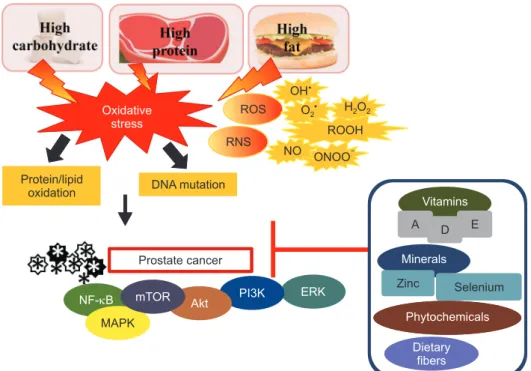
The androgen receptor interacts with androgen recep- tor elements in the promoter regions and thus regu- lates cellular proliferation [3]. In this regard, inappro- priate activation of estrogen receptor alpha signaling concomitantly with the elevation of testosterone has been demonstrated to trigger prostate cancer and pros- tate hyperplasia in mice [27]. Fig. 1. shows the effect of oxidative stress on prostate cancer.
3. Macronutrients-mediated oxidative stress
1) High carbohydratesEmerging evidence revealed that high consumption
NF- B MAPK
mTOR Akt PI3K ERK
Prostate cancer Protein/lipid
oxidation DNA mutation Oxidative
stress
ROS
RNS NO ONOO
ROOH H O2 2 OH
O2
Vitamins
A D E
Minerals
Zinc Selenium
Phytochemicals
Dietary fibers
Fig. 1. The effect of oxidative stress on prostate cancer. Consumption of high carbohydrate, high protein, and high-fat diets may induce oxidative stress. Oxidative damage is not only induced by the unrestricted reactive oxygen species (ROS) production but also due to other oxidants, such as reactive nitrogen species (RNS). Overproduction of reactive species, including hydroxyl radical (•OH), superoxide anion (O2•), hydrogen peroxide (H2O2), hydroperoxides (ROOH), nitric oxide (NO), and peroxynitrite (ONOO−) may lead to protein/lipid oxidation and DNA mutation. Elevation of ROS can result in the progression and development of prostate cancer. The onset and progression of prostate cancer could be prevented by changing dietary habits by modulating nuclear factor-kappa B (NF-κB), mammalian target of rapamycin (mTOR), mitogen-activated protein kinase (MAPK), Akt, extracellular signal-regulated kinase (ERK), and phosphoinositide 3-kinases (PI3K) signaling pathways. Consumption of diet high in vitamins A, D, and E, minerals (selenium and zinc), phytochemicals, and dietary fibers may reduce the risk of prostate cancer.
of macronutrients can increase oxidative stress and thus trigger inflammation [9]. Dietary carbohydrate is crucial to discuss here because it may lead to the long- term consequences of nutritionally mediated oxida- tive stress [28]. Intakes of carbohydrates have drawn a great deal of interest among researchers due to an association between high glycemic load (GL) or glyce- mic index diet with prostate cancer [29]. High GL diet has been characterized as a common characteristic of Western culture, in which they are high in refined car- bohydrates and added sugars [30]. Data from an earlier study have revealed that insulin is a proliferation fac- tor for prostate cancer; conversely, decreased carbohy- drates intake may slow down prostate cancer prolifera- tion and reduce serum insulin [31]. Despite study has found a positive relationship between carbohydrate and prostate cancer, not all data showed such a link. A recent meta-analysis study did not identify an associa- tion between prostate cancer risk and carbohydrate intake [29].
Intake of carbohydrates can disturb the insulin secretion and glycemic response of an individual, in which the effect of blood glucose response depends on the amount of carbohydrates ingested [32]. Hyperin- sulinemia is linked to various cancers, including pros- tate cancer. Insulin resistance leads to reduced insulin sensitivity in peripheral tissues, and thereby caused in hyperinsulinemia. Insulin is a potent growth factor linked to an increase of prostate tumor proliferation [33]. Higher grade of prostate tumors was shown to have high expression of insulin receptor-A (IR-A) iso- form and increased number of IR both in vivo and in vitro studies [34,35]. Stimulation of IR upregulates both mitogen-activated protein (MAP)/extracellular signal- regulated kinase and phosphoinositide 3-kinases/Akt/
mammalian target of rapamycin (PI3K/Akt/mTOR) signaling pathways and subsequently leading to in- hibiting of apoptosis and promoting cell migration and proliferation [33].
2) High animal-based protein
Animal meat is a source of protein in the Western diet, composes of 20% of daily fat, 40% of daily protein, and 15% of daily energy intake [36]. Excessive protein intake could increase ROS production in the digestive gland, impair oxidation of amino acids in the digestive system, and promote instability of antioxidants. Sev- eral metabolites are produced such as hydrogen sulfide
(H2S) and ammonia (NH3), compounds are known to induce toxicity of the mucosa during fermentation of excessive proteins in the gut. Although meats are rich in dietary protein, it can be a source of mutagens due to the presence of heterocyclic amines (HCA) and polycyclic aromatic hydrocarbons (PAH) during high- temperature grilling and cooking and N-nitroso com- pounds in processed meats [37]. Evidence from the case- control study demonstrated the consumption of cooked red meat potentially leads to advanced prostate cancer risk [38]. This finding indicates the metabolic changes induced by red meat are more likely initiated by the formation of carcinogenic PAH and HCA.
During cooking, free Fe2+ is markedly increased in uncured meat [39]. However, nitrite curing inhibits the phosphorylation of heme-Fe via modulation of the porphyrin ring [39]. Indeed, heat treatment can lead to a reduction of the antioxidant enzyme, for instance, glutathione peroxidase, and produces oxygen from oxy- myoglobin, which subsequently leads to the production of H2O2. When the oxidative processes are initiated, free Fe2+ catalyzes the Fenton reaction [40]. Taken to- gether, nitrite curing of meat in the colonic was vitally important because it was related to a low amount of malondialdehyde but doubled heptanal levels and pro- portionally increased of hydroxy-2-nonenal levels in the cooked and overcooked meats [39].
3) High fat consumption
When caloric intake surpasses energy expenses, the substrate promotes in citric acid cycle which increases the production of ROS. Administration of high-fat diet promotes the production of ROS, along with the eleva- tion of tumor necrosis factor-alpha (TNF-α) and adipo- kine secretion, thereby resulting in chronic inflamma- tion [41]. Animal feeding with a high-fat diet increased metastasis rates and local invasion of prostate cancer in transgenic adenocarcinoma of mouse prostate mice, suggesting the circulating cytokine and adipokine alterations in response to excess adipose tissue deposi- tion induced by a high-fat diet may enhance prostate cancer progression [42]. Data from in vivo study also re- vealed that a high-fat diet accelerates the proliferation of prostate tumors and increased M2/M1 macrophage ratio and myeloid-derived suppressor cells fraction via interleukin (IL)-6/phospho-signal transducer and acti- vator of transcription 3 (pSTAT3) signaling [43].
Mitochondrial β-oxidation of free fatty acids are
related to the conversion of oxidized cofactors (FAD and NAD+) into reduced cofactors, namely FADH2 and NADH. Subsequently, it was reoxidized and restored back into FAD and NAD+ by the mitochondrial re- spiratory chain. Through the process of reoxidation, FADH2 and NADH transfer the electrons to the first complexes of the respiratory chain. This electron shift to cytochrome c oxidase and interact with protons and oxygen to form water. The intermediates may bind with oxygen to generate superoxide anion and produce ROS. In this regard, sustained chronic fat consump- tion induces mitochondrial β-oxidation of free fatty acids and resulted to excess electron flow using cyto- chrome c oxidase, which promotes ROS levels. High- fat diet-induced ROS may promote proinflammatory signaling and stimulate nuclear factor-kappa B (NF- κB) transcriptional factor, and hence inducing NF-κB- dependent proinflammatory molecules, for instance, TNF-α, interferon-γ, and iNOS [44].
4. Modulation of prostate cancer by dietary components
1) Vitamins
Vitamins are essential for normal metabolism. Most of the vitamins cannot be synthesized by humans, ex- cept vitamin D; therefore, they need to be consumed from the diet to prevent metabolism disorders [45]. The protective effect of vitamins in prostate cancer has
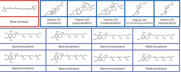
been widely demonstrated in clinical trials and labora- tory experiments [46,47], in which most of the data ex- ists for vitamins D, A or provitamin A carotenoids, and E. Vitamin E (four tocotrienols and four tocopherols) (Fig. 2) is a fat-soluble vitamin found naturally in food.
Both tocotrienols and tocopherols are divided into α-, β-, γ-, and δ- based on the hydroxyl and methyl substitu- tion in their phenolic rings [48]. The predominant bio- logical roles of vitamin E in response to prostate cancer have been reported and the positive impact of oxida- tive damage has been evaluated. The proposed aspects of vitamin E including inhibiting cell proliferation, cell adhesion, and protein kinase C activity, improving im- munity, and modulating anti-angiogenic and antioxida- tion properties [49]. Data from a cohort study (18,541 controls and 11,239 cases) revealed that α-tocopherol reduces the risk of prostate cancer [50]. These findings are in line with an earlier study reported by Antwi et al [51], who found that high α-tocopherol levels are neg- atively linked to serum PSA concentrations in patients with recurrent prostate cancer. Kyriakopoulos et al [52]
further revealed that APC-100, an antioxidant moiety of α-tocopherol, has shown the median progression-free survival of 2.8 months in patients with castrate-resis- tant prostate cancer. Intriguingly, several studies have demonstrated that vitamin E increased prostate cancer.
Evidence from National Institutes of Health (NIH)- AARP Diet and Health Study revealed that a high fre- quency of vitamin E (>7 times/wk) increased the risk
HO H H
H
Vitamin D1 (lumisterol)
HO CH2
CH3 CH3
CH3 CH3 CH3
Vitamin D2 (ergocalciferol)
HO CH2
CH3 CH3
CH3 CH3
Vitamin D3 (cholecalciferol)
HO H
Vitamin D4 (22-dihydroergocalciferol)
HO
Vitamin D5 (sitocalciferol)
Alpha-tocopherol Beta-tocopherol Gamma-tocopherol Delta-tocopherol
Alpha-tocotrienol Beta-tocotrienol Gamma-tocotrienol Delta-tocotrienol
H C3 O
HO CH3 CH3
CH3
CH3 CH3 CH3 CH3
O CH3
CH3 CH3 CH3 CH3 CH3
CH3 HO
O CH3
CH3
CH3 CH3 CH3 CH3 H3C
HO
O CH3
CH3
CH3 CH3 CH3 CH3 HO
H C3 O
HO CH3 CH3
CH3
CH3 CH3 CH3 CH3
O
HO CH3 CH3
CH3
CH3 CH3 CH3 CH3
O H C3
HO CH3
CH3
CH3 CH3 CH3 CH3
O HO
CH3 CH3
CH3 CH3 CH3 CH3
Beta-carotene
Fig. 2. Molecule structures of beta-carotene, vitamin D including lumisterol (D1), ergocalciferol (D2), cholecalciferol (D3), 22-dihydroergocalciferol (D4), and sitocalciferol (D5), and vitamin E congeners including tocopherols (α-tocopherol, β-tocopherol, γ-tocopherol, and δ-tocopherol) and tocotrienols (α-tocotrienol, β-tocotrienol, γ-tocotrienol, and δ-tocotrienol).
of prostate cancer [53]. The study further showed that supplementation of vitamin E (800 IU/d) was linked to the prostate cancer risk, regardless of frequency.
High levels of α-tocopherol in plasma have also been related to high-grade prostate cancer [54]. Although the data show a positive relationship between vitamin E and prostate cancer, several large population studies showed null findings in relation to prostate cancer and benign prostatic hyperplasia (BPH) [55,56].
Vitamin A or provitamin A carotenoids play a criti- cal role in maintaining healthy vision [57]. Carotenoids (Fig. 2) are naturally organic pigments produced in the fungi, bacteria, and plastids of algae and plants. In general, carotenoids absorb wavelengths 400–550 nm, thereby the compounds appear in red, orange, or yellow color. Carotenoids are not only served as an efficient radical scavenger but also exerted anticancer proper- ties by regulating cell signaling pathways, improving immune system, and modulating cell differentiation, apoptosis, and cell cycle [58]. Vitamin A such as retinol exerts antineoplastic properties including suppres- sion of cellular growth and induction of apoptosis [59]. Nonetheless, Peehl and Feldman [59] revealed that retinoids enhanced tumor proliferation, possibly via sex steroids or insulin-like growth factor I recep- tor [50]. Data from Alpha-Tocopherol, Beta-Carotene Cancer Prevention (ATBC) study further demon- strated that supplementation of 20 mg β-carotene per day [60] increased the incidence of prostate cancer in men. Intriguingly, some research has emerged to sug- gest that carotenoids (β-cryptoxanthin, α-carotene, and β-carotene) were not linked to prostate cancer [50]. Cook et al [61] and Omenn et al [62] also demonstrated that supplementation with 30 and 50 mg β-carotene was not related to prostate cancer.
Vitamin D is a fat-soluble vitamin, consists of five isoforms including ergocalciferol with lumisterol (D1), sitocalciferol (D5), 22-dihydroergocalciferol (D4), cho- lecalciferol (D3), ergocalciferol (D2) (Fig. 2), and usu- ally found in fortified food products, flour, and dairy.
Vitamin D not only can be obtained from foods, it can also be synthesized by skin in response to sunlight [63]. Vitamin D is predominantly circulated as 25-hy- droxyvitamin D (25(OH)D) and thus converted by 1α-hydroxylase into its active form, 1,25-dihydroxyvi- tamin D [1,25(OH)2D]. Adequate consumption of vita- min D is essential to regulate bone mineralization and maintain calcium homeostasis [64]. Vitamin D deficien-
cy is linked to the development of prostate cancer. Low vitamin D intake (<20 ng/mL) was demonstrated to be a significant risk factor for the progression of prostate cancer in certain subpopulations [65]. Data from two large nested case-control studies showed that vitamin D reduced the aggressive prostate cancer [66]. In sup- port of this, high vitamin D serum levels were also de- creased the risk of prostate cancer in men [67].
High levels of 25(OH)D decrease the inflamma- tory markers, for instance, IL-8 and serum C-reactive protein in patients with prostate cancer [68]. Evi- dence from an in vitro study has suggested that 1α,25- dihydroxyvitamin D3 upregulated the expression of tu- mor suppressor candidate 3 (TUSC-3) in prostate cancer cells. In fact, a low level of TUSC-3 was demonstrated to correspond with poor prostate cancer prognosis in patients, and TUSC-3 silencing promotes cell prolifera- tion and migration [69]. Intriguingly, data from ATBC study has demonstrated that high circulating 25(OH)D increased the risk of prostate cancer [70]. Such finding highlights the association of high dosages of vitamin D-binding protein in the elevation of prostate cancer among men [71]. Collectively, there are inconsistent findings supporting the clinical use of vitamins E, A, and D in delaying or preventing the progression or onset of prostate cancer, and most of the studies are limited in their duration of exposure and sample size.
In particular, how vitamins may detrimental to the development of prostate cancer and whether low con- centrations of vitamins can be preventive measures are warranted in a long-term study. Table 1 [50-56,60- 62,65-68,70,72-82] shows the clinical studies of dietary components in relation to prostate cancer.
2) Minerals
Minerals are chemical elements required by all or- ganisms. It facilitates the modulation of heartbeat, hormones synthesis, and bone formation. In humans, minerals can be obtained from drinking water and food. However, mineral supplements are essential for those who did not meet the daily intake of mineral [83]. Dietary minerals can be classified into two groups, namely trace minerals and macrominerals. Macromin- erals such as chloride, potassium, magnesium, sodium, calcium, and phosphorus are required in large amounts by the body. Conversely, trace elements are dietary minerals that are needed in minimal levels to main- tain regular cellular function, including iron, fluoride,
iodine, zinc, selenium, and copper. Among all minerals stated above, zinc and selenium have been extensively studied in relation to prostate cancer. Indeed, both zinc and selenium exert antioxidant properties. Zinc serves as a cofactor for SOD enzyme, modulation of metallo- thionein expression, suppression of NADPH oxidase, and regulation of glutathione metabolism in the body [84]. Dietary zinc showed a protective effect in patients with advanced prostate cancer susceptibility. Dietary administration of zinc inhibits the proangiogenic and invasive capabilities of metastatic prostate cancer cells.
Inhibition of Krebs cycle in normal prostate cells lead- ing to accumulation and secretion of citrate, which is
predominantly obtained via the suppression of m-acon- itase, due to an accumulation of zinc in normal pros- tate cells [85]. High levels of zinc in the mitochondria truncate the Krebs cycle by suppressing m-aconitase and determining the peculiar metabolism of normal prostate cells [85]. It has been demonstrated that zinc concentrations are markedly decreased in prostate cancer cells [86], as well as inducing the reactivation of m-aconitase of the Kreb cycle. Besides inhibiting m- aconitase, zinc also induces apoptosis in prostate cancer cells via inhibition of NF-κB, stimulation of caspases cascade, and induction of the cytochrome c from the mitochondria [87]. However, several studies have dem- onstrated either null or/and detrimental effects of di- etary zinc and prostate cancer [55,88]. Data from NIH- AARP Diet and Health Study revealed that excessive consumption of zinc was linked to a 4.36-fold increase of developing fatal prostate cancer in men (1,476 ad- vanced and 8,765 localized cases) [53].
In addition to the effects observed in zinc, the previ- ous study has revealed the role of selenium in relation to prostate cancer. The activity of selenium is modu- lated by antioxidant enzymes leading to a reduction of ROS. Changes in the physiological levels of selenium may affect the metabolic and biochemical processes in prostate cancer. Compared to BPH specimens, malig- nant prostate tissue has relatively low levels of sele- nium [89]. A meta-analysis of 17 western population- based studies demonstrated that selenium in serum levels was inversely associated with prostate cancer susceptibility [72]. Likewise, data from a meta-analysis involving 15 prospective studies (6,021 controls and 4,527 cases) found that high selenium plasma levels were linked to the reduced risk of aggressive prostate cancer [73]. Conversely, in the Italian cohort for the Procomb trial, dietary selenium for two years did not lead to any significant changes in PSA levels or pros- tate cancer susceptibility [74]. Data from the Phase III clinical trial showed that supplementation selenium in a concentration of 200–400 μg for 3–5 years did not lead to any adverse outcomes such as prostate cancer mortality or PSA velocity in patients with a high risk of prostate cancer [75]. Taken together, several, but not all, observational studies show that zinc and sele- nium may possess anti-prostate cancer properties. The preventive role of zinc and selenium against cancer is more likely due to its antioxidant activity. In this regard, there has been a tremendous interest in the Table 1. Clinical studies conducted in some dietary components in
prostate cancer Dietary
component Finding Reference
Vitamin E ↓Risk of prostate cancer and aggressive disease
[50]
↓PSA serum levels in recurrent prostate cancer patients
[51]
Maintain stable disease and median progression-free survival of 2.8 months in patients with castrate-resistant pros- tate cancer
[52]
↑Risk of prostate cancer (>7 times/wk) [53]
↑High-grade of prostate cancer [54]
No effect on prostate cancer and BPH [55,56]
Carotenoids ↑Incidence of prostate cancer (20 mg/d) [60]
Not associated with prostate cancer (30 mg or 50 mg)
[61,62]
Not associated with prostate cancer [50]
Vitamin D ↑Prostate cancer in certain subpopula-
tions (<20 ng/mL) [65]
↓Aggressive prostate cancer [66]
↓Risk of prostate cancer [67]
↓IL-8 and CRP in patients with prostate cancer
↓CRP in prostate cancer patients [68]
↑Risk of prostate cancer [70]
Zinc ↑4.36-fold of developing fatal prostate cancer
[53]
Selenium ↓Prostate cancer susceptibility [72]
↓Risk of aggressive prostate cancer [73]
No effect [74,75]
Flavonoids ↓Prostate cancer [76]
Dietary fiber ↓Total and advanced prostate cancer [77-80]
No effect [81,82]
PSA: prostate-specific antigen, BPH: benign prostatic hyperplasis, IL:
interleukin, CRP: C-reactive protein.
study of zinc and selenium in this disease. The crucial role played by selenium and zinc is nonetheless worth study in-depth in prostate cancer.
3) Phytochemicals
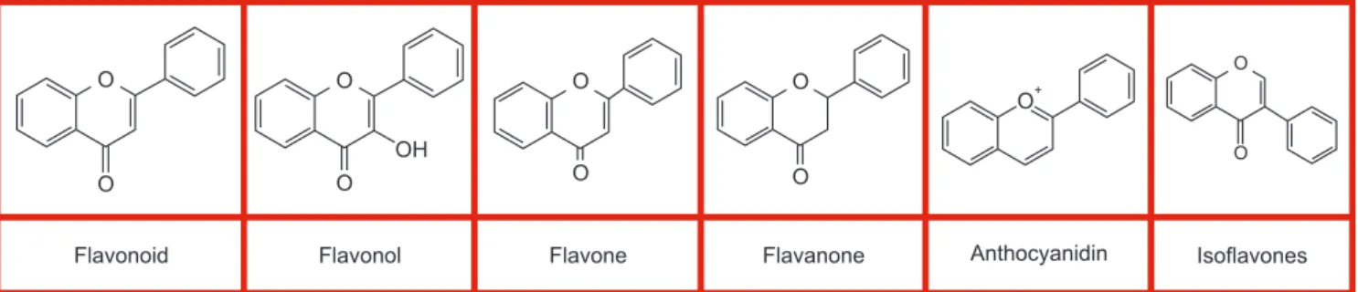
Phytochemicals are plant-derived non-nutritive com- pounds found in herbs and crops. In addition to the vi- tamins and minerals stated above, plants contain large amounts of phytochemicals. Polyphenols are secondary metabolites that are extensively present in vegetables, grains, traditional medicinal herbs, and fruits. Among all polyphenolic compounds, the flavonoids are the most common classes of polyphenol. Flavonoids are divided into different subgroups, namely chalcones, an- thocyanins, catechins, flavanones, flavanonols, flavones, and flavonols, based on the degree of unsaturation and the carbon (Fig. 3).
Flavonoids have beneficial antioxidants and bio- chemical effects against cancer including prostate cancer [90]. Dietary administration of flavonoid tan- geretin suppresses the growth of prostate cancer cells via downregulation of PI3K/Akt/mTOR pathways [90].
Tangeretin is found abundantly in peel of citrus fruits.
Praud et al [91] found that a high intake of proantho- cyanidin is inversely linked to prostate cancer. Impor- tantly, the favorable effect could be attributed to the capacity of some oligomeric forms to influence the pro- liferation of androgen-resistant and androgen-sensitive human prostate cancer cell lines [92]. Study by Neuwirt et al [93] has shown an ability of proanthocyanidins extracted from grape seeds to suppress the growth of androgen-sensitive prostate cancer cell lines via a few mechanisms including regulating the expression of MAP kinase and activator protein 1. Furthermore, the consumption of red wine, a vital source of proanthocy- anidins, was negatively linked to prostate cancer, dem- onstrated in a United States case-control study [94]. In
vitro study showed that resveratrol sensitizes the pros- tate cancer cell lines to chemotherapeutic drugs such as methotrexate, taxol, actinomycin D, cytarabine, and doxorubicin by inducing apoptosis and downregulating survivin levels [95]. These findings are in parallel with an earlier study reported in the preclinical study for resveratrol, largely present in red wine, in which it has chemopreventive effects against prostate cancer [96].
The recent study suggests that resveratrol upregulates DUSP1 expression in androgen-independent prostate cancer cells through suppression of cyclooxygenase-2 and NF-κB activity [97]. Based on the evidence, it was evident that the administration of naturally-occurring phytochemicals may have a protective role in the de- velopment of prostate cancer. Nonetheless, there are several limitations such as the concentration of phy- tochemicals and bioavailability in relation to prostate cancer. Further studies are required to elucidate the role of these phytochemicals or in combination with other agents in prostate cancer prevention and treat- ment.
4) Dietary fibers
Dietary fibers are dietary components that are not degraded enzymatically to absorbable fractions in the stomach and small intestine. Many studies support the roles of dietary fiber in preventing the progression and development of prostate cancer. The previous study showed that insoluble fiber was inversely correlated to total and advanced prostate cancer [77]. This study further demonstrated that very low fiber intake was positively linked to prostate cancer in Japanese men [77]. In line with the study reported by Sawada et al [77], fiber intake has also been reported to have antiprolif- erative activity against prostate cancer [78]. Compared to those who consume fiber (less than 15 g/d), men who consume fiber (more than 15 g/d) can reduce the risk of
Flavonoid Flavonol Flavone Flavanone Anthocyanidin Isoflavones
O
O
O
O OH
O
O
O
O
O+
O
O
Fig. 3. Molecule structures of polyphenols (flavonoid, flavonol, flavone, flavanone, anthocyanidin, and isoflavones).
prostate cancer by 40% [79]. Likewise, data from a pro- spective study in the United States showed the intake of dietary fiber reduced aggressive prostate cancer among European Americans and African Americans [80]. However, some prospective studies from Western countries have found that consumption of fiber was not related to prostate cancer [81,82].
Indeed, the anticancer ability of dietary fiber is me- diated via a few mechanisms including (1) affecting steroid hormone level by reducing circulating andro- gens and estrogens; (2) increasing sex hormone-binding globulin levels; (3) reducing IGF bioactivity and en- hancing insulin sensitivity [98]. Dietary fibers, particu- larly from legume and insoluble fiber, were inversely linked to prostate cancer risk [99]. Importantly, dietary fiber in vegetables and fruits will undergo fermenta- tion by gut microbiota, and thereby lead to the pro- duction of short-chain fatty acids including propionic, butyrate, and acetate acids [100]. Overall, the intake of food rich in dietary fiber regularly may decrease the risk of prostate cancer.
CONCLUSIONS
Evidence from clinical and preclinical studies showed that ROS signaling plays a crucial role in the progres- sion and development of prostate cancer. Several envi- ronmental factors, for example, inflammation, diet, and changes in cellular functions like mitochondrial DNA mutations, androgen signaling, and redox imbalance are all contributed to the elevation of ROS. Increased ROS may enhance genetic instability, promote cell pro- liferation, and alter somatic DNA mutations. Research evidence suggests that a diet high in animal proteins and carbohydrates and excessive fat consumption can generate ROS and thereby results in oxidative stress.
The previous study suggests that a diet rich in vita- mins (A, D, and E) and minerals, dietary fibers, and phytochemicals are likely to alleviate or prevent pros- tate cancer. Despite the beneficial effects of these com- ponents for reducing the risk of prostate cancer have been reported in both preclinical studies and clinical trials, not all studies supported an association between these dietary patterns and risk of prostate cancer.
Therefore, future research is warranted to evaluate the molecular link to better elucidate the possible role of these dietary patterns on prostate cancer preven- tion and treatment. Indeed, the best dietary advice
for prostate cancer management or prevention seems to include: reducing carbohydrate intakes, consuming moderate amounts of calories, decreasing overcooked meats intakes, reducing saturated and total fats, sub- stituting refined carbohydrates with whole grains, and increasing vegetables and fruits.
ACKNOWLEDGEMENTS
This research was supported by Putra Grant (UPM/700-2/1/GPB/2017/9549900).
Conflict of Interest
The authors have nothing to disclose.
Author Contribution
Conceptualization: BLT. Writing – original draft: BLT. Writ- ing – review & editing: BLT, MEN.
REFERENCES
1. World Health Organization. Cancer [Internet]. Geneva: World Health Organization; c2019 [cited 2019 Sep 9]. Available from: https://www.who.int/health-topics/cancer#tab=tab_1.
2. International Agency for Research on Cancer. Prostate (source: Globocan 2018) [Internet]. Geneva: World Health Organization; c2018 [cited 2019 Sep 9]. Available from:
https://gco.iarc.fr/today/data/factsheets/cancers/27-Prostate- fact-sheet.pdf.
3. Fujita K, Nonomura N. Role of androgen receptor in prostate cancer: a review. World J Mens Health 2019;37:288-95.
4. World Cancer Research Fund International. Prostate cancer statistics: prostate cancer is the second most common cancer in men worldwide [Internet]. London: World Cancer Re- search Fund International; c2019 [cited 2019 Sep 9]. Available from: https://www.wcrf.org/dietandcancer/cancer-trends/
prostate-cancer-statistics.
5. Elder K, Dixon JM, Blackmur JP, Laurie J. Endocrine therapy for cancer. Surgery 2018;36:128-33.
6. Taplin ME, Antonarakis ES, Ferrante KJ, Horgan K, Blumen- stein B, Saad F, et al. Androgen receptor modulation opti- mized for response-splice variant: a phase 3, randomized trial of galeterone versus enzalutamide in androgen receptor splice variant-7-expressing metastatic castration-resistant prostate cancer. Eur Urol 2019;76:843-51.
7. Tang J, Xiao L, Cui R, Li D, Zheng X, Zhu L, et al. CX3CL1
increases invasiveness and metastasis by promoting epithelial- to-mesenchymal transition through the TACE/TGF-α/EGFR pathway in hypoxic androgen-independent prostate cancer cells. Oncol Rep 2016;35:1153-62.
8. Miller DR. The role of ROS in the progression and treatment of castration-resistant prostate cancer [thesis]. Omaha: Uni- versity of Nebraska Medical Center; 2019.
9. Tan BL, Norhaizan ME, Liew WP. Nutrients and oxida- tive stress: friend or foe? Oxid Med Cell Longev 2018;2018:
9719584.
10. Ahmed Amar SA, Eryilmaz R, Demir H, Aykan S, Demir C.
Determination of oxidative stress levels and some antioxidant enzyme activities in prostate cancer. Aging Male 2019;22:198- 206.
11. Long MD, Singh PK, Russell JR, Llimos G, Rosario S, Rizvi A, et al. The miR-96 and RARγ signaling axis governs an- drogen signaling and prostate cancer progression. Oncogene 2019;38:421-44.
12. Shi Y, Han JJ, Tennakoon JB, Mehta FF, Merchant FA, Burns AR, et al. Androgens promote prostate cancer cell growth through induction of autophagy. Mol Endocrinol 2013;27:280-95.
13. DiNicolantonio JJ, Lucan SC, O'Keefe JH. The evidence for saturated fat and for sugar related to coronary heart disease.
Prog Cardiovasc Dis 2016;58:464-72.
14. Biobaku F, Ghanim H, Batra M, Dandona P. Macronutrient- mediated inflammation and oxidative stress: relevance to in- sulin resistance, obesity, and atherogenesis. J Clin Endocrinol Metab 2019;104:6118-28.
15. Laurent V, Toulet A, Attané C, Milhas D, Dauvillier S, Zaidi F, et al. Periprostatic adipose tissue favors prostate cancer cell invasion in an obesity-dependent manner: role of oxidative stress. Mol Cancer Res 2019;17:821-35.
16. Csiszar A, Podlutsky A, Podlutskaya N, Sonntag WE, Merlin SZ, Philipp EE, et al. Testing the oxidative stress hypothesis of aging in primate fibroblasts: Is there a correlation between species longevity and cellular ROS production? J Gerontol A Biol Sci Med Sci 2012;67:841-52.
17. Ham HJ, Park JW, Bae YS. Defect of SIRT1-FoxO3a axis is associated with the production of reactive oxygen species during protein kinase CK2 downregulation-mediated cellular senescence and nematode aging. BMB Rep 2019;52:265-70.
18. Dias V, Junn E, Mouradian MM. The role of oxidative stress in Parkinson's disease. J Parkinsons Dis 2013;3:461-91.
19. Salisbury D, Bronas U. Reactive oxygen and nitrogen species:
impact on endothelial dysfunction. Nurs Res 2015;64:53-66.
20. Barreiro E. Role of protein carbonylation in skeletal muscle mass loss associated with chronic conditions. Proteomes
2016;4:E18.
21. Frijhoff J, Winyard PG, Zarkovic N, Davies SS, Stocker R, Cheng D, et al. Clinical relevance of biomarkers of oxidative stress. Antioxid Redox Signal 2015;23:1144-70.
22. Reynaert NL, Gopal P, Rutten EPA, Wouters EFM, Schalkwijk CG. Advanced glycation end products and their receptor in age-related, non-communicable chronic inflammatory dis- eases; overview of clinical evidence and potential contribu- tions to disease. Int J Biochem Cell Biol 2016;81(Pt B):403-18.
23. Gonthier K, Poluri RTK, Audet-Walsh É. Functional genomic studies reveal the androgen receptor as a master regulator of cellular energy metabolism in prostate cancer. J Steroid Bio- chem Mol Biol 2019;191:105367.
24. Kowluru RA, Mishra M. Oxidative stress, mitochondrial damage and diabetic retinopathy. Biochim Biophys Acta 2015;1852:2474-83.
25. Pradedova EV, Nimaeva OD, Salyaev RK. Redox processes in biological systems. Russ J Plant Physiol 2017;64:822-32.
26. Bansal A, Simon MC. Glutathione metabolism in cancer pro- gression and treatment resistance. J Cell Biol 2018;217:2291- 8.
27. Bonkhoff H. Estrogen receptor signaling in prostate cancer:
implications for carcinogenesis and tumor progression. Pros- tate 2018;78:2-10.
28. Zhao T, Wu K, Hogstrand C, Xu YH, Chen GH, Wei CC, et al.
Lipophagy mediated carbohydrate-induced changes of lipid metabolism via oxidative stress, endoplasmic reticulum (ER) stress and ChREBP/PPARγ pathways. Cell Mol Life Sci 2019.
doi: 10.1007/s00018-019-03263-6 [Epub].
29. Fan LL, Su HX, Gu XJ, Chen YH, Nan CJ. Carbohydrate intake and the risk of prostate cancer. Clin Chim Acta 2018;484:60-71.
30. Schulte EM, Avena NM, Gearhardt AN. Which foods may be addictive? The roles of processing, fat content, and glycemic load. PLoS One 2015;10:e0117959.
31. Lubik AA, Gunter JH, Hendy SC, Locke JA, Adomat HH, Thompson V, et al. Insulin increases de novo steroidogenesis in prostate cancer cells. Cancer Res 2011;71:5754-64.
32. Gonzalez JT, Betts JA. Dietary sugars, exercise and hepatic carbohydrate metabolism. Proc Nutr Soc 2019;78:246-56.
33. Arcidiacono B, Iiritano S, Nocera A, Possidente K, Nevolo MT, Ventura V, et al. Insulin resistance and cancer risk: an overview of the pathogenetic mechanisms. Exp Diabetes Res 2012;2012:789174.
34. Cox ME, Gleave ME, Zakikhani M, Bell RH, Piura E, Vickers E, et al. Insulin receptor expression by human prostate can- cers. Prostate 2009;69:33-40.
35. Perks CM, Zielinska HA, Wang J, Jarrett C, Frankow A,
Ladomery MR, et al. Insulin receptor isoform variations in prostate cancer cells. Front Endocrinol (Lausanne) 2016;7:132.
36. Food and Agriculture Organization of the United Nations.
Food balance sheets: a handbook [Internet]. Rome: Food and Agriculture Organization of the United Nations; c2001 [cited 2019 Sep 9]. Available from: http://www.fao.org/3/X9892E/
X9892E00.htm.
37. Ledesma E, Rendueles M, Díaz M. Contamination of meat products during smoking by polycyclic aromatic hydrocar- bons: Processes and prevention. Food Control 2016;60:64-87.
38. Joshi AD, Corral R, Catsburg C, Lewinger JP, Koo J, John EM, et al. Red meat and poultry, cooking practices, genetic suscep- tibility and risk of prostate cancer: results from a multiethnic case-control study. Carcinogenesis 2012;33:2108-18.
39. Van Hecke T, Vossen E, Hemeryck LY, Vanden Bussche J, Vanhaecke L, De Smet S. Increased oxidative and nitrosative reactions during digestion could contribute to the association between well-done red meat consumption and colorectal can- cer. Food Chem 2015;187:29-36.
40. Bokare AD, Choi W. Review of iron-free Fenton-like systems for activating H2O2 in advanced oxidation processes. J Haz- ard Mater 2014;275:121-35.
41. Maurizi G, Della Guardia L, Maurizi A, Poloni A. Adipocytes properties and crosstalk with immune system in obesity- related inflammation. J Cell Physiol 2018;233:88-97.
42. Hu MB, Xu H, Zhu WH, Bai PD, Hu JM, Yang T, et al. High- fat diet-induced adipokine and cytokine alterations promote the progression of prostate cancer in vivo and in vitro. Oncol Lett 2018;15:1607-15.
43. Hayashi T, Fujita K, Nojima S, Hayashi Y, Nakano K, Ishi- zuya Y, et al. High-fat diet-induced inflammation accelerates prostate cancer growth via IL6 signaling. Clin Cancer Res 2018;24:4309-18.
44. Dalvi PS, Chalmers JA, Luo V, Han DY, Wellhauser L, Liu Y, et al. High fat induces acute and chronic inflammation in the hypothalamus: effect of high-fat diet, palmitate and TNF-α on appetite-regulating NPY neurons. Int J Obes (Lond) 2017;41:149-58.
45. Thomas-Valdés S, Tostes MDGV, Anunciação PC, da Silva BP, Sant'Ana HMP. Association between vitamin deficiency and metabolic disorders related to obesity. Crit Rev Food Sci Nutr 2017;57:3332-43.
46. Hernáandez J, Syed S, Weiss G, Fernandes G, von Merveldt D, Troyer DA, et al. The modulation of prostate cancer risk with alpha-tocopherol: a pilot randomized, controlled clinical trial.
J Urol 2005;174:519-22.
47. Huang H, He Y, Cui XX, Goodin S, Wang H, Du ZY, et al.
Potent inhibitory effect of δ-tocopherol on prostate cancer cells cultured in vitro and grown as xenograft tumors in vivo.
J Agric Food Chem 2014;62:10752-8.
48. Joshi YB, Praticò D. Vitamin E in aging, dementia, and Al- zheimer's disease. Biofactors 2012;38:90-7.
49. Lee GY, Han SN. The role of vitamin E in immunity. Nutri- ents 2018;10:E1614.
50. Key TJ, Appleby PN, Travis RC, Albanes D, Alberg AJ, Barri- carte A, et al.; Endogenous Hormones Nutritional Biomarkers Prostate Cancer Collaborative Group. Carotenoids, retinol, tocopherols, and prostate cancer risk: pooled analysis of 15 studies. Am J Clin Nutr 2015;102:1142-57.
51. Antwi SO, Steck SE, Zhang H, Stumm L, Zhang J, Hurley TG, et al. Plasma carotenoids and tocopherols in relation to prostate-specific antigen (PSA) levels among men with bio- chemical recurrence of prostate cancer. Cancer Epidemiol 2015;39:752-62.
52. Kyriakopoulos CE, Heath EI, Eickhoff JC, Kolesar J, Yayehy- irad M, Moll T, et al. A multicenter phase 1/2a dose-escalation study of the antioxidant moiety of vitamin E 2,2,5,7,8-penta- methyl-6-chromanol (APC-100) in men with advanced pros- tate cancer. Invest New Drugs 2016;34:225-30.
53. Lawson KA, Wright ME, Subar A, Mouw T, Hollenbeck A, Schatzkin A, et al. Multivitamin use and risk of prostate cancer in the National Institutes of Health-AARP Diet and Health Study. J Natl Cancer Inst 2007;99:754-64.
54. Albanes D, Till C, Klein EA, Goodman PJ, Mondul AM, Weinstein SJ, et al. Plasma tocopherols and risk of prostate cancer in the Selenium and Vitamin E Cancer Prevention Trial (SELECT). Cancer Prev Res (Phila) 2014;7:886-95.
55. Lane JA, Oliver SE, Appleby PN, Lentjes MA, Emmett P, Kuh D, et al. Prostate cancer risk related to foods, food groups, macronutrients and micronutrients derived from the UK Dietary Cohort Consortium food diaries. Eur J Clin Nutr 2017;71:274-83.
56. Sarre S, Määttänen L, Tammela TL, Auvinen A, Murtola TJ. Postscreening follow-up of the Finnish Prostate Cancer Screening Trial on putative prostate cancer risk factors: vita- min and mineral use, male pattern baldness, pubertal devel- opment and non-steroidal anti-inflammatory drug use. Scand J Urol 2016;50:267-73.
57. Gilbert C. The eye signs of vitamin A deficiency. Community Eye Health 2013;26:66-7.
58. Karadas F, Erdogan S, Kor D, Oto G, Uluman M. The effects of different types of antioxidants (Se, vitamin E and carot- enoids) in broiler diets on the growth performance, skin pig- mentation and liver and plasma antioxidant concentrations.
Rev Bras Cienc Avic 2016;18:101-16.
59. Peehl DM, Feldman D. The role of vitamin D and retinoids in controlling prostate cancer progression. Endocr Relat Cancer 2003;10:131-40.
60. Alpha-Tocopherol, Beta Carotene Cancer Prevention Study Group. The effect of vitamin E and beta carotene on the in- cidence of lung cancer and other cancers in male smokers. N Engl J Med 1994;330:1029-35.
61. Cook NR, Le IM, Manson JE, Buring JE, Hennekens CH. Ef- fects of beta-carotene supplementation on cancer incidence by baseline characteristics in the Physicians' Health Study (United States). Cancer Causes Control 2000;11:617-26.
62. Omenn GS, Goodman GE, Thornquist MD, Balmes J, Cullen MR, Glass A, et al. Risk factors for lung cancer and for inter- vention effects in CARET, the Beta-Carotene and Retinol Ef- ficacy Trial. J Natl Cancer Inst 1996;88:1550-9.
63. Abboud M, Rybchyn MS, Rizk R, Fraser DR, Mason RS.
Sunlight exposure is just one of the factors which influence vitamin D status. Photochem Photobiol Sci 2017;16:302-13.
64. Goltzman D. Functions of vitamin D in bone. Histochem Cell Biol 2018;149:305-12.
65. Nelson SM, Batai K, Ahaghotu C, Agurs-Collins T, Kittles RA. Association between serum 25-hydroxy-vitamin D and aggressive prostate cancer in African American men. Nutri- ents 2017;9:E12.
66. Batai K, Murphy AB, Ruden M, Newsome J, Shah E, Dixon MA, et al. Race and BMI modify associations of calcium and vitamin D intake with prostate cancer. BMC Cancer 2017;17:64.
67. Mondul AM, Weinstein SJ, Layne TM, Albanes D. Vitamin D and cancer risk and mortality: state of the science, gaps, and challenges. Epidemiol Rev 2017;39:28-48.
68. Xie DD, Chen YH, Xu S, Zhang C, Wang DM, Wang H, et al.
Low vitamin D status is associated with inflammation in pa- tients with prostate cancer. Oncotarget 2017;8:22076-85.
69. Lin VC, Huang SP, Ting HJ, Ma WL, Yu CC, Huang CY, et al.
Vitamin D receptor-binding site variants affect prostate can- cer progression. Oncotarget 2017;8:74119-28.
70. Albanes D, Mondul AM, Yu K, Parisi D, Horst RL, Virtamo J, et al. Serum 25-hydroxy vitamin D and prostate cancer risk in a large nested case-control study. Cancer Epidemiol Biomark- ers Prev 2011;20:1850-60.
71. Weinstein SJ, Mondul AM, Kopp W, Rager H, Virtamo J, Albanes D. Circulating 25-hydroxyvitamin D, vitamin D- binding protein and risk of prostate cancer. Int J Cancer 2013;132:2940-7.
72. Cui Z, Liu D, Liu C, Liu G. Serum selenium levels and pros- tate cancer risk: a MOOSE-compliant meta-analysis. Medi- cine (Baltimore) 2017;96:e5944.
73. Allen NE, Travis RC, Appleby PN, Albanes D, Barnett MJ, Black A, et al. Selenium and prostate cancer: analysis of indi- vidual participant data from fifteen prospective studies. J Natl Cancer Inst 2016;108:djw153.
74. Morgia G, Voce S, Palmieri F, Gentile M, Iapicca G, Giannan- toni A, et al. Association between selenium and lycopene supplementation and incidence of prostate cancer: results from the post-hoc analysis of the procomb trial. Phytomedi- cine 2017;34:1-5.
75. Lü J, Zhang J, Jiang C, Deng Y, Özten N, Bosland MC. Cancer chemoprevention research with selenium in the post-SELECT era: promises and challenges. Nutr Cancer 2016;68:1-17.
76. Reale G, Russo GI, Di Mauro M, Regis F, Campisi D, Giudice AL, et al. Association between dietary flavonoids intake and prostate cancer risk: a case-control study in Sicily. Comple- ment Ther Med 2018;39:14-8.
77. Sawada N, Iwasaki M, Yamaji T, Shimazu T, Sasazuki S, Inoue M, et al. Fiber intake and risk of subsequent prostate cancer in Japanese men. Am J Clin Nutr 2015;101:118-25.
78. Lewis JE, Soler-Vilá H, Clark PE, Kresty LA, Allen GO, Hu JJ. Intake of plant foods and associated nutrients in prostate cancer risk. Nutr Cancer 2009;61:216-24.
79. Walker AR, Walker BF, Tsotetsi NG, Sebitso C, Siwedi D, Walker AJ. Case-control study of prostate cancer in black pa- tients in Soweto, South Africa. Br J Cancer 1992;65:438-41.
80. Tabung F, Steck SE, Su LJ, Mohler JL, Fontham ET, Bensen JT, et al. Intake of grains and dietary fiber and prostate cancer aggressiveness by race. Prostate Cancer 2012;2012:323296.
81. Bradbury KE, Appleby PN, Key TJ. Fruit, vegetable, and fiber intake in relation to cancer risk: findings from the European Prospective Investigation into Cancer and Nutrition (EPIC).
Am J Clin Nutr 2014;100 Suppl 1:394S-8S.
82. Sheng T, Shen RL, Shao H, Ma TH. No association between fiber intake and prostate cancer risk: a meta-analysis of epide- miological studies. World J Surg Oncol 2015;13:264.
83. Schwalfenberg GK, Genuis SJ. Vitamin D, essential minerals, and toxic elements: exploring interactions between nutrients and toxicants in clinical medicine. ScientificWorldJournal 2015;2015:318595.
84. Marreiro DD, Cruz KJ, Morais JB, Beserra JB, Severo JS, de Oliveira AR. Zinc and oxidative stress: current mechanisms.
Antioxidants (Basel) 2017;6:E24.
85. Costello LC, Franklin RB. The clinical relevance of the me- tabolism of prostate cancer; zinc and tumor suppression: con- necting the dots. Mol Cancer 2006;5:17.
86. Tsui KH, Chang PL, Juang HH. Zinc blocks gene expression of mitochondrial aconitase in human prostatic carcinoma cells. Int J Cancer 2006;118:609-15.
87. Feng P, Liang JY, Li TL, Guan ZX, Zou J, Franklin R, et al.
Zinc induces mitochondria apoptogenesis in prostate cells.
Mol Urol 2000;4:31-6.
88. Eriksen KT, Halkjær J, Meliker JR, McElroy JA, Sørensen M, Tjønneland A, et al. Dietary cadmium intake and risk of pros- tate cancer: a Danish prospective cohort study. BMC Cancer 2015;15:177.
89. Singh BP, Dwivedi S, Dhakad U, Murthy RC, Choubey VK, Goel A, et al. Status and interrelationship of zinc, copper, iron, calcium and selenium in prostate cancer. Indian J Clin Biochem 2016;31:50-6 .
90. Zhu WB, Xiao N, Liu XJ. Dietary flavonoid tangeretin induc- es reprogramming of epithelial to mesenchymal transition in prostate cancer cells by targeting the PI3K/Akt/mTOR signal- ing pathway. Oncol Lett 2018;15:433-40.
91. Praud D, Parpinel M, Guercio V, Bosetti C, Serraino D, Fac- chini G, et al. Proanthocyanidins and the risk of prostate can- cer in Italy. Cancer Causes Control 2018;29:261-8.
92. Kampa M, Theodoropoulou K, Mavromati F, Pelekanou V, Notas G, Lagoudaki ED, et al. Novel oligomeric proanthocy- anidin derivatives interact with membrane androgen sites and induce regression of hormone-independent prostate cancer. J Pharmacol Exp Ther 2011;337:24-32.
93. Neuwirt H, Arias MC, Puhr M, Hobisch A, Culig Z. Oligo- meric proanthocyanidin complexes (OPC) exert anti-pro- liferative and pro-apoptotic effects on prostate cancer cells.
Prostate 2008;68:1647-54.
94. Schoonen WM, Salinas CA, Kiemeney LA, Stanford JL. Alco- hol consumption and risk of prostate cancer in middle-aged men. Int J Cancer 2005;113:133-40.
95. Gupta SC, Kannappan R, Reuter S, Kim JH, Aggarwal BB.
Chemosensitization of tumors by resveratrol. Ann N Y Acad Sci 2011;1215:150-60.
96. Kumar A, Dhar S, Rimando AM, Lage JM, Lewin JR, Zhang X, et al. Epigenetic potential of resveratrol and analogs in preclinical models of prostate cancer. Ann N Y Acad Sci 2015;1348:1-9.
97. Martínez-Martínez D, Soto A, Gil-Araujo B, Gallego B, Chi- loeches A, Lasa M. Resveratrol promotes apoptosis through the induction of dual specificity phosphatase 1 and sensi- tizes prostate cancer cells to cisplatin. Food Chem Toxicol 2019;124:273-9.
98. Johnston KL, Thomas EL, Bell JD, Frost GS, Robertson MD.
Resistant starch improves insulin sensitivity in metabolic syn- drome. Diabet Med 2010;27:391-7.
99. Deschasaux M, Pouchieu C, His M, Hercberg S, Latino- Martel P, Touvier M. Dietary total and insoluble fiber intakes are inversely associated with prostate cancer risk. J Nutr 2014;144:504-10.
100. Yang Y, Nirmagustina DE, Kumrungsee T, Okazaki Y, Tomo- take H, Kato N. Feeding of the water extract from Ganoderma lingzhi to rats modulates secondary bile acids, intestinal mi- croflora, mucins, and propionate important to colon cancer.
Biosci Biotechnol Biochem 2017;81:1796-804.