www.krspine.org
Predictive Factors for the Short-Term Efficacy of Epidural Injections in Lumbar Disc Herniation
Treatment
Eugene J. Park, M.D., Seong-Min Kim, M.D., Seungho Chung, M.D., Woo-Kie Min, MD, Ph.D.
J Korean Soc Spine Surg 2020 Decemberr;27(4):115-124.
Originally published online December 31, 2020;
https://doi.org/10.4184/jkss.2020.27.4.115
Korean Society of Spine Surgery
SMG-SNU Boramae Medical Center, 20, Boramae-ro 5-gil, Dongjak-gu, Seoul 07061, Korea Tel: +82-2-831-3413 Fax: +82-2-831-3414
©Copyright 2017 Korean Society of Spine Surgery pISSN 2093-4378 eISSN 2093-4386
The online version of this article, along with updated information and services, is located on the World Wide Web at:
http://www.krspine.org/DOIx.php?id=10.4184/jkss.2020.27.4.115
This is an Open Access article distributed under the terms of the Creative Commons Attribution Non-Commercial License (http://
creativecommons.org/licenses/by-nc/4.0) which permits unrestricted non-commercial use, distribution, and reproduction in any medium, provided the original work is properly cited.
Spine Surgery
© Copyright 2020 Korean Society of Spine Surgery
Journal of Korean Society of Spine Surgery. www.krspine.org. pISSN 2093-4378 eISSN 2093-4386
This is an Open Access article distributed under the terms of the Creative Commons Attribution Non-Commercial License (http://creativecommons.org/licenses/by-nc/4.0/) which permits unrestricted non-commercial use, distribution, and reproduction in any medium, provided the original work is properly cited.
115
J Korean Soc Spine Surg. 2020 Dec;27(4):115-124. https://doi.org/10.4184/jkss.2020.27.4.115
Original Article
Clinical Outcomes of Cervical Transforaminal Epidural Block Using Local Anesthetics with or without a Steroid for Cervical Spondylotic Radiculopathy
Eugene J. Park, M.D., Seong-Min Kim, M.D., Seungho Chung, M.D., Woo-Kie Min, MD, Ph.D.
Department of Orthopedic Surgery, Kyungpook National University Hospital, Kyungpook National University School of Medicine, Daegu, Korea
Study Design: A retrospective chart review.
Objectives: To evaluate and compare the clinical outcomes of cervical transforaminal epidural block (CTEB) using local anesthetics with or without a steroid for cervical spondylotic radiculopathy (CSR).
Summary of Literature Review: The typical mixture for a CTEB is a combination of local anesthetics with a non-particulate steroid.
However, there are potential complications related to steroid injections such as steroid-induced osteoporosis, hypothalamus-pituitary- adrenal gland axis suppression, and hyperglycemia.
Materials and Methods: From January 2018 to October 2019, 35 patients who underwent CTEB for CSR were enrolled in this study.
Cases with arm pain over 4 on a visual analog scale (VAS) were included. In the first 19 cases, a combination of 1 mL of 1% lidocaine and 1 mL of dexamethasone was used (group A), and in the next 16 cases, 1 mL of 1% lidocaine mixed with 1 mL of normal saline was used (group B). Arm pain VAS and the Neck Disability Index (NDI) were obtained perioperatively.
Results: Baseline characteristics were not significantly different between the two groups. In both groups, the arm pain VAS significantly decreased at 30 minutes, 2 weeks, and 6 weeks post-injection compared to pre-injection values. However, the arm pain aggravated 12 weeks post-injection. The NDI of both groups significantly improved 6 weeks post-injection compared to pre-injection. The clinical outcomes of arm pain VAS and NDI at 30 minutes, 2 weeks, and 6 weeks post-injection, as well as the amounts of change, were not significantly different between both groups.
Conclusions: CTEB for CSR without a steroid improved symptoms by 6 weeks. The degree of improvement was similar to when CTEB was performed with a steroid in terms of VAS and NDI.
Key words: Nerve block, Cervical vertebrae, Dexamethasone, Lidocaine
Received: September 3, 2020 Revised: September 9, 2020 Accepted: November 3, 2020 Published Online: December 31, 2020
Corresponding author: Woo-Kie Min, M.D., Ph.D.
ORCID ID: Eugene J. Park: https//orcid.org/0000-0002-3974-9460 Seong-Min Kim: https//orcid.org/0000-0001-6786-6739 Seungho Chung: https//orcid.org/0000-0001-7432-724X Woo-Kie Min: https//orcid.org/0000-0003-2079-5085 Department of Orthopedic Surgery, Kyungpook National University Hospital 130 Dongdeok-ro, Jung-gu, Daegu, 41944 Korea
TEL: +82-53-200-5638, FAX: +82-53-422-6605 E-mail: [email protected]
Introduction
Cervical spondylotic radiculopathy (CSR) is a compression of the nerve root due to the degeneration process.1) The pathologic changes of CSR include hypertrophy of facet joints, ligamentum flavum, periradicular fibrous tissue, disc bulging, and inflammation by local cytokine and hypoxia.2) Fortunately, the natural history of symptoms of cervical spondylosis is reported as benign and can be managed by nonoperative treatments.3) A cervical nerve block is one of the common conservative treatment modalities for treating
cervical radiculopathy when refractory to other conservative treatments, including lifestyle modification, oral medication, physical therapy, etc.
As one of the cervical nerve block methods, a cervical epidural block can be divided into interlaminar and transforaminal in terms of approach.4) Cervical interlaminar epidural block (CIEB) is performed through the interlaminar space using the loss of resistance technique. Complications of CIEB include stiff neck with accompanied neck pain, flushing, and dural puncture, which can lead to catastrophic results such as an epidural abscess or hematoma.5-8) Although the use of fluoroscope enhances proper needle placement, the failure rate was still high, and improper placement of the needle led to severe complications.9,10) The effect of CIEB for CSR is also limited due to the blocked flow of medication to the exact pathology. On the other hand, cervical transforaminal epidural block (CTEB) is reported to be more effective, deliver medication more accurately, and has less serious complication compared to CIEB.4,11,12)
A fluoroscope-assisted CTEB using steroids for CSR showed significant improvement in pain along with accurate positioning of the needle.13,14) The most common mixture for a cervical epidural block is a combination of steroids with local anesthetics.11) Multiple mechanisms of steroids such as anti-inflammatory effect, neural membrane stabilization, and blocking C-fiber activity support the usage of steroids for pain control.4,15,16) Early studies of cervical nerve block used particulate steroids as the injectate.13,14) Since using particulate steroids were related to a higher and severe complication,17) and non-particulate steroids showed similar clinical results compared with particulate steroids,18-20) we have routinely used non-particulate steroids (dexamethasone) for CTEB. However, the administration of steroids is theoretically known to have complications such as suppression of the pituitary-adrenal axis, osteoporosis, avascular necrosis of bone, hyperglycemia, and etc.17) In addition, some studies showed that short- term benefit of steroids for cervical radiculopathy was not as significant as it is known to be, and lidocaine itself has anti- inflammatory action.11,21) Therefore, we hypothesized that the clinical outcome of CTEB with lidocaine with steroids might not differ from using lidocaine alone. To our knowledge, there was no report comparing the clinical effect of CTEB using local anesthetics with or without steroids confined to CSR. Although
the herniated disc is the most common cause of cervical radiculopathy, due to the benign course with conservative measurement even without nerve block,22) we have excluded soft disc herniation and involved only CSR.
Material and Methods
This study was a retrospective chart review approved by the institutional review board of our institute. (IRB no. 2020- 05-044) A total of 80 patients underwent CTEB for single- level CSR from January 2018 to October 2019. CSR was comprehensively diagnosed by the senior author (W.M.) using clinical symptoms of upper extremity radiating pain, physical findings of positive Spurling’s test or shoulder abduction relief sign, and radiographic studies including X-ray, MRI, or CT (Fig. 1). In ambiguous cases, additional oblique sagittal MRI or nerve conduction velocity/electromyographic study was obtained to support the diagnosis. Inclusion criteria were as follows: unilateral symptoms showing dermatomal distribution, radiologically single-level lesion, specifically, X-ray showing disc space narrowing on lateral view or spur formation in the foramen on oblique view, sagittal CT image showing spur formation in the foramen, and MRI showing nerve root compression. CTEB was performed when conservative treatment methods of rest, activity modification, oral medication, and physical therapy had failed, and the patient had persisting upper extremity pain of more than VAS 4.
We have excluded patients with a history of cervical surgery or injections within 6 months, myelopathic symptoms or motor deficits, soft disc herniation without other spondylotic change, trauma-induced radiculopathy, and absence of follow up clinical data (Fig. 2).
Fluoroscope guidance was used during CTEB in all cases.
The patient was in a supine position with the ipsilateral shoulder padded beneath. With the head externally rotated to the contralateral side of the pathology, the injection site was draped. An oblique view showing the intervertebral foramen was obtained. A 22-gauge needle was inserted inside the foramen as close to the anterior aspect of the superior facet on the oblique fluoroscopic view. The fluoroscopic view was changed to anteroposterior and lateral view, and the needle tip position was finely adjusted. The ideal location of the needle tip on the anteroposterior and lateral fluoroscopic view was
CTEB with or without Steroid for CSR Journal of Korean Society of Spine Surgery
www.krspine.org 117 the lateral half of the lateral mass and the anterior edge of
the lateral mass, respectively. After a negative aspiration test,
1.0cc of contrast medium (Lopamiro-300, iopamidol, Ilsung Pharmaceuticals, Seoul, Republic of Korea) was injected, and adequacy of spread was confirmed on the anteroposterior and lateral fluoroscopic view (Fig. 3). Finally, after another negative aspiration test, the injection of the local anesthetics with or without steroids was performed slowly. Immediately after the procedure, the pain and mental status of the patient were intermittently checked.
Patients that underwent CTEB using local anesthetic with steroids were designated to Group A, and patients that underwent CTEB using local anesthetic with normal saline were designated to Group B. One-percent lidocaine (Jeil Pharmaceutical, Daegu, Republic of Korea) and dexamethasone (Daewon Pharmaceutical, Seoul, Republic of Korea) were used for local anesthetic and steroids, respectively.
Fig. 1. Radiographic findings of cervical spondylotic radiculopathy. A 47-year old man complaining of radiating pain on the left upper extremity. (A-C) Plain cervical films, showing spondylotic changes of mild sclerosis, disc space narrowing, and uncoverte- bral spur formation on the C6-7 level. Black arrows indicate the index level. (D, E) Magnetic resonance images. The sagittal and axial views of the C6-7 level show relatively normal findings. (F) Oblique-sagittal view perpendicular to the nerve root shows C7 nerve root compression with loss of perineural fat. The white arrow indicates the narrowed intervertebral foramina.
A
D
B
E
C
F
Fig. 2. Patient selection flow chart.
In short, the amount of injectate was a mixture of 1ml of 1%
lidocaine and 1ml (5 mg) of dexamethasone for Group A and a mixture of 1ml of 1% lidocaine and 1ml of normal saline for Group B.
For evaluation of clinical outcome, Visual Analog Scale (VAS) was checked pre-injection, 30 minutes, 2 weeks, 6 weeks, and 12 weeks post-injection, and Neck Disability Index (NDI) was checked pre-injection and 6 weeks post-injection. Statistical analysis was performed using SPSS version 20 (IBM, Armonk, New York). Due to the limited number of cases, VAS and NDI were compared using Mann-Whitney U test, and Fisher’
s exact test was used to compare demographic and surgical data. The p-value below 0.05 was considered to be statistically significant.
Results
Among the 80 patients that underwent CTEB for CSR, 35 patients were eligible in this study. Nineteen patients were designated to Group A, and the other 16 patients were designated to Group B. The mean age of Group A and Group B was 63.8±9.7 years and 64.0±9.7 years, respectively.
Gender, level of CTEB, symptom duration, affected side, follow-up length were not different between the two groups.
The baseline demographic data are summarized in Table 1.
The baseline arm pain VAS and NDI were not different between the two groups. In both groups, VAS was significantly lower at 30 minutes post-injection and was more decreased at 6 weeks post-injection. However, both groups significantly showed aggravation of arm pain VAS at 12 weeks post- injection, compared to 6 weeks post-injection (Fig. 4A).
The amount of VAS change at each time point compared to
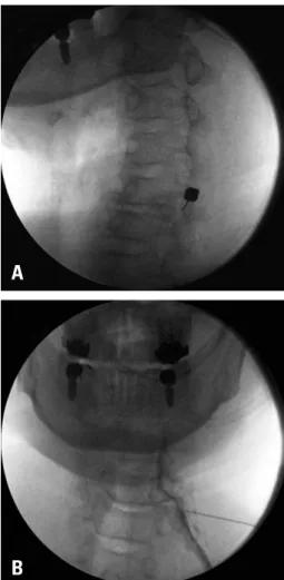
Fig. 3. C-arm images during a cervical transforaminal epidural block of left C6. (A) Oblique fluoroscopic view. The needle tip is positioned on the posterior end of the foramen. (B) Anteroposterior fluoroscopic view, showing the spread of the contrast medium along the nerve root and into the epidural space.
A
B
Fig. 4. Changes in arm pain VAS scores and NDI of both groups. (A)Arm pain VAS of both groups significantly decreased until 6 weeks post- injection in comparison to the pre-injection values, and then increased at 3 months post-injection. (B) The NDI in both groups improved at 6 weeks post-injection compared to pre-injection. VAS, visual analog scale; NDI, Neck Disability Index; N/S, normal saline).
A
B
CTEB with or without Steroid for CSR Journal of Korean Society of Spine Surgery
www.krspine.org 119 pre-injection was not significantly different between the two
groups. (Table 2) VAS was decreased considerably from pre- injection to 6 weeks post-injection, but increased significantly from 6 weeks post-injection to 12 weeks post-injection. Still, the VAS at 12 weeks post-injection was significantly lower compared to pre-injection in both groups. NDI showed substantial improvement in 6 weeks post-injection compared to pre-injection in both groups (Fig. 4B). Fortunately, no injection-related complication was experienced in all cases.
Discussion
Cervical radiculopathy is usually caused by acute soft disc herniation or chronic ‘hard disc’, which is a degenerative process. The degenerative process of disc bulging, hypertrophy of uncovertebral joint, and enlargement of the facet joint can cause narrowing of the foraminal space and pressure on the nerve root. While soft disc herniation generally has a benign natural course even without invasive procedures, CSR occurred
Table 1. Demographics and surgical data
Variables Group A (with steroid) (n=19) Group B (without steroid) (n=16) p-value1)
Age (years) 63.8 ± 9.7 64.0 ± 9.7 0.964
Gender Male 12 6
0.130
Female 7 10
CTEB level
C5 1 1
0.545
C6 9 10
C7 8 4
C8 1 0
Symptom duration (months) 14.6 ± 27.2 12.9 ± 2.0 0.698
Affected side Right 10 6
0.922
Left 9 10
Operation (no, %) 5 (26.3) 1 (6.3) 0.187
Follow-up (months) 5.3 ± 4.3 4.0 ± 2.2 0.756
CTEB: cervical transforaminal epidural block.
*Mann-Whitney test and Fisher’s exact test were used for statistical analysis.
†p-value below 0.05 was considered statistically significant.
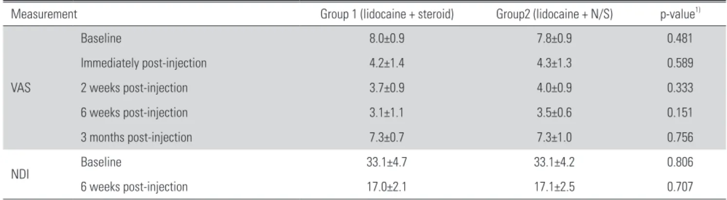
Table 2. Comparison of perioperative clinical outcomes of both groups
Measurement Group 1 (lidocaine + steroid) Group2 (lidocaine + N/S) p-value1)
VAS
Baseline 8.0±0.9 7.8±0.9 0.481
Immediately post-injection 4.2±1.4 4.3±1.3 0.589
2 weeks post-injection 3.7±0.9 4.0±0.9 0.333
6 weeks post-injection 3.1±1.1 3.5±0.6 0.151
3 months post-injection 7.3±0.7 7.3±1.0 0.756
NDI Baseline 33.1±4.7 33.1±4.2 0.806
6 weeks post-injection 17.0±2.1 17.1±2.5 0.707
VAS: Visual Analog Scale, NDI: Neck Disability Index.
*Mann-Whitney test was used for statistical analysis.
†p-value below 0.05 was considered statistically significant.
by the degenerative process is a more chronic pathology with an ambiguous natural history. Our studies show that the clinical outcome of CTEB for CSR was improved until 6 weeks but aggravated at 3 months with similar results regardless of the use of steroids.
Carette et al. performed interlaminar epidural steroid injections using methylprednisolone or isotonic saline for sciatica due to lumbar herniated nucleus pulposus.
Methylprednisolone was superior on the 6 weeks after injection in terms of leg pain. However, the other outcome measures at 3 weeks, 6 weeks, 3 months, and 12 months after injection showed no difference. They concluded that using epidural steroids does not offer significant benefits.25) Another randomized controlled study showed that using steroids in CTEB for cervical radiculopathy showed no significant difference in clinical outcomes. In addition, the use of steroids did not show superior short-term results compared to not using it. However, due to the heterogenic disease entity of cervical radiculopathy, including soft disc herniation, which majority is known to be self-limiting, the exact effect of CTEB is inconclusive.11) The operation rate of previous studies regarding CTEB ranged from 30-75%.13,14)
In general, CTEB is considered a safe technique in experienced hands.12,25) The method developed by Larkin et al. might be safe and useful in performing a cervical nerve block.26) However, there have been case reports of serious complications even with correct needle placement and contrast spread, such as headache, transient neurologic deficit, anterior cord syndrome, and even death. Possible mechanisms include injury of the vertebral artery or injection through the radicular artery.12,15,27-29) Ma et al. suggested that placing the needle as posterior as possible to the lateral mass on the lateral fluoroscopic view to avoid complications.12) Using non- particulate steroids such as dexamethasone, short-acting local anesthetics, and blunt needles were recommended to avoid complications related to CTEB.19,30) Woo et al. reported that using a low concentration of local anesthetics below the seizure threshold can achieve a similar clinical outcome as the 1-2% of lidocaine with less complication.31) Since advertent intra-arterial injection of lidocaine could cause a seizure attack, CTEB should be carefully performed even without steroids.30,32) Jee et al. reported the usefulness of ultrasound- guided CTEB. Although fluoroscopic-guided or ultrasound-
guided CTEB both did not show a significant difference in terms of inadvertent intravascular injections, the ultrasound image revealed vessels in the pathway of routine needle placement under fluoroscopic-guidance. They concluded that ultrasound might help identify atypical vessels during CTEB.33) In our cases, since no cases experienced injection-related complications in both groups, we could not conclude the result of safety issues using steroids.
A systematic review suggested that about half of the patients undergoing CTEB show pain improvement for up to 3 months.34) In our study, CTEB for CSR improved clinical outcomes significantly until 6 weeks post-injection but aggravated at 3 months post-injection. We used lidocaine with or without steroids as the injectate of CTEB and compared the two groups. Both groups showed clinical improvement in arm pain VAS and NDI until 6 weeks. However, unlike the systematic review, the arm pain VAS of our cases was aggravated 3 months post-injection. Interestingly, the two groups did not show a significant difference at all checkpoints.
Our results support that performing CTEB without steroids could achieve similar clinical outcomes compared to CTEB with steroids.
We presume that such pain improvement by local anesthetics, lidocaine, alone may be achieved by the anti- inflammatory effect. Inflammatory change is a physiologic process responding to injury and is divided into acute and chronic. Local anesthetics reversibly block nerve conduction through the sodium channel and show analgesic effect. It also decreases acute inflammatory reaction by vasoconstrictor effect, preservation of endothelial barrier function, and inhibit release and activity of cytokine. However, the detailed mechanisms of these effects are poorly understood.35) In addition, calcium- signaling G protein-coupled receptors (GPCRs) have been reported as the target for lidocaine. Epidural injection of lidocaine can elevate its plasma level enough to inhibit calcium- signaling GPCR. Such action of lidocaine is assumed to be responsible for not only acute but also chronic pain relief.36) The study by Yabuki et al. concluded that lidocaine itself has an effect of reducing inflammation. Exposure of the lidocaine of the inflamed cauda equina showed decreased inflammatory reaction both grossly and histologically, and increased conduction velocity in lidocaine applied group compared to the control group.21)
CTEB with or without Steroid for CSR Journal of Korean Society of Spine Surgery
www.krspine.org 121 There are limitations to our study. First, this was not a
prospective study with small sample size. Thus, it is difficult to generalize the results of our study. Second, the exact mechanism of using local anesthetics, lidocaine, which we used in this study, alone for the pain improvement, was not histologically evaluated. Future studies comparing the histological changes of the inflamed nerve root could be useful. Finally, the follow- up period was relatively short to generate significant results in terms of operation rate. However, our study’s strength is that this is the first study that compares the clinical effect of steroids for CTEB only for CSR by excluding soft disc herniation cases.
Conclusions
CTEB with lidocaine without steroid for CSR improved early clinical symptoms. The degree of improvement was similar to CTEB using lidocaine with steroids in terms of VAS and NDI.
Although long-term studies with larger samples should be performed, considering the clinical effect and complications of steroids, we suggest using local anesthetics alone for CTEB.
Acknowledgment
We appreciate Dr. Young-Soo Jung and Dr. Jin-An Jang for the data collection and organization of the raw data of this study.
REFERENCES
1. Abbed KM, Coumans JV. Cervical radiculopathy: patho- physiology, presentation, and clinical evaluation. Neurosur- gery. 2007 Jan;60(1 Supp1 1):S28-34. DOI: 10.1227/01.
NEU.0000249223.51871.C2.
2. Carette S, Fehlings MG. Clinical practice. Cervical radicu- lopathy. N Engl J Med. 2005 Jul 28;353(4):392-9. DOI:
10.1056/NEJMcp043887.
3. Lees F, Turner JA. Natural history and prognosis of cer- vical spondylosis. British medical journal. 1963 Dec 28;2(5373):1607-10. DOI: 10.1136/bmj.2.5373.1607.
4. Huston CW. Cervical epidural steroid injections in the man- agement of cervical radiculitis: interlaminar versus trans- foraminal. A review. Curr Rev Musculoskelet Med. 2009 Mar;2(1):30-42. DOI: 10.1007/s12178-008-9041-4.
5. Cicala RS, Westbrook L, Angel JJ. Side effects and compli- cations of cervical epidural steroid injections. J Pain Symp- tom Manage. 1989 Jun;4(2):64-6. DOI: 10.1016/0885- 3924(89)90024-9.
6. Waldman SD. Cervical epidural abscess after cervical epidural nerve block with steroids. Anesth Analg. 1991 May;72(5):717-8. DOI: 10.1213/00000539-199105000- 00033.
7. Stoll A, Sanchez M, Pawl RP. Epidural hematoma after epidural block: Implications for its use in pain management.
Surgical Neurology. 2002 Apr;57(4):235-40. DOI: Pii S0090-3019(02)00639-0
8. Johnson BA, Schellhas KP, Pollei SR. Epidurography and therapeutic epidural injections: technical considerations and experience with 5334 cases. AJNR Am J Neuroradiol. 1999 Apr;20(4):697-705.
9. Stojanovic MP, Vu TN, Caneris O, et al. The role of fluo- roscopy in cervical epidural steroid injections: an analysis of contrast dispersal patterns. Spine (Phila Pa 1976). 2002 Mar 1;27(5):509-14. DOI: 10.1097/00007632-200203010- 00011.
10. Hogan QH. Epidural anatomy examined by cryomicro- tome section. Influence of age, vertebral level, and disease.
Reg Anesth. 1996 Sep-Oct;21(5):395-406.
11. Anderberg L, Annertz M, Persson L, et al. Transforaminal steroid injections for the treatment of cervical radiculopa- thy: a prospective and randomised study. Eur Spine J. 2007 Mar;16(3):321-8. DOI: 10.1007/s00586-006-0142-8.
12. Ma III DJ, Gilula LA, Riew KD. Complications of fluoro- scopically guided extraforaminal cervical nerve blocks: an analysis of 1036 injections. JBJS. 2005 May;87(5):1025-30.
DOI: 10.2106/JBJS.D.02139.
13. Slipman CW, Lipetz JS, Jackson HB, et al. Therapeutic selective nerve root block in the nonsurgical treatment of atraumatic cervical spondylotic radicular pain: a retrospec- tive analysis with independent clinical review. Arch Phys Med Rehabil. 2000 Jun;81(6):741-6. DOI: 10.1016/
s0003-9993(00)90104-7.
14. Kolstad F, Leivseth G, Nygaard OP. Transforaminal ste- roid injections in the treatment of cervical radiculopathy. A prospective outcome study. Acta Neurochir (Wien). 2005 Oct;147(10):1065-70; discussion 70. DOI: 10.1007/
s00701-005-0542-2.
15. Baker R, Dreyfuss P, Mercer S, et al. Cervical transforaminal injection of corticosteroids into a radicular artery: a possible mechanism for spinal cord injury. Pain. 2003 May;103(1- 2):211-5. DOI: Pii S0304-3959(02)00343-3
16. Rhee JM, Yoon T, Riew KD. Cervical radiculopathy. J Am Acad Orthop Surg. 2007 Aug;15(8):486-94. DOI:
10.5435/00124635-200708000-00005.
17. Abdi S, Datta S, Lucas LF. Role of epidural steroids in the management of chronic spinal pain: a systematic review of effectiveness and complications. Pain Physician. 2005 Jan;8(1):127-43.
18. Dreyfuss P, Baker R, Bogduk N. Comparative effectiveness of cervical transforaminal injections with particulate and nonparticulate corticosteroid preparations for cervical ra- dicular pain. Pain Med. 2006 May-Jun;7(3):237-42. DOI:
10.1111/j.1526-4637.2006.00162.x.
19. Scanlon GC, Moeller-Bertram T, Romanowsky SM, et al.
Cervical transforaminal epidural steroid injections: more dangerous than we think? Spine. 2007 May 15;32(11):1249- 56. DOI: 10.1097/BRS.0b013e318053ec50.
20. Hwang B, Lee J, Park B. Neurological Complication Rates of Epidural Injections and Selective Nerve Blocks:
A Comparison of Steroid Use Patterns. The Clinical Jour- nal of Pain. 2020 Jun;36(6):449-457. DOI: 10.1097/
AJP.0000000000000816.
21. Yabuki S, Kawaguchi Y, Nordborg C, et al. Effects of li- docaine on nucleus pulposus-induced nerve root injury.
A neurophysiologic and histologic study of the pig cauda equina. Spine (Phila Pa 1976). 1998 Nov 15;23(22):2383- 9; discussion 9-90. DOI: 10.1097/00007632-199811150- 00004.
22. Saal JS, Saal JA, Yurth EF. Nonoperative management of herniated cervical intervertebral disc with radiculopathy.
Spine (Phila Pa 1976). 1996 Aug 15;21(16):1877-83. DOI:
10.1097/00007632-199608150-00008.
23. Park HJ, Kim SS, Lee SY, et al. A practical MRI grad- ing system for cervical foraminal stenosis based on oblique sagittal images. Br J Radiol. 2013 May;86(1025):20120515.
DOI: 10.1259/bjr.20120515.
24. Carette S, Leclaire R, Marcoux S, et al. Epidural cor- ticosteroid injections for sciatica due to herniated nucleus pulposus. New England Journal of Medicine.
1997 Jun 5;336(23):1634-40. DOI: Doi 10.1056/
Nejm199706053362303.
25. Derby R, Lee SH, Kim BJ, et al. Complications following cervical epidural steroid injections by expert interventional- ists in 2003. Pain Physician. 2004 Oct;7(4):445-9.
26. Larkin TM, Carragee E, Cohen S. A novel technique for delivery of epidural steroids and diagnosing the level of nerve root pathology. J Spinal Disord Tech. 2003 Apr;16(2):186- 92. DOI: 10.1097/00024720-200304000-00011.
27. Brouwers PJ, Kottink EJ, Simon MA, et al. A cervical ante- rior spinal artery syndrome after diagnostic blockade of the right C6-nerve root. Pain. 2001 Apr;91(3):397-9. DOI:
10.1016/s0304-3959(00)00437-1.
28. Rozin L, Rozin R, Koehler SA, et al. Death during trans- foraminal epidural steroid nerve root block (C7) due to perforation of the left vertebral artery. American Journal of Forensic Medicine and Pathology. 2003 Dec;24(4):351-5.
DOI: 10.1097/01.paf.0000097790.45455.45.
29. Karasek M, Bogduk N. Temporary neurologic deficit af- ter cervical transforaminal injection of local anesthetic.
Pain Med. 2004 Jun;5(2):202-5. DOI: 10.1111/j.1526- 4637.2004.04028.x.
30. Schellhas KP, Pollei SR, Johnson BA, et al. Selective cervi- cal nerve root blockade: Experience with a safe and reliable technique using an anterolateral approach for needle place- ment. American Journal of Neuroradiology. 2007 Nov- Dec;28(10):1909-14. DOI: 10.3174/ajnr.A0707.
31. Woo JH, Park HS. Cervical Transforaminal Epidural Block Using Low-Dose Local Anesthetic: A Prospective, Randomized, Double-Blind Study. Pain Medicine. 2015 Jan;16(1):61-7. DOI: 10.1111/pme.12582.
32. Chung SG. Convulsion caused by a lidocaine test in cervi- cal transforaminal epidural steroid injection. PM R. 2011 Jul;3(7):674-7. DOI: 10.1016/j.pmrj.2011.02.005.
33. Jee H, Lee JH, Kim J, et al. Ultrasound-guided selective nerve root block versus fluoroscopy-guided transforaminal block for the treatment of radicular pain in the lower cervi- cal spine: a randomized, blinded, controlled study. Skeletal Radiol. 2013 Jan;42(1):69-78. DOI: 10.1007/s00256- 012-1434-1.
34. Conger A, Cushman DM, Speckman RA, et al. The Effec- tiveness of Fluoroscopically Guided Cervical Transforaminal Epidural Steroid Injection for the Treatment of Radicular Pain; a Systematic Review and Meta-analysis. Pain Med.
CTEB with or without Steroid for CSR Journal of Korean Society of Spine Surgery
www.krspine.org 123 2020 Jan 1;21(1):41-54. DOI: 10.1093/pm/pnz127.
35. Swanton BJ, Shorten GD. Anti-inflammatory effects of local anesthetic agents. Int Anesthesiol Clin. 2003 Win- ter;41(1):1-19. DOI: 10.1097/00004311-200341010- 00003.
36. Amir R, Argoff CE, Bennett GJ, et al. The role of sodium channels in chronic inflammatory and neuropathic pain.
J Pain. 2006 May;7(5 Suppl 3):S1-29. DOI: 10.1016/
j.jpain.2006.01.444.
경추 퇴행성 신경근증의 치료로 시행하는 경추 경추간공 경막외 신경 차단술에 있어서 스테로이드 사용 유 무에 따른 임상 결과
박유진재진 • 김성민 • 정승호 • 민우기 경북대학교 의과대학 정형외과학교실
연구 계획: 후향적 의무 기록 연구
목적: 경추 퇴행성 신경근증에 대해 국소 마취제를 이용한 경추 경추간공 경막외 신경 차단술을 시행하는 데 있어서 스테로이드 혼용 유무에 따른 임상 결과의 비교 분석
선행 연구문헌의 요약: 일반적인 경추 추간공 경막외 신경 차단술에 이용되는 약제는 국소 마취제와 비입자성 스테로이드이다. 하지만, 스테로이드는 이 차성 골다공증, 시상하부-뇌하수체-부신 축 억제 및 고혈당증 등의 잠재적 부작용을 가지고 있다.
대상 및 방법: 2018년 1월부터 2019년 10월까지 경추 퇴행성 신경근증에 대해서 경추 경추간공 경막외 차단술을 시행받은 35예를 대상으로 연구를 진 행하였다. 상지의 통증이 VAS 4점 초과인 환자들만 포함하였다. 첫 19예는 1% 리도카인 1 ml와 덱사메타손 1 ml를 혼용하였고(A군), 이후의 16예는 1%
리도카인 1 ml와 1 ml 생리 식염수를 혼용하였다(B군). 상지의 VAS (Visual Analog Scale) 점수와 Neck Disability Index (NDI)를 조사하였다.
결과: 기본 환자 정보는 두 군 사이에 차이가 없었다. VAS 점수는 두 군 모두에서 차단술 30분 후, 2주 후, 6주 후에서 술전에 비해 유의미한 감소를 보였 다. 하지만, 두 군 모두 12주 후에는 6주 후에 비해 악화되었다. 차단술 6주 이후 NDI는 술전보다 유의미하게 호전되었다. 차단술 30분 후, 2주 후, 6주 후 의 VAS 점수와 NDI는 두 군 사이에 유의미한 차이가 관찰되지 않았다.
결론: 경추 경추간공 경막외 차단술을 경추 퇴행성 신경근증에 시행한 경우 6주까지 호전시켰다. 하지만, 스테로이드 사용 유무는 VAS 점수와 NDI 등의 임상적 결과에 영향을 끼치지 못하였다.
색인 단어: 신경 차단술, 경추, 덱사메타손, 리도카인
약칭 제목: 경추 경추간공 신경 차단술 시 스테로이드 사용 유무의 임상적 차이
접수일: 2020년 9월 3일 수정일: 2020년 9월 9일 게재확정일: 2020년 11월 3일 교신저자: 민우기
대구광역시 중구 동덕로 130 경북대학교병원 정형외과학교실
TEL: 053-200-5638 FAX: 053-422-6605 E-mail: [email protected]