Vestibular Neuritis versus Stroke
Ji-Soo Kim, MD, PhD
1,21
Department of Neurology, Seoul National University College of Medicine, Seoul;
2Dizziness Center, Clinical Neuroscience Center, Department of Neurology, Seoul National University Bundang Hospital, Seongnam, Korea
Both vestibular neuritis (VN) and stroke may present acute prolonged spontaneous vertigo. VN is ascribed to acute unilateral loss of vestibular function, probably due to reactivation of Herpes simplex virus in the vestibular ganglia. The diagnostic hallmarks of VN are spontaneous horizontal-torsional nystagmus beating away from the lesion side, abnormal head impulse test for the in- volved semicircular canals, ipsilesional caloric paresis, decreased responses of vestibular-evoked myogenic potentials during stim- ulation of the affected ear, and unsteadiness with a falling tendency toward the lesion side. Differentiation of stroke is important due to its potential grave prognosis and different treatment modalities. The findings of bedside ocular motor examination, such as HINTS (negative head impulse tests, direction-changing nystagmus, and skew deviation) are more sensitive than brain imaging for detection of strokes in patients acute prolonged (≥ 24 hours) spontaneous vertigo. Even in patients with the typical patterns of spontaneous nystagmus observed in VN, brain imaging is indicated when the patients had unprecedented headache, positive HINTS, severe unsteadiness, or no recovery within 1-2 days.
Keywords: Vestibular neuritis; Stroke; Vertigo; Nystagmus; Head impulse test
Vestibular neuritis (VN) refers to a disorder characterized by acute, isolated, spontaneous vertigo due to unilateral vestibular de-afferentia- tion.
1,2VN accounts for 3.2-9% of the patients visiting a dizziness unit, and has an incidence of ~3.5 per 100,000 population. Characteristic symptoms of acute VN include vertigo, nausea/vomiting, oscillopsia, and unsteadiness.
3Patients with VN mostly present with subacute or acute spontaneous vertigo with nausea/vomiting, and unsteadiness. The symptoms may develop suddenly or evolve over several hours. Prodro- mal dizziness may be reported in 8.6-24% of the patients. Most (74%) of these preceding episodes are described as non-vertiginous dizziness, of- ten associated with a feeling of nausea or unsteadiness. The episodes may have abrupt or gradual onset, and may last a few minutes to days. Patients may have precedent or concurrent viral illnesses.
The vertigo in VN may gradually increase over several hours and reach a peak within the first day. Vertigo is usually described as rotation- al, and is markedly increased by head movements. Patients usually prefer to lie in bed with their eyes closed in a side position with the healthy ear
down. Most patients suffer from severe nausea and vomiting. Severe ver- tigo improves markedly over a day or two.
Since stimulation of single semicircular canal induces eye movements approximately in the plane of that canal, the patterns of spontaneous nystagmus in VN would depend on relative involvement of each semi- circular canal. Accordingly, when all three canals are completely dam- aged, the nystagmus is mixed horizontal-torsional and the fast compo- nent of the spontaneous nystagmus is away from the affected side. How- ever, the spontaneous nystagmus invariably accompanies a vertical com- ponent that is mostly upbeat since the anterior semicircular canal (AC) is more commonly affected than posterior semicircular canal (PC). As the nystagmus is markedly suppressed by visual fixation, proper observation of the nystagmus requires a removal of visual fixation using Frenzel glasses or infrared video goggles. The nystagmus increases during the gaze in the direction of the nystagmus and decreases when looking into the other direction (Alexander’s law), but never change directions. The nystagmus is mostly augmented by horizontal head shaking, vibratory
Correspondence to: Ji-Soo Kim, MD, PhD
Department of Neurology, Seoul National University Bundang Hospital, 173-82 Gumi-ro, Bundang-gu, Seongnam 13620, Korea Tel: +82-31-787-7463, E-mail: jisookim@snu.ac.kr
Received: Apr. 5, 2019 / Accepted: Apr. 30, 2019
REVIEW
stimuli on the mastoids or the brow, or hyperventilation.
The spontaneous nystagmus gives rise to illusion of apparent move- ment of the environment (oscillopsia). Since the illusory visual percep- tion of rotation is produced by the slow phase of the nystagmus in the op- posite direction, right beating spontaneous nystagmus in left VN induc- es contraversive rightward (clockwise) rotation of the surroundings.
Patients tend to fall toward the affected side on standing with the feet together or on attempted walking. However, they should maintain sit- ting balance or stand unaided with the feet apart since the brain can still utilize the information for balance control via the visual and somatosen- sory systems.
The vestibular labyrinth may be subdivided into the superior and in- ferior divisions (Fig. 1). The superior vestibular labyrinth comprises AC and horizontal semicircular canals (HC), and the utricle, and their affer- ents. In contrast, the inferior vestibular labyrinth consists of PC and sac- cule, and their afferents. With the aids of head impulse test (HIT) that enables us to evaluate the function of each semicircular canal,
4and ves-
tibular evoked myogenic potentials (VEMPs) that reflect the function of the utricle and saccule,
5we are now able to securely diagnose the 3 distinc- tive patterns of VN, the superior, inferior, and total (superior+inferior) types. Since VN preferentially affect the superior division, superior VN is most common (55-100%), followed by total (15-30%) and inferior VN (3.7-15%).
6During the acute phase, the spontaneous nystagmus may be seen even with visual fixation, but better be appreciated with removal of visual fixa- tion. Nystagmus can be recorded and quantitatively analyzed using ocu- lography. Three-dimensional video-oculography is commonly used, which can measure the direction and slow phase velocity of spontaneous nystagmus. Nystagmus may be induced or modulated by various ma- neuvers such as head-shaking, vibration, or hyperventilation.
HIT can evaluate the function of each semicircular canal bedside.
4Bedside HIT is feasible, and the sensitivity is clinically acceptable. How- ever, bedside HIT may be negative, especially when the vestibular deficits are partial, or the corrective saccades are covert, i.e. the corrective sac- Fig. 1. Divisional configuration of the labyrinth and 3 distinctive types of vestibular neuritis (VN). (A) The vestibular labyrinth may be subdivided into the superior and
inferior divisions. The superior vestibular labyrinth comprises the anterior (AC) and horizontal semicircular canals (HC), and the utricle, and their afferents. In contrast, the inferior vestibular labyrinth consists of the posterior semicircular canal (PC) and saccule, and their afferents. (B-D) According to the divisions involved, VN may be classified into distinctive types, the superior (B), inferior (C), and total (D).A
B
C
D
cades occur only during head impulse rather than after the head rota- tion. Furthermore, bedside HITs in the planes of the vertical canals are more difficult to interpret than the horizontal HIT. In such cases, the quantitative HIT using video oculography would aid in detecting the deficits of the vestibulo-ocular reflex (VOR).
7HIT has been suggested a predictor of symptom recovery in a previous study, in which 80% of pa- tients with persistent dizziness had a positive HIT while only 10% of pa- tients without dizziness showed a positive HIT.
Unilateral caloric paresis has been the diagnostic hallmark of VN.
However, caloric test can only evaluate the function of HC only in the lower frequency range (~ 0.003 Hz), and would be normal in VN sparing HC, likewise in inferior VN.
2Evaluation for the OTR should include measurements of head tilt, skew deviation, and ocular torsion. Ocular torsion can be determined on fundus photos. The sensory manifestation of ocular torsion can be mea- sured as a deviation of the subjective visual vertical (SVV). The SVV tilt can be determined bedside using a simple, self-made bucket, or be mea- sured using a computerized program in the laboratory.
Even though the origins of cervical and ocular VEMPs remain con- troversial, cervical and ocular VEMPs appear to be valuable tools in eval- uating the otolithic function in VN.
8Furthermore, the dissociated pat- terns of abnormalities in cervical and ocular VEMPs may provide im- portant clues for determining the involved vestibular division in VN. In patients with VN and intact cervical VEMP evoked by air-conducted sounds (ACS), the severity of ACS-induced ocular VEMP abnormality correlated with HC dysfunction on caloric or head impulse testing. In a recent study, patients with superior VN showed abnormal ocular VEMP and normal cervical VEMP in response to ACS. In contrast, patients with inferior VN have normal ocular VEMP and abnormal cervical VEMP induced by ACS.
8This study supports the use of ocular and cer- vical VEMPs in determining each subtype of VN.
9Rotatory chair test has a limited diagnostic value in VN since whole body rotation modulates the activities of both labyrinths simultaneously, which renders evaluation of unilateral vestibular dysfunction difficult.
The horizontal VOR elicited by sinusoidal rotation is asymmetrical, and the VOR gain is decreased during velocity step rotation toward the af- fected ear, especially during the acute phase. In contrast, the VOR gain during velocity step rotation toward the contralateral ear remains un- changed. The VOR time constants are also decreased during the acute phase in both directions.
In VN, direct visualization of the affected vestibular nerve has been reported with gadolinium-enhanced 3T MRI. However, imaging the af- fected nerve is of more academic interest than of practical utility in VN.
In VN, neuroimaging is mostly indicated when a central cause is sus- pected.
Since no confirmatory diagnostic tests are available, VN is primarily a diagnosis of exclusion. Diagnosis of VN is mostly based on constellation of the bedside and laboratory findings (Table 1). Even though the detailed findings may differ according to the vestibular divisions affected, the key symptoms and signs of VN generally include acute onset of isolated sus- tained vertigo with nausea/vomiting, unidirectional horizontal-torsion- al nystagmus beating away from the lesion side, impaired semicircular function documented by head impulse or caloric tests, ipsiversive OTR and SVV tilt, decreased or abolished VEMP responses during stimula- tion of the affected ear, and unsteadiness with a falling tendency to the lesion side. Recent development of HIT and VEMPs has enabled us to evaluate the function all three semicircular canals, utricle, and saccule, and to securely define the subtypes of VN (Table 2).
Table 1. Clinical features of vestibular neuritis
• Subacute or acute onset of spontaneous vertigo with nausea/vomiting.
• Oscillopsia: spinning of surroundings in the direction of nystagmus quick phase.
• Horizontal-torsional spontaneous nystagmus beating toward the unaffected side.
• Impaired function of the semicircular canals as revealed by head impulse tests or caloric testing.
• Ipsiversive ocular tilt reaction (head tilt, skew deviation, and ocular torsion) and ipsiversive tilt of the subjective visual vertical/horizontal.
• Decreased or absent responses of vestibular-evoked myogenic potentials during stimulation of the affected ear.
• Falling tendency toward the lesion side.
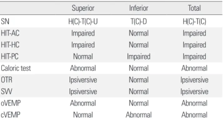
Table 2. Comparison of the findings among the subtypes of vestibular neuritis
Superior Inferior Total
SN H(C)-T(C)-U T(C)-D H(C)-T(C)
HIT-AC Impaired Normal Impaired
HIT-HC Impaired Normal Impaired
HIT-PC Normal Impaired Impaired
Caloric test Abnormal Normal Abnormal
OTR Ipsiversive Normal Ipsiversive
SVV Ipsiversive Normal Ipsiversive
oVEMP Abnormal Normal Abnormal
cVEMP Normal Abnormal Abnormal
AC, anterior semicircular canal; C, contraversive (quick phase); cVEMP, cervical ves-
tibular-evoked myogenic potential; D, downbeat; H, horizontal; HC, horizontal semi-
circular canal; HIT, head impulse test; OTR, ocular tilt reaction; oVEMP, ocular vestib-
ular-evoked myogenic potential; PC, posterior semicircular canal; SN, spontaneous
nystagmus; T, torsional; U, upbeat; SVV, subjective visual vertical.
Superior VN
Superior VN is the most common type. The spontaneous nystagmus is mostly contraversive horizontal-torsional and upbeating (Fig. 2A). In- deed, three-dimensional recording of eye movements in VN showed that the rotational axes of spontaneous nystagmus clustered along the axis of HC or between the axes of HC and AC (Fig. 2B). Patients also show posi- tive HIT for the involved AC and HC. Laboratory findings include ip- siversive ocular torsion and tilt of the SVV, ipsilesional caloric paresis, and abnormal ocular VEMP with preservation of hearing and cervical VEMP (Fig. 2C-G, Table 2). HIT may be quantified using a video-based equipment.
Inferior VN
VN rarely involves the inferior division only.
9Diagnosis of inferior VN is challenging since the usual signs of VN are absent in this disorder.
As a result, isolated inferior VN may erroneously be ascribed to a central pathology if there is no scrutinized evaluation for the inferior vestibular function. The spontaneous nystagmus is contraversive torsional and downbeat (Fig. 3A). The rotation axis of the spontaneous nystagmus is best aligned with that of the involved PC (Fig. 3B). Other findings include positive HIT only for the affected PC and abnormal cervical VEMP in response to ACD in the presence of normally functioning HC and AC, as determined by normal HIT and caloric test (Table 2). Ocular torsion, SVV, and ocular VEMP are mostly within the normal range (Fig. 3C-G).
Some patients may have tinnitus and hearing loss in the involved side.
Total VN
The spontaneous nystagmus is mostly torsional-horizontal beating away from the affected ear. Otherwise, the patients with total VN would show all the abnormalities observed in superior and inferior VN (Table 2).
Severe vertigo and static vestibular imbalance markedly improve over a few days in most patients with a gradual resolution over the following weeks. Improvement of the initial symptoms occurs by virtue of central compensation for the vestibular tone imbalance rather than by recovery of the function in the affected ear. One of the of central compensation signs is the reduction of spontaneous nystagmus, which usually takes 3 weeks to subside. Neural recordings in animals recovering from unilat- eral vestibular deafferentation show that central compensation depends on restoration of normal spontaneous activity in the ipsilesional vestibu- lar nucleus, thereby rebalancing the activity between the left and right
vestibular nuclei. Later, when the vestibular function is restored in the damaged ear, mild spontaneous nystagmus may beat in the opposite di- rection (recovery nystagmus). While the static symptoms invariably re- solve, albeit often not totally, the dynamic symptoms may last longer or persist. In a previous study, the static vestibular imbalances (spontaneous nystagmus, ipsilesional SVV tilt, and ocular torsion) had mostly resolved by 3 months after symptoms onset, whereas the signs of dynamic vestib- ular imbalances (HIT, head-shaking nystagmus, vibration-induced nys- tagmus, and caloric test) can still be observed in more than 30% of the patients at one year from the symptom onset.
10Tests of the VOR function using a rotary chair yielded similar results. When tested 3-5 days after the onset of VN, the responses to low acceleration whole body rotation in the lesion side was decreased to 50% and, in the healthy side, to 75% of nor- mal controls. Of note, the VOR responses to rotations in the healthy side were also decreased, which may be ascribed to central compensation to improve the response asymmetry. The VOR responses to the rotation in the affected side increase with time. This improvement may result from either central compensation or periphery recovery, observed as improve- ment in caloric responses by an average of 30%. When tested 3 months later, the majority of patients show symmetrical responses with an in- crease of the responses during rotation toward the involved side. This pattern is similar, but less pronounced in the pitch plane. In view of more rapid resolution of the static vestibular imbalances after VN, evaluation of the dynamic vestibular imbalance may provide more useful informa- tion for underlying vestibulopathy, especially in the compensated phase.
In a few patients with limited restoration of the labyrinthine function, oscillopsia may persist during rapid head movements. This is caused by a persisting deficit of the VOR in higher frequency range, which cannot be compensated for centrally. The persistent loss of balance that some pa- tients experience after acute VN may be due to inadequate central com- pensation or due to incomplete peripheral recovery. Vestibular rehabili- tation has a role in treating both.
VN is known to recur only in 2-11%. In 10-15% of patients with VN, a typical BPPV develops in the affected ear within a few weeks, which sug- gests that the otoconia may be loosened due to inflammation of the laby- rinth. The second most important complication is phobic postural verti- go, which refers to persistent dizziness and unsteadiness associated with fear of falling without any real falls or vestibular dysfunction that can ex- plain the symptoms.
Although VN itself is not life threatening, distinguishing VN from
Fig. 2. Clinical features of superior vestibular neuritis (VN). (A) The patient with right VN involving the superior division shows left beat, upbeat, and counterclockwise
(from the patient’s perspective) torsional nystagmus. (B) The rotational axes of the spontaneous nystagmus cluster between those of anterior and horizontal semicircu- lar canal. (C) Rightward ocular torsion is noted on fundus photos (normal range: 0°-12.6°, Negative values indicate intorsion.). (D-G) Patients also shows right caloric paresis (E) and decreased ampulitude of ocular vestibular-evoked myogenic potential (VEMP) during right ear stimulation with air-conducted sounds (ACS) in the pres- ence of normal hearing (D) and cervical VEMP to ACS. LH, horizontal position of the left eye; LV, vertical position of the left eye; LT, torsional position of the left eye;RHC, right horizontal semicircular canal; RAC, right anterior semicircular canal; RPC, right posterior semicircular canal semicircular canal.
A B
C
D
F
E
G
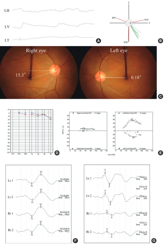
Fig. 3. Clinical features of inferior vestibular neuritis (VN). (A) The patient with right inferior VN shows counterclockwise (from the patient’s perspective) torsional and
downbeat nystagmus. (B) The rotational axes of the spontaneous nystagmus cluster around that of posterior semicircular canal. (C-G) The patient shows no wave for- mation of right cervical vestibular-evoked myogenic potential (VEMP) in response to air-conducted sounds (F) while the findings of fundus photos (C), audiometry (D), bithermal caloric tests (E), and ocular VEMP induced by ACS are normal. LH, horizontal position of the left eye; LV, vertical position of the left eye; LT, torsional position of the left eye; RH, horizontal position of the right eye; RV, vertical position of the right eye; RT, torsional position of the right eye; RHC, right horizontal semicircular ca- nal; RAC, right anterior semicircular canal; RPC, right posterior semicircular canal semicircular canal.A B
C
D
F
E
G
other debilitating disorders such as strokes is essential. Acute unilateral peripheral vestibulopathy may also be caused by vascular compromise of the peripheral vestibular labyrinth.
11,12Diagnosis of isolated labyrin- thine infarction remains a diagnostic challenge since current imaging techniques cannot detect it. However, isolated labyrinthine infarction is exceedingly rare, and usually accompanies cochlear damage and resul- tant hearing loss. Rarely, isolated labyrinthine infarction may progress to involve the brainstem or cerebellar territory supplied by the anterior in- ferior cerebellar artery (AICA). Vestibular pseudoneuritis has also been described in infarctions involving the vestibular nucleus or inferior cer- ebellum.
13Occasionally, serial evaluation is necessary since even the dif- fusion-weighted MRI may fail to detect small infarction involving the brainstem or cerebellum during the acute phase. Plaques of multiple sclerosis or lacunar infarctions involving the root entry zone of the eight nerve may mimic VN. From a clinical point of view, the first question to be answered in patients with acute vertigo and nystagmus is whether the symptoms are caused by VN or by central ‘vestibular pseudoneuritis’.
14It is not always easy to differentiate isolated vascular vertigo from acute pe- ripheral vestibulopathy at the bedside.
15However, a rather simple neu- rotological examination including normal horizontal head impulse test, direction-changing nystagmus, and skew deviation (HINTS) can reli- ably detect central vertigo with a high sensitivity and specificity.
16Even these bedside tests are more sensitive for stroke than early MRIs while maintaining a high specificity. Indeed, initial diffusion-weighted MRI may be false negative in 12-20% of the stroke patients within the first 48 hour. Since mild degree of skew deviation is hard to detect in the pres- ence of spontaneous nystagmus, and gaze-evoked nystagmus may be absent in cerebellar stroke, bedside HIT appears to be the best tool for differentiating isolated vascular vertigo from acute VN.
17,18However, since bedside HIT may be negative in patients with covert corrective sac- cades and may be inconclusive in patients with nystagmus, recording of HIT using a video-based equipment may be helpful in patients with equivocal results. Of course, positive HIT does not necessarily eliminate the possibility of central lesions. Since recurrence is rare in VN, an alter- native diagnosis should be considered whenever patients report more than one episode.
CONCLUSION
Careful history taking and focused neurological examination are usu-
ally enough for diagnosis of VN. With the aids of HIT and cervical and ocular VEMPs, each subtype of VN can be secured diagnosed as superi- or, inferior or total VN. Even though very rare, inferior VN should be considered in patients with acute spontaneous vertigo and torsional downbeat nystagmus. Imaging should be considered whenever there is any finding inconsistent with VN since it is a diagnosis of exclusion.
Management during the acute phase is primarily supportive while long- term treatment should be designed to improve vestibular compensation.
REFERENCES
1. Baloh RW, Kerber KA. Clinical neurophysiology of the vestibular system.
4th ed. New York: Oxford University Press; 2011.
2. Dix MR, Hallpike CS. The pathology, symptomatology and diagnosis of certain common disorders of the vestibular system. Ann Otol Rhinol Lar-
yngol 1952;61(4):987-1016.
3. Strupp M, Brandt T. Vestibular neuritis. Seminars in Neurology 2009;29(5):
509-519.
4. Halmagyi GM, Curthoys IS. A clinical sign of canal paresis. Arch Neurol 1988;45(7):737-739.
5. Rosengren SM, McAngus Todd NP, Colebatch JG. Vestibular-evoked ex- traocular potentials produced by stimulation with bone-conducted sound.
Clin Neurophysiol 2005;116(8):1938-1948.
6. Fetter M, Dichgans J. Vestibular neuritis spares the inferior division of the vestibular nerve. Brain 1996;119:755-763.
7. Aw ST, Fetter M, Cremer PD, Karlberg M, Halmagyi GM. Individual semi- circular canal function in superior and inferior vestibular neuritis. Neu-
rology 2001;57(5):768-774.
8. Shin BS, Oh SY, Kim JS, Kim TW, Suh MW, Lee H, et al. Cervical and oc- ular vestibular-evoked myogenic potentials in acute vestibular neuritis.
Clin Neurophysiol 2012;123(2):369-375.
9. Kim JS, Kim HJ. Inferior vestibular neuritis. J Neurol 2012;259(8):1553- 1560.
10. Choi KD, Oh SY, Kim HJ, Koo JW, Cho BM, Kim JS. Recovery of vestib- ular imbalances after vestibular neuritis. Laryngoscope 2007;117(7):1307- 1312.
11. Kim JS, Lee H. Inner ear dysfunction due to vertebrobasilar ischemic stroke.
Seminars in Neurology 2009;29(5):534-540.
12. Choi KD, Lee H, Kim JS. Vertigo in brainstem and cerebellar strokes. Curr
Opin Neurol 2013;26(1):90-95.
13. Lee H, Sohn SI, Cho YW, Lee SR, Ahn BH, Park BR, et al. Cerebellar in- farction presenting isolated vertigo: frequency and vascular topographi- cal patterns. Neurology 2006;67(7):1178-1183.
14. Hotson JR, Baloh RW. Acute vestibular syndrome. N Engl J Med 1998;
339(10):680-685.
15. Baloh RW. Clinical practice. Vestibular neuritis. N Engl J Med 2003;
348(11):1027-1032.
16. Kattah JC, Talkad AV, Wang DZ, Hsieh YH, Newman-Toker DE. HINTS to diagnose stroke in the acute vestibular syndrome: three-step bedside oculomotor examination more sensitive than early MRI diffusion-weight- ed imaging. Stroke 2009;40(11):3504-3510.
17. Newman-Toker DE, Kattah JC, Alvernia JE, Wang DZ. Normal head im- pulse test differentiates acute cerebellar strokes from vestibular neuritis.
Neurology 2008;70(24 Pt 2):2378-2385.
18. Cnyrim CD, Newman-Toker D, Karch C, Brandt T, Strupp M. Bedside differentiation of vestibular neuritis from central “vestibular pseudoneu- ritis”. J Neurol Neurosurg Psychiatry 2008;79(4):458-460.