• 검색 결과가 없습니다.

. These aggressive biological

N/A
N/A
Protected

Academic year: 2021

Share ". These aggressive biological "

Copied!
7
0
0

로드 중.... (전체 텍스트 보기)

전체 글

(1)

Ⅰ. INTRODUCTION

Biological characteristics such as rapid growth rate, invasive capability, and metastatic potential of cancer cells affect the aggressiveness of cancer and are major factors in determining the prognosis of cancer patients

9)

. These aggressive biological

characteristics are known to be promoted (strengthened) during the process of epithelial- mesenchymal transition (EMT), which is characterized by a decreased expression of epithelial markers and/or an increased expression of mesenchymal markers. Cells that have acquired a mesenchymal phenotype through EMT display resistance to chemotherapeutic and apoptotic reagents, as well as increased motility and invasiveness

13)

. Thus, EMT may be one of the most important events that contribute to aggravating the prognosis of cancer. EMT is not only intrinsically acquired through multiple genetic and epigenetic changes, but is also induced by

Korean Journal of Oral and Maxillofacial Pathology 2019;43(6):237-243 ISSN:1225-1577(Print); 2384-0900(Online) Available online at http://journal.kaomp.org https://doi.org/10.17779/KAOMP.2019.43.6.001

* Correspondence: Hae Ryoun Park, Department of Oral Pathology, School of Dentistry, Pusan National University, 49 Busandaehak-Ro, Yangsan-Si, Kyeongsangnam-Do 50612, South Korea

Tel: +82-51-510-8250, Fax: +82-51-510-8249 E-mail: parkhr@pusan.ac.kr

ORCID: 0000-0003-1908-0824

저산소성 조건에 노출된 구강암세포의 상피간엽전환관련 유전자 발현 프로파일

박대근, 김소라, 우복희, 이지혜, 박혜련*

부산대학교 치의학전문대학원 구강병리학교실, BK21 Plus, 치주질환신호네트워크연구센터

<Abstract>

Epithelial-Mesenchymal Transition-Related Gene-Expression Profile in Hypoxic Oral Cancer Cells

Dae-Gun Park, So Ra Kim, Bok Hee Woo, Ji Hye Lee, Hae Ryoun Park

*

Department of Oral Pathology, BK21 Plus Project, Periodontal Disease Signaling Network Research Center (MRC), School of Dentistry, Pusan National University, 49 Busandaehak-Ro, Yangsan 50612, South Korea

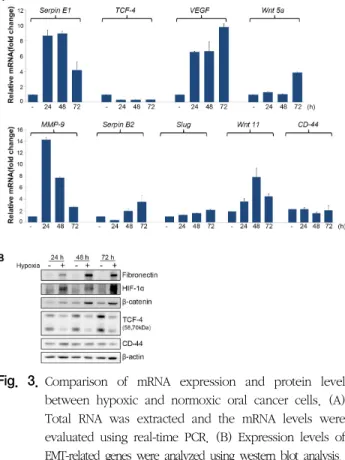
Hypoxia is one of the most common features of cancer. It is also associated with cancer progression and the acquisition of aggressiveness, which includes invasion and metastasis. Oral squamous cell carcinoma accounts for 90% of all oral cancers, and its 5-year survival rate is about 50%. Despite various attempts and trials, its prognosis has not improved. Among numerous adverse prognostic factors, hypoxia is suspected as one of the most important factors, as it increases the aggressiveness of oral cancer cells. We attempted to observe the effect of hypoxia on the expression of epithelial-mesenchymal transition markers in oral cancer cells. We analyzed and compared both the mRNA and protein expression levels of epithelial-mesenchymal markers using qRT-PCR and western blotting in both normoxic and hypoxic YD10B oral squamous cell carcinoma cells. Eighty-six genes were analyzed through real-time PCR using commercial microarray plates, performed in triplicate. Among the 86 genes, the expression of 24 were increased (≥ 2 fold) by hypoxia, while that of three genes was decreased (≥ 2 fold). Hypoxia significantly affects epithelial-mesenchymal transition-related genes. Further studies on the regulation of these genes may help to develop more efficient therapeutic modalities for oral cancer and to improve prognosis of oral cancer patients.

Key words : Hypoxia, Oral cancer, Epithelial-mesenchymal transition, TCF-4

(2)

non-genetic extrinsic factors derived from the tumor microenvironment, such as hypoxia

18)

.

Hypoxia is commonly observed in cancer tissues because the formation of blood vessels cannot keep up with the growth rate of anaplastic cancer cells, especially in highly malignant tumors. Though cancer malignancy is promoted by selective genetic mutations, growing data corroborate the role of hypoxia in the transformation of cancer cells into a more aggressive phenotype, and these changes may be mediated by EMT

6,7)

.

There has been no significant progression in the prognosis of oral cancer, mostly oral squamous cell carcinoma (OSCC), in the past decades, when compared with the other types of cancers such as leukemia, lung, and breast cancers

1,11,19)

. Prognosis of these cancers has been significantly improved by the development of a variety of chemotherapeutic reagents, as well as the enhanced efficacy of radiotherapy. No effective chemotherapeutic reagent has been found for oral cancer, whereas numerous medications have been found and effectively utilized for other types of cancers

14,15)

. Oral cancer cells exhibit resistance to most chemotherapeutic reagents that are effective in other types of cancers, even though the factors involved in the acquisition of aggressive behavior of OSCC cells have been continuously studied.

Taken together, identifying genes involved in the acquisition of EMT characteristics in oral cancer cells under hypoxic conditions may be critical for developing effective cancer therapies. However, few studies on the functional link between hypoxia and EMT-related genes have been conducted, and the information on the link remains limited, especially in oral cancer cells

6)

. Through studies identifying the contributing factors that exacerbate the prognosis of OSCC, we can get insights into potential therapeutic strategies for oral cancer.

In this study, we investigated hypoxia- induced changes in mRNA expression profiles that can influence EMT in OSCC cells. In addition, the present study described and discussed

the functional significance of a particular gene in the pathogenesis of OSCC.

Ⅱ. MATERIALS & METHODS

1. Cell culture and hypoxic treatment

The human oral squamous cell carcinoma (OSCC) cell line, YD10B, gifted from Professor JI Yook (College of Dentistry, Yonsei University, South Korea), was used. They were grown in a 3:1 mixture of Dulbecco’s Modified Eagle’s Medium and Ham’s nutrient mixture F12 (Hyclone, Logan, UT) supplemented with 10% fetal bovine serum (FBS; Atlas Biologicals, Fort Collins, CO) and 1% penicillin-streptomycin (GIBCO-BRL, Rockville, MD). The cells were maintained at 37°C in a humidified 5% CO

2

-95% air incubator. For hypoxic treatment, YD10B OSCC cells were incubated in a multi-gas incubator (MCO-5M, Sanyo, Japan) that was equilibrated at 1% O

2

, 5% CO

2

, and balanced N

2

.

2. Cell viability assay

Cells were seeded in 96-well plates and then incubated overnight. On the next day, the plates were further incubated for various periods in normoxic or hypoxic chambers. After incubation, the medium was removed, and 100 μL of MTT (5 mg/mL) was added to each well. The formazan crystals that formed were solubilized in 200 μL of DMSO. The colored solution was quantified at 570 nm absorbance using a spectrophotometer. Cell viability was determined as percent of the control. Each condition was performed in triplicate, and data were obtained from at least 3 separate experiments.

3. Gene expression by the EMT PCR array

RNA was isolated from oral cancer cells using the RNeasy

Mini Kit (Qiagen, Hilden, Germany) and converted to first

(3)

to RT

2

qPCR Master Mix, which contains SYBR Green and a reference dye. RT

2

Profiler PCR array panel was used to profile the changes in the gene expression of 84 EMT- related key markers. Real-time PCR thermal cycling was performed using the ABI 7500 thermal cycler. Fold changes in mRNA expression were determined with the

ΔΔ

Ct method.

Differences between normoxic and hypoxic OSCC cells were analyzed by the Array Data Analysis Web Portal (http://www.sabiosciences.com/pcrarraydataanalysis).

4. Western blot analysis

Cells were harvested, pelleted by centrifugation, washed with ice-cold PBS, and lysed on ice for 20 min with RIPA buffer.

The cell lysates were analyzed using antibodies against HIF-1 α, β-catenin, fibronectin, TCF-4 (Cell Signaling, Beverly, MA), and β-actin (Santa Cruz Biotechnology, Santa Cruz, CA).

5. Transient Transfections and luciferase assay.

All transient transfections were performed using the Lipofectamine Plus reagent (Invitrogen, Carlsbad, CA). The Tcf/Lef reporter plasmid, TOP-flash, was used as a reporter construct. In brief, cells were co-transfected with 2 μg of the reporter construct pTOP-flash and/or a mutant β-catenin vector, a constitutive active form of β-catenin. To serve as an internal control for transfection, cells were co-transfected with a Renilla luciferase vector. Luciferase activities were measured 48 h after transfection.

6. Statistical analysis

Data were obtained from at least 3 independent experiments, and the results were presented as the mean values ± SD.

Comparisons between two groups was performed using Student’s t-tests (GraphPad Prism 5.03, GraphPad Software, Inc., La Jolla, CA). P values less than 0.05 were considered statistically significant.

Ⅲ. RESULTS

1. Hypoxia induces changes in EMT-related mRNA expression profiles of OSCC cells

To determine whether hypoxia affects the cellular proliferation of OSCC, cells were grown in a hypoxic chamber. As shown in Fig. 1, hypoxia significantly suppressed the growth of YD10B OSCC cells in a time-dependent manner.

Fig. 1. Differential mRNA expression profile of normoxic and hypoxic oral cancer cells. Gene expression was assessed using RT

2

Profiler PCR array from Qiagen to compare gene expression in normoxic and hypoxic YD10B oral squamous cell carcinoma cells.

Normoxic and hypoxic oral cancer cells were compared using mRNA expression profile arrays that profiled the expression of 84 EMT-related genes. Twenty-four EMT-related genes had expressions that were at least twofold higher in the hypoxic OSCC cells than that in the normoxic cells (Fig.

2). Hypoxic OSCC cells were found to overexpress Il1rn by

25.9-fold and have a 2.79-fold downregulation of TCF-4 relative

to the normoxic cells. In addition to the overexpression of

Fn1 (6.1 fold), Snai1 (4.7 fold), Bmp1 (3.9 fold), Itga5 (3.1

fold), and Snai2 (2.0 fold), Tmeff1 (-2.08 fold) and Krt14

(-2.43 fold) were downregulated (Table 1 and 2).

(4)

2. Hypoxia downregulated transcription and translation of TCF-4 in OSCC cells

Both hypoxia and EMT are known to be related to the aggressiveness of cancer cells, including invasive capability and chemoresistance

6)

. We, thus, hypothesized that hypoxia promotes the expression of EMT-related genes in oral cancer cells. However, the PCR array study revealed TCF-4, a major transcription factor involved in EMT-related changes in cancer cells, to be the most significantly downregulated gene. The suppressed mRNA expression of TCF-4 in hypoxic oral cancer cells was further validated by real-time PCR. The hypoxic status of the cells was confirmed by observing the

dramatically induced by hypoxia. In addition, other hypoxia- inducible genes, such as PAI-1 and Wnt-5a, were also upregulated in hypoxic oral cancer cells. To determine whether the suppressed expression of TCF-4 was reflected at the translational level, the protein expression of TCF-4 was examined using western blot analysis. TCF-4 protein was significantly downregulated by hypoxia in a time-dependent manner, whereas HIF-1α, a representative protein that accumulates under hypoxic conditions, as well as fibronectin and β-catenin, was upregulated.

3. Hypoxia suppressed β-catenin signaling despite the upregulation of β-catenin under hypoxic conditions in OSCC cells

Though TCF-4 is a key downstream effector of the Wnt/β -catenin signaling pathway and a transcription factor that modulates the expression of a diverse set of genes by forming Fig. 2. A clustergram of normoxic and hypoxic cells. Gene

expression was constructed using SABiosciences online software.

Table 1. Upregulated genes

Table 2. Downregulated genes

(5)

a large complex with β-catenin, target gene regulation is mainly controlled by the level of Wnt and its binding to the cell surface, which results in the stabilization and translocation of β-catenin

4)

. The activity of the Wnt/β-catenin signaling pathway was monitored using TOP- flash luciferase assay;

transcriptional activity was found to be significantly suppressed in hypoxic oral cancer cells (Fig. 4). Interestingly, expression of the transcriptional target genes of Wnt/β-catenin signaling, such as CD44, showed no change, even in the presence of the constitutively active form of β-catenin and decreased transcriptional activity (Fig. 3).

Ⅳ. DISCUSSION

Hypoxia, one of the major environmental factors encountered by malignant tumor cells, primarily inhibits the growth of cells by restricting numerous molecules and organelles that are essential for cell proliferation. However, some cells can survive and adapt to this harsh condition by modulating numerous factors that are involved in promoting cellular malignancy

20)

. These cells display higher durability and resistance to therapeutic modalities compared to normoxic cells. Studies reveal that hypoxia can further perturb the expression levels of many mRNAs that are already dysregulated in cancer, especially genes that promote the aggressiveness of cancer cells

10,12)

. These hypoxia-responsive mRNAs are critical not only for tumor invasion and metastasis, but also for tumor progression and development of chemoresistance, based on EMT-related changes

5)

. In this study, we found significant differences in the EMT-related mRNA expression profiles of normoxic and hypoxic oral cancer cells, including the genes Snai1 and Snai2, which are major transcription factors involved in EMT. Expression levels of various Wnt genes were also increased in hypoxic cells compared to that in normoxic cells.

Wnt signaling is one of the key pathways that are involved Fig. 3. Comparison of mRNA expression and protein level

between hypoxic and normoxic oral cancer cells. (A) Total RNA was extracted and the mRNA levels were evaluated using real-time PCR. (B) Expression levels of EMT-related genes were analyzed using western blot analysis.

Fig. 4. Relative luciferase activity in cells transfected with TOP- flash vectors in normoxic and hypoxic oral cancer cells.

Cells were co-transfected with TOP-flash firefly luciferase

reporter plasmid and either mutant β-catenin of constitutive

form, or empty vector (Mock). Constitutively-expressing

Renilla luciferase vector was also used in a separate setup

to serve as an internal control for transfection efficiency,

and the results are expressed as relative firefly/Renilla

luciferase signals at normoxic and hypoxic conditions.

(6)

in the development of cancer malignancy, including EMT.

Wnt binding to receptors on the cell surface initiates the signaling cascade and triggers the stabilization and nuclear translocation of β-catenin

4)

. Then, the translocated β-catenin forms a complex with nuclear TCF-4 and activates the expression of EMT-specific transcription factors, as well as the transcription of numerous target genes that are related to malignant transformation

21)

. The increased expression of various Wnt ligands in hypoxic OSCC cells in the present study suggests that hypoxia may contribute to tumor progression by activating Wnt signaling, as well as HIF-1 signaling, which is known as a classical signal transduction pathway in hypoxic tumor cells

2,17)

. However, hypoxia suppressed the transcriptional activity of β-catenin in oral cancer cells by downregulating TCF-4 expression transcriptionally and translationally, even though Wnt ligands and β-catenin were significantly increased.

It reported that hypoxia suppresses the transcriptional regulation of the β-catenin/ TCF-4 complex via the competitive binding of HIF-1α with TCF-4, emphasizing the importance of the HIF-1 pathway

8)

.

On the contrary, it is interesting that the expression of the downstream target genes of the β-catenin/TCF-4 complex was not decreased, even with the significant decrease in TCF-4 expression and the suppression of β-catenin/TCF-4 signaling in hypoxic oral cancer cells. This may be partly explained by the findings that TCF-4 acts as a repressor of transcription of those target genes by binding to corepressors such as Groucho in the absence of β-catenin

3)

. It. supported the repressive role of TCF-4 by observing the significant downregulation of TCF-4 in breast cancer tissue relative to that in normal breast tissue

16)

. In this study, hypoxia-induced reduction of TCF-4 expression did not exacerbate the transcription of growth- controlling genes in oral cancer. In addition, a reduction in the amount of TCF-4 may not reduce the overall response to Wnt-dependent gene transcription by its association with β-catenin; however, it is not clear

whether the reduction of TCF-4 is enough to contribute to EMT changes.

In conclusion, hypoxia can modulate the biological behaviors of oral cancer cells and enhance their malignant characteristics by changing EMT-related gene expression profiles through various pathways. It is plausible that the hypoxia-induced modulation of EMT-related genes may underlie the acquisition of aggressiveness such as chemoresistance, as well as invasion and metastasis. Thus, further advancement of our knowledge on hypoxia-induced regulation of mRNAs and cancer-susceptible genes would facilitate the development of a new therapeutic strategy aimed to prevent hypoxia- induced progression of malignant tumors, including cellular adaptation and resistance to therapeutic remedies.

ACKNOWLEDGMENTS

This work was supported by a 2-year Research Grant of Pusan National University

CONFLICT OF INTEREST The authors declare no conflict of interest.

REFERENCES

1. Caswell-Jin JL, Plevritis SK, Tian L, Cadham CJ, Xu C, Stout NK, Sledge GW, Mandelblatt JS, Kurian AW: Change in Survival in Metastatic Breast Cancer with Treatment Advances: Meta- Analysis and Systematic Review. JNCI Cancer Spectrum 2018;

2(4) 1-10.

2. Balamurugan K: HIF-1 at the crossroads of hypoxia, inflammation, and cancer. Int J Cancer 2016;138:1058-1066

3. Cavallo RA, Cox RT, Moline MM, Roose J, Polevoy GA,

(7)

interact to repress Wingless signalling activity. Nature 1998;

395:604-608.

4. Clevers H, Nusse R: Wnt/β-catenin signaling and disease. Cell 2012;149:1192-1205.

5. Gonzalez DM, Medici D: Signaling mechanisms of the epithelial- mesenchymal transition. Sci Signal 2014;7(344):1-38 6. Joseph JP, Harishankar MK, Pillai AA, Devi A: Hypoxia induced

EMT: A review on the mechanism of tumor progression and metastasis in OSCC. Oral Oncol 2018;80:23-32.

7. Jung HY, Fattet L, Yang J: Molecular pathways: linking tumor microenvironment to epithelial-mesenchymal transition in metastasis. Clin Cancer Res 2015;21:962-968

8. Kaidi A, Williams AC, Paraskeva C: Interaction between beta- catenin and HIF-1 promotes cellular adaptation to hypoxia.

Nat Cell Biol 2007; 9:210-217.

9. Larsen SR, Johansen J, Sørensen JA, Krogdahl A: The prognostic significance of histological features in oral squamous cell carcinoma. J Oral Pathol Med 2009;38(8): 657-662.

10. Liu Z, Tu K, Wang Y, Yao B, Li Q, Wang L, Dou C, Liu Q, Zheng X: Hypoxia Accelerates Aggressiveness of Hepatocellular Carcinoma Cells Involving Oxidative Stress, Epithelial- Mesenchymal Transition and Non-Canonical Hedgehog Signaling. Cell Physiol Biochem 2017;44:1856-1868 11. Lu S, Yu Y, Chen Z, Ye X, Li Z, Niu X: Maintenance Therapy

Improves Survival Outcomes in Patients with Advanced Non-small Cell Lung Cancer: A Meta-analysis of 14 Studies.

Lung 2015;193:805-814.

12. Lv X, Li J, Zhang C, Hu T, Li S, He S, Yan H, Tan Y, Lei M, Wen M, Zuo J: The role of hypoxia-inducible factors in tumor angiogenesis and cell metabolism. Genes Dis 2016;

4:19-24

13. Naik PP, Das DN, Panda PK, Mukhopadhyay S, Sinha N, Praharaj PP, Agarwal R, Bhutia SK: Implications of cancer stem cells in developing therapeutic resistance in oral cancer. Oral Oncol 2016;62:122-135

14. Reers S, Pfannerstill AC, Maushagen R, Pries R, Wollenberg B: Stem cell profiling in head and neck cancer reveals an Oct-4 expressing subpopulation with properties of chemoresistance.

Oral Oncol 2014;50:155-162.

15. Sannigrahi MK, Singh V, Sharma R, Panda NK, Khullar M: Role of autophagy in head and neck cancer and therapeutic resistance. Oral Dis 2015;21:283-291.

16. Shulewitz M, Soloviev I, Wu T, Koeppen H, Polakis P, Sakanaka C: Repressor roles for TCF-4 and Sfrp1 in Wnt signaling in breast cancer. Oncogene 2006;25:4361-4369.

17. Soni S, Padwad YS : HIF-1 in cancer therapy: two decade long story of a transcription factor. Acta Oncol 2017;56:503-515.

18. Thompson PA, Kantarjian HM, Cortes JE: Diagnosis and Treatment of Chronic Myeloid Leukemia in 2015. Mayo Clin Proc 2015;90:1440-54.

19. Yang L, Shang Z, Long S, Wang N, Shan G, Zhang R: Roles of genetic and microenvironmental factors in cancer epithelial- to-mesenchymal transition and therapeutic implication. Exp Cell Res 2018;370:190-197

20. Yeldag G, Rice A, Del Río Hernández A: Chemoresistance and the Self-Maintaining Tumor Microenvironment. Cancers (Basel) 2018;10(12):1-33

21. Zhan T, Rindtorff N, Boutros M: Wnt signaling in cancer.

Oncogene 2017;36:1461-1473.

수치

Fig.  1.  Differential  mRNA  expression  profile  of  normoxic  and  hypoxic  oral  cancer  cells
Table  2.  Downregulated  genes
Fig.  4.  Relative  luciferase  activity  in  cells  transfected  with  TOP-  flash  vectors  in  normoxic  and  hypoxic  oral  cancer  cells.

참조

관련 문서

(A-E) Overexpression of HOXC6 induce the downregulation of FOXN2. HEK293 cells were transfected with HONC6 overexpression vector for 48 hours prior isolation

(B), (C) HeLa EJ5-GFP cells were transfected with control, RIF1, RBPMS , both. RIF1 and RBPMS siRNA for 5 hours and then transfected

Control- and ASTRIN- depleted HeLa cells were untreated or treated with 5 Gy. IR and were then fixed at

In contrast, cells expressing wild-type form or constitutively active form of RapC (GFP-RapC and GFP-RapC G13V ) showed decreased cell area and cell adhesion, whereas rapC

Sesn2-luciferase transactivation was determined in the lysates of HEK293 cells transfected with an Nrf2 expression construct along with either a Sesn2 (pGL4-phSESN2)

이 연구에서도 DNA 서열 분석 결과 β-catenin 유전자의 세 번째 codon에서 돌연변이가 발견되었는데 (ACT→ TCT), 이는 β-catenin codon 3를 threonine 에서

protein levels in control or PIG3 depleted HCT116 and HeLa cells were tested. by

4 &gt; Effects of Taro on COX-2 expression and iNOS expression(hot water) in human thyroid cancer cells. The cells were pretreated for 48hours with either