12
Molecular Pathogenesis of Non-Small Cell Lung Carcinomas
Non-small cell lung carcinoma (NSCLC) from histological and biological perspectives is a complex neoplasm. The sequential preneoplastic changes have been defined for centrally arising squamous cell carcinomas (SCCs) of the lung, and they are less documented for peripherally arising adeno- carcinomas. The main morphologic forms of preneoplastic lesions recognized in the lung are squamous dysplasias for SCC, and atypical adenomatous hyperplasia for adenocarcinoma. Several studies have provided information regarding the molecular characterization of lung preneoplastic changes, especially for SCC. These molecular changes have been detected in the histologically normal and abnormal respiratory epithelium of smokers and patients with lung cancer, phenomenon known as field of cancerization. Our improved understanding of the changes and origins of the field of cancerization can be applied clinically to improve early detection of lung cancer. In the last decade, significant progress has been made in the characterization of molecular abnormalities in NSCLC tumors that are being used as molecular targets and predictive biomarkers for patients’ selection for targeted therapy. As our understanding of the biology of the molecular pathogenesis of lung cancer evolves, there is an opportunity to use this knowledge for the development of novel chemoprevention strategies using those molecularly targeted agents used to treat advanced lung cancer, a concept coined as reverse migration. The rapid development of technologies for large-scale molecular analysis, includeing microarrays and next-generation sequencing will facilitate high-throughput molecular analysis of lung cancer preneoplastic lesions and the field of cancerization. (J Lung Cancer 2012;
11(1):12 20)
Key Words: Lung neoplasms, Non-small-cell lung carcinoma, Adenocarcinoma, Squamous cell carcinoma, Epidermal growth factor receptor, Precancerous conditions
Ignacio I. Wistuba, M.D.
Departments of Pathology and Tho- racic/Head and Neck Medical On- cology, The University of Texas MD Anderson Cancer Center, Houston, TX, USA
Received: June 12, 2012 Revised: June 14, 2012 Accepted: June 14, 2012
Address for correspondence Ignacio I. Wistuba, M.D.
Departments of Thoracic/Head and Neck Medical Onclogy and Path- ology, The University of Texas MD Anderson Cancer Center, Houston, TX 77030, USA
Tel: 1-713-563-9184 Fax: 1-713-730-0309
E-mail: iiwistuba@mdanderson.org
INTRODUCTION
Lung cancer is the leading cause of cancer deaths worldwide (1). The vast majority of the lung cancers are diagnosed at advanced stages when the options for treatment are not curative. The majority of patients when diagnosed have either locally advanced unresectable lung cancer or metastatic lung cancer (2). From histopathological and biological perspectives, lung cancer is a highly complex neoplasm. Accurate histo- logical classification of lung cancer is essential for patients to receive appropriate therapy (3). Although classification of the
vast majority of lung cancers is straightforward, areas of controversy and diagnostic challenges remain (3). Lung cancer consists of several histological types, being the most frequent the non-small cell lung carcinomas (NSCLC) types squamous cell carcinoma (SCC, 30%) and adenocarcinoma (45%) (3).
The advances in molecular methodologies are providing new insights into the biology involved in the pathogenesis of NSCLC (4). It has been shown that clinically evident lung cancers are the results of the accumulation of a number of genetic and epigenetic changes, including abnormalities for the inactivation of tumor suppressor genes and the activation of oncogenes (2,4). All those molecular abnormalities involve the
Molecular Pathogenesis of NSCLC 13
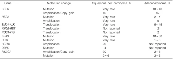
Table 1. Summary of the Frequency of Molecular Abnormalities on Oncogenes Associated with the Lung Adenocarcinoma and Squamous Cell Carcinoma Histologies
Gene Molecular change Squamous cell carcinoma % Adenocarcinoma %
EGFR Mutation Very rare 10∼40
Amplification/Copy gain 40 15
HER2 Mutation Very rare 2∼4
Amplification Very rare 5
EML4-ALK Translocation Very rare 5∼15
KIF5B-RET Translocation Not reported 2
ROS1-FIG Translocation Not reported 2
KRAS Mutation Very rare 10∼30
BRAF Mutation Very rare 1∼3
FGFR1 Amplification 20 Not reported
DDR2 Mutation 4 Not reported
PIK3CA Amplification/Copy gain 30 2∼6
Mutation 2∼6 2∼6
“hallmarks of cancer,” including abnormalities in self-suffi- ciency of growth signals, insensitivity to anti-growth signals, sustained angiogenesis, evading apoptosis, limitless replicative potential, and tissue invasion and metastasis (5). The recent molecular advances in our understanding of the pathogenesis of NSCLC will provide a unique opportunity to improve the prevention and treatment of lung cancer (6).
In this review, the molecular abnormalities associated to the early pathogenesis of the two most common histological types of lung cancer, SCC and adenocarcinoma, will be described.
Two important concepts of the pathogenesis of lung cancer (field of cancerization) and the development of novel chemo- prevention strategies (reverse migration) will be discussed.
Further research in the early pathogenesis of lung cancer may provide new methods for assessing the likelihood of developing invasive lung cancer in smokers and allow for early detection and monitoring of their response to chemoprevention regimens using molecularly targeted agents.
MOLECULAR ABNORMALITIES OF SQUAMOUS CELL CARCINOMAS AND
ADENOCARCINOMAS OF THE LUNG
1) Squamous cell carcinoma (SCC)
This tumor type frequently harbors multiple genetic abnor- malities, including activation of several oncogenes (e.g., SOX2, FGFR1, PIK3CA, EGFR, and DDR2), and inactivation of tumor suppressor genes (e.g., TP53, and p16INK4a) (Table 1, Fig. 1) (7,8). The sex determining region Y-box 2 (SOX2) gene has
been suggested to be a lineage survival oncogene in lung SCC (9). SOX2 is a transcription factor coded by a gene located at 3q26.33. This protein is implicated in the maintenance of embryonic stem cells and the induction of pluripotent stem cells from various different cell types, and plays important roles in multiple developmental processes, including lung branching (9).
SOX2 protein is expressed in normal bronchial epithelium and preneoplastic squamous lesions (10), and is more frequently detected in SCC (90%) compared with adenocarcinoma (20%) (9,10). SOX2 gene is amplified in 20% SCC of the lung, and correlates with the protein overexpression (9). SOX2 ampli- fication has not been detected in lung tumors with adeno- carcinoma histology.
Recently, two clinically relevant molecular abnormalities have been identified to occur in SCC: amplification of FGFR1 gene (chromosome 8p11-12), and mutation of the DDR2 gene.
The FGFR1 gene encodes for a transmembrane tyrosine kinase (TK) and is amplified in ∼20% of SCCs and only in 1∼3%
of adenocarcinomas (11). Mutations of another TK, DDR2 gene, occur in ∼4% of SCCs, and it has not been reported in adenocarcinomas of the lung (12). Importantly, lung tumors with FGFR1 amplification respond to FGFR TK inhibitors (TKIs) (13), and lung cancers with DDR2 mutation are sensitive to dasatinib and nilotinib treatments (12).
2) Adenocarcinoma
Lung adenocarcinomas are histologically and biologically heterogeneous tumors (14,15). Most adenocarcinomas (∼90%) have mixed histological growth patterns, including acinar,
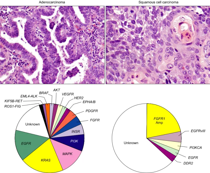
Fig. 1. Paradigm changes in the classification of non-small cell lung carcinoma. Targetable abnormalities of multiple oncogenes, particularly mutations, translocations and gene amplifications have been recently discovered in both adenocarcinoma and squamous cell carcinoma histologies. Most abnormalities are referred to gene mutations, except EML4-ALK, KIF5B-RET and ROS1-FIG rearrangement and FGFR1 amplification (Amp).
papillary, lepidic (former bronchioloalveolar carcinoma, BAC), micropapillary, and solid patterns. Attempts to establish cor- relations between histological features and molecular abnor- malities have been unsuccessful (3).
Over the past decade, significant progress has been made in the characterization of molecular abnormalities of lung adeno- carcinoma (4). Similarly to SCC, adenocarcinomas of the lung show frequent inactivation of tumor suppressor genes, include- ing, among others, TP53, and p16INK4a (7,8). DNA sequencing of 188 lung adenocarcinomas including 623 genes with known or potential relationships to cancer revealed more than 1,000 somatic mutations across the samples, and 26 genes were
mutated at significantly high frequencies (16). The frequently mutated oncogenes included TKs HER2, multiple ephrin receptor genes (notably EPHA3), vascular endothelial growth factor receptor KDR, and neurotropic TK (NTRK) genes (16).
In the last decade, lung adenocarcinomas arising in never or light smokers have shown to be characterized by significantly higher frequencies of a series of targetable oncogenes abnor- malities (4), including EGFR and HER2 TK domain activating mutations, and EML4-ALK translocation (Table 1, Fig. 1) (4).
Recently, two additional potentially targetable gene translo- cations, KIF5B-RET (10p;11q)(p11.22; q11-21) (17-19) and ROS1-FIG (chromosome 6q22) (20), have been identified in
Molecular Pathogenesis of NSCLC 15
lung adenocarcinoma from never and ever smokers. Other potentially targetable genetic abnormalities have been detected in both major NSCLC histologies, including among others, PIK3CA mutation and amplification, MET amplification (7q21- q31), AKT1 and MAP2K1 mutations (4,16).
EGFR mutations in lung cancer are limited to the first four exons of the TK domain (exons 18∼21), being the most frequent mutations in-frame deletions in exon 19 (44% of all mutations) and missense mutations in exon 21 (41% of all mutations) (21,22). In addition, in-frame duplications/insertions occurring in exon 20 have been described in about 5% of the mutant cases, and rare missense mutations occur in multiple sites (21,22). EGFR mutations occur predominantly in adeno- carcinoma (∼15% to 48% vs. other NSCLC histologies ∼2%), and are more frequent in never-smokers (54% vs. ever-smokers 16%) and female patients (49% vs. male patients 19%) (21,22).
Activating EGFR mutations strongly correlate with clinical response to TKIs, and currently is the most important criterion to select patients for EGFR TKI therapy in lung cancer (23).
HER2 mutations have been described predominantly in adeno- carcinoma histology (∼2 to 4%) and patients with East-Asian ethnic background and never-smoker history (21,22).
EML4-ALK translocation (2;5)(p23q35), with subsequent activation of ALK, has been detected in ∼6% of lung adeno- carcinomas (24,25). This translocation occurs particularly in patients with never or light smoking history, and associated with young onset of tumor. Tumors with EML4-ALK trans- location usually lack EGFR and KRAS mutations (25-27).
Histologically, EML4-ALK-rearranged adenocarcinomas have been described to have a frequently a solid pattern with signet ring cells (27), but this translocation can been identified in all histological subtypes of lung adenocarcinomas.
KRAS mutations are most common in lung adenocarcinoma than other NSCLC histology types, and are more frequently found in tumors from patients with smoking history (∼30%) (28). In lung cancer, KRAS mutations are found in codons 12, 13 and 61, and they (42% of all mutations) are mainly GGT to TGT transversions producing a Glycine to Cysteine amino acid changes (29,30). KRAS mutations are usually not detected in EGFR-mutant tumors, and have been associated to low response rates to EGFR-TKI therapies (31). BRAF oncogene can be activated in NSCLC, particularly adenocarcinoma histology (1∼3%), by gene point mutations affecting exons 11
and 15 (4).
Most (70∼80%) lung adenocarcinomas express the thyroid transcription factor-1 (TTF-1) protein encoded by the gene TITF-1, also termed NKX2-1 (32). TTF-1 is a homeodo- main-containing transcription factor expressed at the onset of lung morphogenesis in epithelial cells (33). In the postnatal lung, TTF-1 expression is restricted primarily to alveolar type II cells and subsets of non-ciliated bronchiolar epithelial cells (33). The TITF-1 gene has been suggested to be a lineage- specific oncogene, and gene amplification has been detected in
∼11% of lung adenocarcinomas, which correlated with poor survival rates in surgically resected tumors (32,34). However, conversely to the putative oncogene properties, high level of TTF-1 protein expression in adenocarcinomas has been cor- related with smaller tumor size, lower pathologic tumor-nodes- metastasis (TNM) stage, and better survival rates (15,34).
PRECURSOR LESIONS OF NSCLC
Lung cancers are believed to arise after a series of pro- gressive pathological changes (preneoplastic or precursor les- ions) in the respiratory mucosa (Fig. 2). For NSCLC, the 2004 World Health Organization (WHO) histological classification of preinvasive lesions of the lung lists two main morphologic forms of preneoplastic lesions (14): a) squamous dysplasia and carcinoma in situ (CIS); and, b) atypical adenomatous hyper- plasia (AAH) (Table 2). While the sequential preneoplastic changes have been defined for SCC, they have been poorly documented for adenocarcinomas and other lung cancer histo- logies (e.g., large carcinomas, small cell lung carcinoma, etc.) (35). Mucosal changes in the large airways that may precede invasive SCC include squamous dysplasia and CIS (35).
Adenocarcinomas may be preceded by morphological changes including AAH in peripheral airway (36).
1) Squamous preneoplastic lesions
The mucosal changes in the large airways that precede invasive SCC include: hyperplasia, squamous metaplasia, squamous dysplasia and CIS (Fig. 2). Dysplastic squamous lesions may be of different intensities (mild, moderate, and severe); however, these lesions represent a continuum of cytologic and histologic atypical changes that may show some overlapping between categories (35). Little is known about the
Fig. 2. Sequential histological abnormalities involved in the pathogenesis of squamous cell carcinoma and adenocarcinoma of the lung. The mucosal changes in the large airways that precede invasive squamous cell carcinoma include: hyperplasia, squamous metaplasia, squamous dysplasia and carcinoma in situ. Dysplastic squamous lesions may be of different intensities: mild, moderate and severe. Atypical adenomatous hyperplasia (AAH) is considered a putative precursor of adenocarcinoma, particularly those with extensive lepidic pattern (former bronchioloalveolar [BAC] pattern). AAH is a discrete parenchymal lesion arising in the alveoli close to terminal and respiratory bronchioles.
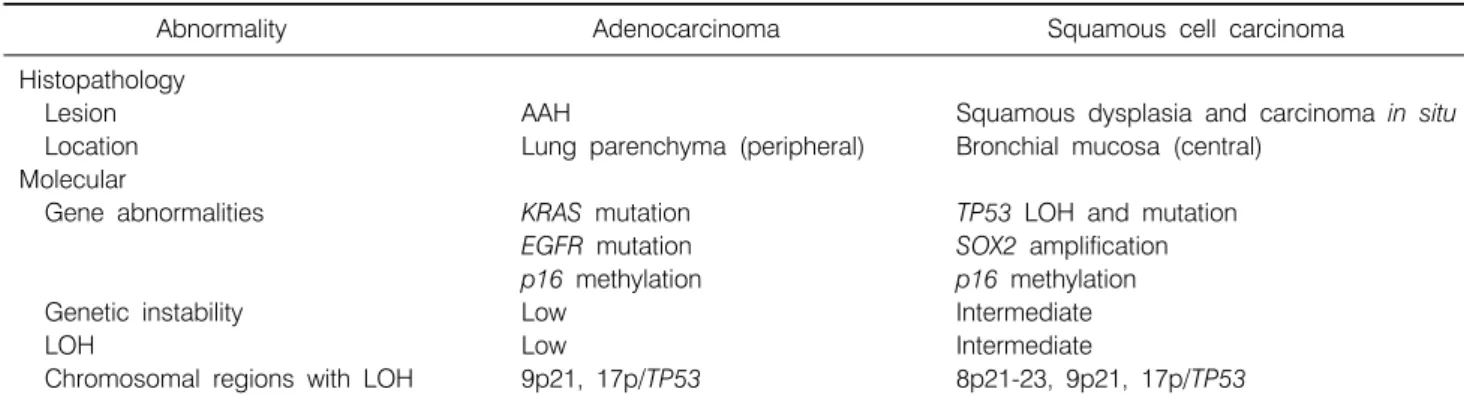
Table 2. Histopathological and Molecular Abnormalities of NSCLC Precursor Lesions
Abnormality Adenocarcinoma Squamous cell carcinoma
Histopathology
Lesion AAH Squamous dysplasia and carcinoma in situ
Location Lung parenchyma (peripheral) Bronchial mucosa (central)
Molecular
Gene abnormalities KRAS mutation TP53 LOH and mutation
EGFR mutation SOX2 amplification
p16 methylation p16 methylation
Genetic instability Low Intermediate
LOH Low Intermediate
Chromosomal regions with LOH 9p21, 17p/TP53 8p21-23, 9p21, 17p/TP53 NSCLC: non-small cell lung carcinoma, AAH: atypical adenomatous hyperplasia, LOH: loss of heterozygosity.
rate and risks of progression of squamous dysplasia to CIS and ultimately to invasive SCC.
The current working model of the sequential molecular
abnormalities in the pathogenesis of SCC indicates that the genetic abnormalities (gene deletions, genetic instability, and other changes) commence in histologically normal epithelium
Molecular Pathogenesis of NSCLC 17
and increase with increasing severity of histologic change, and that the genetic changes follow a sequence (37). Several studies have shown that multiple clonal and subclonal patches of airway cells with molecular abnormalities can be detected in the normal and slightly abnormal bronchial epithelium of smokers and patients with lung cancer (37,38). The molecular abnormalities associated with the early pathogenesis of SCC include allelic losses at multiple regions of chromosome 3p and 9p21 (p16INK4a), subsequent losses of chromosome regions 8p21-23, 13q14 (RB), and 17p13 (TP53) (39) (Table 2).
Additionally, it has been shown that p16INK4a methylation increases during the progression of SCC preneoplastic lesions (39).
Recent studies showed progressive amplification of 3q ampli- fication in the evolution of squamous dysplasias and implicate SOX2 as a key target of this process (40). Interestingly, in a small cohort of squamous dysplasias lesions the degree of 3q amplification associates with clinical progression of the pre- cursor lesions (41). However, despite all these advances, neither histological features nor molecular changes can be reliably used for accurate risk of progression of squamous preneoplastic lesions and be used to assess the risk of developing SCC (42).
2) Adenocarcinoma precursor lesions
It has been suggested that adenocarcinomas may be preceded by AAH in peripheral airway cells (Fig. 2) (36,43); however, the respiratory structures and the specific epithelia cell types involved in the origin of most lung adenocarcinomas are unknown.
AAH is a discrete parenchymal lesion arising in the alveoli close to terminal and respiratory bronchioles. Because of their size, AAH are frequently incidental histological findings, but they may be detected grossly, especially if they are 0.5 cm or larger. The increasing use of high resolution computed tomography scans for screening purposes has led to an increasing awareness of this entity, as it remains one of the most important differential diagnoses of air filled peripheral lesions (called “ground glass opacities”). The origin of AAH is still unknown, but the differentiation phenotype derived from immunohistochemical and ultrastructural features suggests an origin from the progenitor cells of the peripheral airways, such Clara cells and type II pneumocytes (44,45).
Several molecular changes frequently present in lung adeno-
carcinomas are also present in AAH lesions, and they are further evidence that AAH may represent true preneoplastic lesions (44). The most important finding is the presence of KRAS (codon 12) mutations in up to 39% of AAHs, which are also a relatively frequent alteration in lung adenocarcinomas (36,46). Other molecular alterations detected in AAH are overexpression of cyclin D1 (∼70%), p53 (range, 10∼58%), survivin (48%), and HER2/neu (7%) proteins over-expression (36,47,48). Interestingly, EGFR mutations have been detected in some cases of AAH accompanying resected peripheral adenocarcinomas, providing further evidence that they represent precursor lesions of peripheral adenocarcinomas (49).
However, the respiratory structures and the specific epithelial cell types involved in the origin of most lung adenocarcinomas are unknown, and candidates include Clara cells, alveolar type II cells, and an epithelial cell located at the junction between the terminal bronchiole and alveolus, termed bronchioloalveolar stem cell (BASC). In addition, lung adenocarcinoma may arise from bronchial or bronchiolar epithelia, since EGFR activating mutation has been found in normal bronchial epithelium adjacent to EGFR-mutant adenocarcinomas in 43% of patients (50,51).
LUNG CANCER PROGENITOR OR STEM CELLS
Classically, stem cells are located in specialized niche and are preserved from overuse in maintaining tissue homeostasis for their conservation throughout the organism’s life. As such, stem cells undergo infrequent cycling and stem cell com- partments are identified based upon this characteristic. Curren- tly, it is believed that at least four stem cell niches are present in adult lung airway epithelium: gland duct basal cells, inter-cartelagenous zone surface basal cells, bronchiolar Clara cells associated with pulmonary neuroendocrine cell bodies, and bronchioloalveolar duct junction variant Clara cells (52).
From mouse model studies, it has been shown that BASCs are putative stem cell initiators of KRAS induced NSCLC adenocarcinoma (53). In addition, to the identification of BASCs, several stem cell markers for lung tumors have been identified. The most studied lung cancer stem cell marker has been CD133, which has been shown to express in NSCLC and other lung tumors (e.g., small cell carcinoma) (54). Recently,
the stem cell markers aldehyde dehydrogenase 1A1 (ALDH1A1) has been also shown to be frequently expressed in NSCLC associated to patients’ outcome (55). However, despite multiple studies, the progenitor or stem cells for human lung cancers have not been identified yet.
LUNG AIRWAY FIELD CANCERIZATION
It has been shown that human tumors and adjacent histo- logically normal tissue share certain molecular abnormalities, a phenomenon coined as field cancerization (56). The concept of field of cancerization was first introduced in 1953 in the setting of oral cancer (57). Later, field of cancerization involving histological and molecular changes of the lung airway of pa- tients with lung cancer and smokers began to be characterized (56). A detailed analysis of histologically normal, premalignant and malignant epithelia from smokers and patients with SCC, have indicated that multiple, sequentially occurring, allele-spe- cific chromosomal deletions and genomic instability begin in dispersed clonally independent foci early in the pathogenesis of this malignancy, including histologically normal bronchial epithelium (39). In addition, it has been demonstrated that KRAS (58) and EGFR (50,51) mutations are found in per- ipherally located histologically normal lung tissue adjacent to lung adenocarcinomas. The finding of higher frequency of EGFR mutation in normal cells obtained from bronchioles close to the tumor compared to more distant sites, suggested the presence of a localized field effect in the pathogenesis of lung adenocarcinomas (50). These and other observations suggest a compartmentalization of the field of cancerization, and the dissimilarity between SCCs and adenocarcinomas may reflect the differential mechanisms of pathogenesis of these NSCLC subtypes.
Our improved understanding of the changes and origins of the field of cancerization can be applied clinically to improve our ability of risk assessment, screening, diagnosis and treat- ment of patients with or at risk for lung cancer. Recently, global gene expression (mRNA) studies using microarrays and pathway signatures analysis have been derived from cells obtained from normal bronchial epithelium of smokers, and showed to exhibit properties to predict the development of lung cancer (59,60). Global alterations in gene expression differences between normal bronchial epithelium of healthy cancer-free
smokers and that of never smokers have been described (59).
Interestingly, irreversible changes in gene expression in airways of former smokers after years of smoking cessation were described to underlay the still increased risk of developing of lung cancer observed in this individuals (59). Interestingly, an 80-gene expression signature developed from bronchial epithe- lial cells obtained from the large airway can distinguish smok- ers without overt cancer from smokers without lung cancer (60).
TARGETED PREVENTION AND REVERSE MIGRATION IN LUNG CANCER
Positive chemoprevention trials usually involve known tar- gets that can be inhibited effectively by drugs. The significant progress made in the characterization of targetable molecular abnormalities in NSCLC tumors offers a unique opportunity to use this knowledge for the development of novel chemopre- vention strategies using molecularly targeted agents. However, our knowledge of the molecular pathogenesis of lung cancer must continue to evolve.
Recently, it has been proposed to reverse migration of findings on molecular markers, pharmacogenomics and targeted therapy from the advanced NSCLC setting to the chemopre- vention setting (61). For reverse migration to be an effective strategy, several assumptions must be true: 1) pre-malignancy shares molecular properties with malignancy; 2) effective tar- geted therapy of malignancy is applicable to precursor lesions;
and 3) predictive biomarkers can be applied to patients at risk for malignancy. The current knowledge on the early patho- genesis of NSCLC suggests that these assumptions are probably true, therefore, there is an opportunity for the utilization of targeted agents for primary, secondary and tertiary chemopre- vention in lung cancer, particularly NSCLC.
FUTURE DIRECTIONS
Applying advanced high-throughput molecular methodolo- gies currently used to study advanced lung tumors to the molecular analysis of the field of cancerization is expected to expand our understanding of the biology of the lung cancer.
The rapid development of technologies for large-scale mole- cular analysis, including the newest next-generation sequencing
Molecular Pathogenesis of NSCLC 19
(NGS) of RNA (transcriptome) and DNA (whole genome or exome) is facilitating high-throughput molecular studies holding various advantages over traditionally molecular analysis of nucleic acids abnormalities. NGS can simultaneously detect deletions, insertions, copy number alterations, translocations, and exome-wide base substitutions (including known hot-spot mutations) in all known cancer-related genes (62). An important step to apply NGS in lung cancer pathogenesis was a recent study in which RNA of bronchial airway epithelial cells brushings from never-smoker and ever-smoker individuals with and without lung cancer was analyzed by RNA-sequencing providing additional insights besides those provided when using RNA microarrays (63). A comprehensive analysis of the early molecular events in NSCLC pathogenesis utilizing novel methodologies will undoubtedly unravel novel molecular targets and biomarkers that can aid prevention deliver its longstanding promise in lung cancer.
REFERENCES
1. Jemal A, Siegel R, Xu J, Ward E. Cancer statistics, 2010. CA Cancer J Clin 2010;60:277-300.
2. Herbst RS, Heymach JV, Lippman SM. Lung cancer. N Engl J Med 2008;359:1367-1380.
3. Travis WD, Rekhtman N. Pathological diagnosis and classi- fication of lung cancer in small biopsies and cytology: stra- tegic management of tissue for molecular testing. Semin Respir Crit Care Med 2011;32:22-31.
4. Pao W, Girard N. New driver mutations in non-small-cell lung cancer. Lancet Oncol 2011;12:175-180.
5. Hanahan D, Weinberg RA. Hallmarks of cancer: the next generation. Cell 2011;144:646-674.
6. Kadara H, Behrens C, Yuan P, et al. A five-gene and corres- ponding protein signature for stage-I lung adenocarcinoma prognosis. Clin Cancer Res 2011;17:1490-1501.
7. Gold KA, Wistuba II, Kim ES. New strategies in squamous cell carcinoma of the lung: identification of tumor drivers to personalize therapy. Clin Cancer Res 2012;18:3002-3007.
8. Perez-Moreno P, Brambilla E, Thomas R, Soria JC. Squamous cell carcinoma of the lung: molecular subtypes and therapeutic opportunities. Clin Cancer Res 2012;18:2443-2451.
9. Bass AJ, Watanabe H, Mermel CH, et al. SOX2 is an amplified lineage-survival oncogene in lung and esophageal squamous cell carcinomas. Nat Genet 2009;41:1238-1242.
10. Yuan P, Kadara H, Behrens C, et al. Sex determining region Y-Box 2 (SOX2) is a potential cell-lineage gene highly expressed in the pathogenesis of squamous cell carcinomas of the lung. PLoS One 2010;5:e9112.
11. Weiss J, Sos ML, Seidel D, et al. Frequent and focal FGFR1 amplification associates with therapeutically tractable FGFR1
dependency in squamous cell lung cancer. Sci Transl Med 2010;2:62ra93.
12. Hammerman PS, Sos ML, Ramos AH, et al. Mutations in the DDR2 kinase gene identify a novel therapeutic target in squamous cell lung cancer. Cancer Discov 2011;1:78-89.
13. Dutt A, Ramos AH, Hammerman PS, et al. Inhibitor-sensitive FGFR1 amplification in human non-small cell lung cancer.
PLoS One 2011;6:e20351.
14. Travis WD, Brambilla E, Müller-Hermelink HK, Harris CC.
World Health Organization classification of tumours: patho- logy and genetics of tumours of the lung, pleura, thymus and heart. Lyon: IARC Press; 2004. p.9-124.
15. Solis LM, Behrens C, Raso MG, et al. Histologic patterns and molecular characteristics of lung adenocarcinoma associated with clinical outcome. Cancer 2012;118:2889-2899.
16. Ding L, Getz G, Wheeler DA, et al. Somatic mutations affect key pathways in lung adenocarcinoma. Nature 2008;455:1069- 1075.
17. Kohno T, Ichikawa H, Totoki Y, et al. KIF5B-RET fusions in lung adenocarcinoma. Nat Med 2012;18:375-377.
18. Takeuchi K, Soda M, Togashi Y, et al. RET, ROS1 and ALK fusions in lung cancer. Nat Med 2012;18:378-381.
19. Lipson D, Capelletti M, Yelensky R, et al. Identification of new ALK and RET gene fusions from colorectal and lung cancer biopsies. Nat Med 2012;18:382-384.
20. Bergethon K, Shaw AT, Ou SH, et al. ROS1 rearrangements define a unique molecular class of lung cancers. J Clin Oncol 2012;30:863-870.
21. Shigematsu H, Gazdar AF. Somatic mutations of epidermal growth factor receptor signaling pathway in lung cancers. Int J Cancer 2006;118:257-262.
22. Shigematsu H, Lin L, Takahashi T, et al. Clinical and bio- logical features associated with epidermal growth factor recep- tor gene mutations in lung cancers. J Natl Cancer Inst 2005;
97:339-346.
23. Mok TS, Wu YL, Thongprasert S, et al. Gefitinib or carbo- platin-paclitaxel in pulmonary adenocarcinoma. N Engl J Med 2009;361:947-957.
24. Soda M, Choi YL, Enomoto M, et al. Identification of the transforming EML4-ALK fusion gene in non-small-cell lung cancer. Nature 2007;448:561-566.
25. Inamura K, Takeuchi K, Togashi Y, et al. EML4-ALK lung cancers are characterized by rare other mutations, a TTF-1 cell lineage, an acinar histology, and young onset. Mod Pathol 2009;22:508-515.
26. Shaw AT, Yeap BY, Solomon BJ, et al. Effect of crizotinib on overall survival in patients with advanced non-small-cell lung cancer harbouring ALK gene rearrangement: a retro- spective analysis. Lancet Oncol 2011;12:1004-1012.
27. Inamura K, Takeuchi K, Togashi Y, et al. EML4-ALK fusion is linked to histological characteristics in a subset of lung cancers. J Thorac Oncol 2008;3:13-17.
28. Dacic S, Shuai Y, Yousem S, Ohori P, Nikiforova M. Clinic- opathological predictors of EGFR/KRAS mutational status in primary lung adenocarcinomas. Mod Pathol 2010;23:159-168.
29. Siegfried JM, Gillespie AT, Mera R, et al. Prognostic value of specific KRAS mutations in lung adenocarcinomas. Cancer Epidemiol Biomarkers Prev 1997;6:841-847.
30. Riely GJ, Kris MG, Rosenbaum D, et al. Frequency and dis- tinctive spectrum of KRAS mutations in never smokers with lung adenocarcinoma. Clin Cancer Res 2008;14:5731-5734.
31. Massarelli E, Varella-Garcia M, Tang X, et al. KRAS mutation is an important predictor of resistance to therapy with epider- mal growth factor receptor tyrosine kinase inhibitors in non- small-cell lung cancer. Clin Cancer Res 2007;13:2890-2896.
32. Tang X, Kadara H, Behrens C, et al. Abnormalities of the TITF-1 lineage-specific oncogene in NSCLC: implications in lung cancer pathogenesis and prognosis. Clin Cancer Res 2011;17:2434-2443.
33. Whitsett J. A lungful of transcription factors. Nat Genet 1998;
20:7-8.
34. Perner S, Wagner PL, Soltermann A, et al. TTF1 expression in non-small cell lung carcinoma: association with TTF1 gene amplification and improved survival. J Pathol 2009;217:65-72.
35. Kerr KM. Pulmonary preinvasive neoplasia. J Clin Pathol 2001;54:257-271.
36. Westra WH. Early glandular neoplasia of the lung. Respir Res 2000;1:163-169.
37. Wistuba II. Genetics of preneoplasia: lessons from lung cancer.
Curr Mol Med 2007;7:3-14.
38. Park IW, Wistuba II, Maitra A, et al. Multiple clonal abnor- malities in the bronchial epithelium of patients with lung cancer. J Natl Cancer Inst 1999;91:1863-1868.
39. Wistuba II, Mao L, Gazdar AF. Smoking molecular damage in bronchial epithelium. Oncogene 2002;21:7298-7306.
40. McCaughan F, Pole JC, Bankier AT, et al. Progressive 3q amplification consistently targets SOX2 in preinvasive squa- mous lung cancer. Am J Respir Crit Care Med 2010;182:83- 91.
41. McCaughan F, Pipinikas CP, Janes SM, George PJ, Rabbitts PH, Dear PH. Genomic evidence of pre-invasive clonal expansion, dispersal and progression in bronchial dysplasia. J Pathol 2011;224:153-159.
42. Wistuba II. Histologic evaluation of bronchial squamous lesions: any role in lung cancer risk assessment? Clin Cancer Res 2005;11:1358-1360.
43. Colby TV, Wistuba II, Gazdar A. Precursors to pulmonary neoplasia. Adv Anat Pathol 1998;5:205-215.
44. Kitamura H, Kameda Y, Ito T, Hayashi H. Atypical adeno- matous hyperplasia of the lung. Implications for the patho- genesis of peripheral lung adenocarcinoma. Am J Clin Pathol 1999;111:610-622.
45. Osanai M, Igarashi T, Yoshida Y. Unique cellular features in atypical adenomatous hyperplasia of the lung: ultrastructural evidence of its cytodifferentiation. Ultrastruct Pathol 2001;25:
367-373.
46. Westra WH, Baas IO, Hruban RH, et al. K-ras oncogene activation in atypical alveolar hyperplasias of the human lung.
Cancer Res 1996;56:2224-2228.
47. Tominaga M, Sueoka N, Irie K, et al. Detection and discri-
mination of preneoplastic and early stages of lung adenocar- cinoma using hnRNP B1 combined with the cell cycle-related markers p16, cyclin D1, and Ki-67. Lung Cancer 2003;40:
45-53.
48. Nakanishi K, Kawai T, Kumaki F, Hiroi S, Mukai M, Ikeda E. Survivin expression in atypical adenomatous hyperplasia of the lung. Am J Clin Pathol 2003;120:712-719.
49. Yatabe Y, Kosaka T, Takahashi T, Mitsudomi T. EGFR mutation is specific for terminal respiratory unit type adeno- carcinoma. Am J Surg Pathol 2005;29:633-639.
50. Tang X, Shigematsu H, Bekele BN, et al. EGFR tyrosine kinase domain mutations are detected in histologically normal respiratory epithelium in lung cancer patients. Cancer Res 2005;65:7568-7572.
51. Tang X, Varella-Garcia M, Xavier AC, et al. Epidermal growth factor receptor abnormalities in the pathogenesis and progression of lung adenocarcinomas. Cancer Prev Res (Phila) 2008;1:192-200.
52. Rawlins EL, Okubo T, Que J, et al. Epithelial stem/progenitor cells in lung postnatal growth, maintenance, and repair. Cold Spring Harb Symp Quant Biol 2008;73:291-295.
53. Kim CF, Jackson EL, Woolfenden AE, et al. Identification of bronchioalveolar stem cells in normal lung and lung cancer.
Cell 2005;121:823-835.
54. Eramo A, Lotti F, Sette G, et al. Identification and expansion of the tumorigenic lung cancer stem cell population. Cell Death Differ 2008;15:504-514.
55. Sullivan JP, Spinola M, Dodge M, et al. Aldehyde dehy- drogenase activity selects for lung adenocarcinoma stem cells dependent on notch signaling. Cancer Res 2010;70:9937-9948.
56. Kadara H, Wistuba II. Field cancerization in non-small cell lung cancer: implications in disease pathogenesis. Proc Am Thorac Soc 2012;9:38-42.
57. Slaughter DP, Southwick HW, Smejkal W. Field cancerization in oral stratified squamous epithelium; clinical implications of multicentric origin. Cancer 1953;6:963-968.
58. Nelson MA, Wymer J, Clements N Jr. Detection of K-ras gene mutations in non-neoplastic lung tissue and lung cancers.
Cancer Lett 1996;103:115-121.
59. Spira A, Beane J, Shah V, et al. Effects of cigarette smoke on the human airway epithelial cell transcriptome. Proc Natl Acad Sci U S A 2004;101:10143-10148.
60. Spira A, Beane JE, Shah V, et al. Airway epithelial gene expression in the diagnostic evaluation of smokers with suspect lung cancer. Nat Med 2007;13:361-366.
61. Gold KA, Kim ES, Lee JJ, Wistuba II, Farhangfar CJ, Hong WK. The BATTLE to personalize lung cancer prevention through reverse migration. Cancer Prev Res (Phila) 2011;4:
962-972.
62. Meyerson M, Gabriel S, Getz G. Advances in understanding cancer genomes through second-generation sequencing. Nat Rev Genet 2010;11:685-696.
63. Beane J, Vick J, Schembri F, et al. Characterizing the impact of smoking and lung cancer on the airway transcriptome using RNA-Seq. Cancer Prev Res (Phila) 2011;4:803-817.