A radiographic evaluation of graft height changes after maxillary sinus augmentation
전체 글
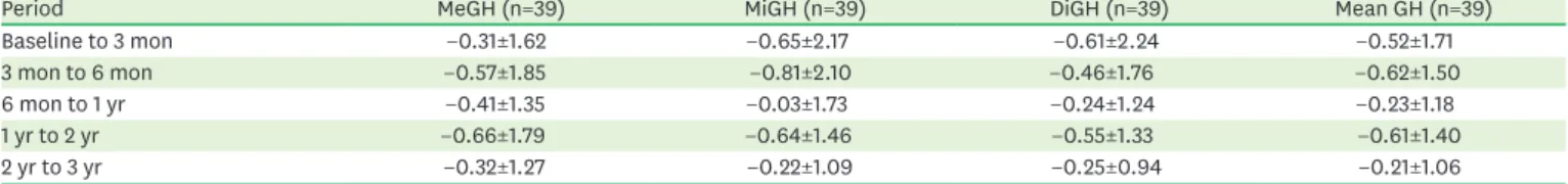
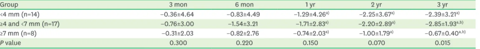
수치

관련 문서
1 John Owen, Justification by Faith Alone, in The Works of John Owen, ed. John Bolt, trans. Scott Clark, "Do This and Live: Christ's Active Obedience as the
3 Mean percentages of bone implant contact ratio in the control group and experimental groups at 6 and 12 weeks after placement of the
The thickness of the maxillary sinus lateral wall according to tooth site and measurement level was measured by using image-processing software and its histologic
The purpose of this study was to evaluate surface changes and abrasion of the implant fixture and TiN coated abutment screw after repeated delivery and
With the increase of the dental implant procedure, there are many cases of Maxillary Sinus Floor Elevation Surgery(MSFES). Additionally, its side effect has
Methods to overcome insufficient bone due to poor bone quality, the pneumatization of a maxillary sinus and other anatomical limitations of implant placement
Histopathologic findings of control group at 4 weeks show little bone- implant contact (BIC) around the implant (asterisks) and new-bone formation in the defect
The OSFE (osteotome sinus floor elevation) technique has been used for maxillary sinus augmentation.. The implants were clinically and radiographically followed
