© 2016 Korean Breast Cancer Society. All rights reserved. http://ejbc.kr | pISSN 1738-6756
INTRODUCTION
Mammography is a sensitive method for the detection of microcalcifications, which may be the only manifestation of early breast cancer [1-3]. Based on imaging characteristics, microcalcifications often cannot be classified as benign or malignant; in these cases, pathological examination by biopsy is recommended [3-6].
Recent advances in ultrasound (US) technology and the re-
finement of breast imaging techniques have enabled radiol- ogists to evaluate breast microcalcifications. Several studies have reported that handheld breast US (HHUS) is useful in identifying malignant microcalcifications and provides effec- tive guidance for percutaneous biopsy [7-12]. US-guided breast biopsy has several advantages over stereotactic biopsy, which include improved patient comfort, real-time visualiza- tion of needle placement, absence of radiation exposure, short- er duration of the procedure, and relatively low cost. However, most radiologists now performing breast US use handheld systems, which has some disadvantages because the scans are operator-dependent and therefore suffer from a lack of repro- ducibility.
Automated whole breast US (AWUS) scanners were origi- nally designed to examine the entire breast and to overcome the operator dependency of HHUS [13,14]. Several studies have concluded that the sensitivity, diagnostic performances,
Detectability and Usefulness of Automated Whole Breast Ultrasound in Patients with Suspicious Microcalcifications on Mammography: Comparison with Handheld Breast Ultrasound
Jae Jeong Choi, Sung Hun Kim1, Bong Joo Kang1, Byung Joo Song2
Department of Radiology, Dongtan Sacred Heart Hospital, Hallym University Medical Center, Hwaseong; Departments of 1Radiology and 2General Surgery, Seoul St. Mary’s Hospital, The Catholic University of Korea College of Medicine, Seoul, Korea
ORIGINAL ARTICLE
Purpose: The purpose of this study was to prospectively evalu- ate the detectability and usefulness of automated whole breast ultrasound (AWUS) and to compare it with handheld breast ultra- sound (HHUS) in cases with suspicious microcalcifications iden- tified by mammography. Methods: Forty-two patients with 43 suspicious microcalcifications (25 malignant and 18 benign) de- tected by mammography underwent AWUS, HHUS, and histol- ogic examination. With knowledge of the mammographic find- ings, HHUS was performed to assess the visibility of the micro- calcifications and the presence of associated masses or ductal changes. Two radiologists reviewed the AWUS images in con- sensus using the same methods employed for HHUS. Detect- ability of AWUS was compared with that of HHUS and was cor- related with histologic and mammographic findings. Results: Of the 43 lesions, 32 (74.4%) were detectable by AWUS and 31 (72.1%) by HHUS. No significant differences in sensitivity were found between the two methods (p=0.998). AWUS detected
96% (24/25) of malignant microcalcifications and 44.4% (8/18) of benign microcalcifications. AWUS was more successful in the detection of malignant vs. benign lesions (96.0% vs. 44.4%, p=0.002), lesions >10 mm vs. ≤10 mm in size (86.7% [26/30]
vs. 46.2% [6/13], p=0.009), lesions with a fine pleomorphic or linear shape vs. a round or amorphous or coarse heterogeneous shape (94.7% [18/19] vs. 58.3% [14/24], p=0.021), and lesions associated with a mass or architectural distortion vs. without ob- vious changes on mammography (100% [19/19] vs. 54.2%
[13/24], p=0.022). Conclusion: Detectability of AWUS was com- parable to that of HHUS in cases where suspicious microcalcifi- cations were identified on mammography. Therefore, AWUS might be helpful in the performance of ultrasound-guided percu- taneous procedures for highly suspicious microcalcifications.
Key Words: Breast neoplasms, Calcinosis, Mammary glands, Mammography, Ultrasonography
Correspondence to: Sung Hun Kim
Department of Radiology, Seoul St. Mary’s Hospital, The Catholic University of Korea College of Medicine, 222 Banpo-daero, Seocho-gu, Seoul 06591, Korea
Tel: +82-2-2258-6253, Fax: +82-2-2258-1457 E-mail: rad-ksh@catholic.ac.kr
This works was supported from the Catholic Medical Center Research Foundation.
Received: June 21, 2016 Accepted: September 19, 2016
Breast
Cancer
and image quality of AWUS and HHUS are equivalent [15- 21]. However, these studies have primarily evaluated mass le- sions in the breast. To date, there have been few studies evalu- ating AWUS findings for suspicious microcalcifications de- tected by mammography.
The purpose of this prospective study was to evaluate the ability of AWUS to detect suspicious microcalcifications origin- ally identified by mammography, to characterize the lesion variables that affect their detectability, and to compare the ef- fectiveness of AWUS with that of HHUS.
METHODS
Patients
Between March and August of 2012, 45 consecutive pa- tients with mammographically detected suspicious microcal- cifications, who were scheduled for US-guided or mammo- graphy-guided biopsy, were enrolled in our prospective study.
Three of these patients were excluded because the biopsy was not performed. Thus, our study ultimately included 42 pa- tients with 43 mammographically detected suspicious micro-
calcifications (Figure 1). The patients, who ranged in age from 28 to 80 years (average age, 53 years), underwent AWUS and HHUS examinations prior to their biopsy procedures. Six pa- tients had a palpable lump in the breast. Institutional Review Board approval (KC12DSSI0235) and informed patient con- sent were obtained prior to the procedures.
All microcalcifications were sampled by US-guided 14- gauge core needle biopsy or mammography-guided (stereo- tactic) 11-gauge vacuum-assisted biopsy. If the microcalcifica- tions were not visible by HHUS, stereotactic 11-gauge vacu- um-assisted biopsy was performed. Two patients with micro- calcifications visible by HHUS underwent stereotactic 11-gauge vacuum-assisted biopsy instead, because the extent of the lesion visualized by HHUS was much smaller than that visualized by mammography. A median number of seven core samples (range, 5–12) per biopsy were obtained by 14-gauge core needle biopsy and a median number of 14 core samples (range, 10–17) per biopsy were obtained by stereotactic 11- gauge vacuum-assisted biopsy. After five core samples were obtained by 14-gauge needle core biopsy and 10 core samples were obtained by 11-gauge vacuum-assisted biopsy, radio-
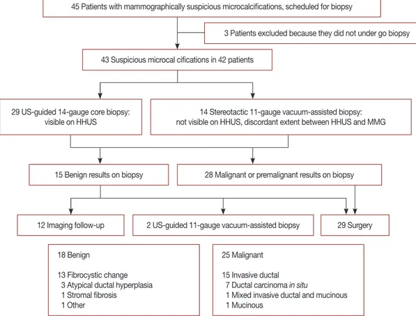
Figure 1. Flow chart shows the study population, inclusion and exclusion criteria, and pathologic findings. Three patients with benign results on biop- sy underwent 11-gauge vacuum-assisted biopsy or surgical excision due to patient anxiety.
US=ultrasound; HHUS=handheld breast ultrasound; MMG=mammography.
45 Patients with mammographically suspicious microcalcifications, scheduled for biopsy
3 Patients excluded because they did not under go biopsy
43 Suspicious microcal cifications in 42 patients
15 Benign results on biopsy
12 Imaging follow-up
18 Benign
13 Fibrocystic change 3 Atypical ductal hyperplasia 1 Stromal fibrosis
1 Other
25 Malignant 15 Invasive ductal 7 Ductal carcinoma in situ
1 Mixed invasive ductal and mucinous 1 Mucinous
2 US-guided 11-gauge vacuum-assisted biopsy 29 Surgery 28 Malignant or premalignant results on biopsy 29 US-guided 14-gauge core biopsy:
visible on HHUS
14 Stereotactic 11-gauge vacuum-assisted biopsy:
not visible on HHUS, discordant extent between HHUS and MMG
graphs were obtained to confirm the presence of representa- tive microcalcifications in the tissue specimen. Additional core samples were obtained if a sufficient number of micro- calcifications were not visualized in the tissue specimen. A ra- diopaque marker was not inserted after the biopsy procedure.
All patients underwent surgery with mammography-guid- ed wire localization if malignancy or atypical ductal hyperpla- sia was diagnosed from the 14-gauge core needle or 11-gauge vacuum-assisted biopsy samples. In those cases, we consid- ered the pathological results obtained from the surgical speci- mens to be the final diagnosis. One case of atypical ductal hy- perplasia was confirmed to be invasive ductal carcinoma fol- lowing surgical excision. Three patients with benign disease diagnosed by core needle biopsy underwent surgical excision or 11-gauge vacuum-assisted biopsy due to patient anxiety.
No histologic underestimation was discovered in these pa- tients. Follow-up imaging was performed on the remaining patients for 24 to 30 months.
Mammographic examinations and assessments
The standard craniocaudal and mediolateral oblique views were obtained using a Mammomat 3000 unit (Siemens Medi- cal Solutions, Solna, Sweden) and a Lorad M3 mammography unit (Hologic Inc., Boston, USA). Lesion size was defined as the maximum diameter on either craniocaudal or mediolater- al oblique images, and the presence of an associated mass or architectural distortion was analyzed on standard mammo- graphic views. Spot compression and magnification images were also available for review in 15 of the 42 patients. The morphology, distribution, and final assessment categories of the microcalcifications and breast density were classified ac- cording to the American College of Radiology Breast Imaging Reporting and Data System (BI-RADS). Subcategories of cat- egory 4 were created according to the probability of malig- nancy: 3%–10% in 4a (low suspicion), 11%–50% in 4b (inter- mediate suspicion), and 51%–94% in 4c lesions (moderate suspicion). Mammographic breast densities were classified as follows: a, the breasts are almost entirely fatty; b, there are scattered areas of fibroglandular density; c, the breasts are het- erogeneously dense, which may obscure small masses; d, the breast area extremely dense, which lowers the sensitivity of mammography.
Handheld breast ultrasound
HHUS examinations were performed by one of three radi- ologists (1, 8, and 10 years of experience with breast HHUS, respectively) with prior knowledge of the mammographic findings using a 7–15 MHz linear transducer (iU22 Ultra- sound System; Philips Ultrasound, Bothell, USA) or a 6–14
MHz linear transducer (EUB-8500 Scanner; Hitachi Medical, Tokyo, Japan).
HHUS examinations focused on regions containing micro- calcifications defined by their positions on a clock, distance from the nipple, and depth as determined by mammography.
The radiologists assessed the visibility of the microcalcifica- tions and the presence of associated masses or ductal changes.
Lesions were considered to be microcalcifications when echo- genic dots not traced to echogenic anatomic structures were discovered by HHUS in regions that had been previously identified by mammography.
Automated whole breast ultrasound
AWUS images were obtained using an ACUSON S2000 Automated Breast Volume Scanner (ABVS; Siemens Medical Solutions, Mountain View, USA) by one trained radiographer.
The AWUS acquired 15.4×16.8×maximum 6-cm volume data sets of the breast in one sweep with a 5–14 MHz wide- aperture linear probe. The breast was initially scanned in the anterior-posterior view, which included the nipple and most parts of the breast with the patient in a supine position. The lateral and medial views, which mainly included the outer breast and the inner breast, were then scanned with the pa- tient in an oblique position. After the acquisition of 3D vol- ume data, it was automatically sent from the ACUSON S2000 ABVS to the workstation and reviewed in multiple orienta- tions using a multi-planar reconstruction display. The images were displayed at a scan thickness of 1 mm without overlap.
Image review and data analysis for automated whole breast ultrasound
Two radiologists, who had 8 and 6 years of experience with breast imaging, respectively, analyzed all of the AWUS data in consensus. Each radiologist had more than 12 months of ex- perience with AWUS prior to participating in this study. As one of the radiologists also performed the HHUS examina- tions, analysis of AWUS data was performed at 6-month in- tervals to avoid bias. After reviewing the mammographic im- ages, the visibility of microcalcifications, associated masses, and ductal changes were assessed on the AWUS images. The radiologists focused on regions containing microcalcifications defined by using their positions with respect to a clock, dis- tance from the nipple, and depth as determined by mammo- graphy. Lesions were considered to be microcalcifications when echogenic dots not traced to echogenic anatomic structures were discovered by AWUS in regions that had been previously identified by mammography. We did not assess the diagnostic performance of the two US modalities because it was difficult to characterize microcalcifications according to BI-RADS.
USA) and MedCalc® version 11.2.1.0 (MedCalc Software, Mariakerke, Belgium) software programs. We used Fisher ex- act test for categorical variables and the Mann-Whitney U- test for continuous variables. In addition, simple logistic re- gression was performed to identify the association between risk factors and dichotomous outcomes. A p-value of less than 0.05 was considered statistically significant.
RESULTS
Of the 43 mammographically suspicious microcalcifica- tions, 25 lesions (58.1%) were malignant (15 invasive ductal carcinomas, one mixed invasive ductal and mucinous carci- noma, one mucinous carcinoma, and seven ductal carcinoma in situ [DCIS]) and the remaining 18 (41.9%) were benign (three atypical ductal hyperplasia, 13 fibrocystic changes, one massive histiocytic infiltration with multinucleated giant cells, and one stromal fibrosis).
On mammograms, the lesion size varied from 0.5 to 10.9 cm. The size and breast density did not differ significantly be- tween benign and malignant histology. The mammographic findings for the 43 lesions are summarized in Table 1.
Of the 43 lesions, 32 (74.4%) were detectable on AWUS and 31 (72.1%) were detectable on HHUS (Table 2). No significant differences were found in the detection rates between AWUS Statistical analyses
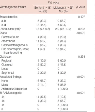
The detection rates of the mammographically identified microcalcifications by AWUS and HHUS were calculated. To determine whether detection by US was affected by pathology (benign vs. malignant lesions) or mammographic findings (size, shape, distribution, presence of an associated mass, BI- RADS category, and breast density), statistical analysis was performed using SAS version 9.1 (SAS Institute Inc., Cary, Table 1. Mammographic features of 43 suspicious microcalcifications
Mammographic feature
Pathology
p-value Benign (n=18)
No. (%)
Malignant (n=25) No. (%)
Breast densities 0.407
a, b 5 (33.3) 10 (66.7)
c, d 13 (46.4) 15 (53.6)
Lesion extent (cm)* 1.3 (0.5–6.6) 2.0 (0.6–10.9) 0.232
Shape <0.001
Punctate/round 4 (80.0) 1 (20.0)
Amorphous 11 (68.7) 5 (31.3)
Coarse heterogeneous 2 (66.7) 1 (33.3) Fine pleomorphic, linear,
linear-branching
1 (5.3) 18 (94.7)
Distribution 0.234
Regional 4 (40.0) 6 (60.0)
Clustered 12 (52.2) 11 (47.8)
Linear 0 0
Segmental 2 (20.0) 8 (80.0)
Associated findings <0.001
None 16 (66.7) 8 (33.3)
Mass 2 (11.1) 16 (88.9)
Architectural distortion 0 1 (100.0)
BI-RADS categories <0.001
4a 14 (87.5) 2 (12.5)
4b 4 (33.3) 8 (66.7)
4c 0 8 (100.0)
5 0 7 (100.0)
BI-RADS=Breast Imaging Reporting and Data System.
*Median (range).
Table 2. Comparison of detectability for suspicious microcalcifications between AWUS and HHUS
Pathology No. of detections (detection rate)
p-value
AWUS HHUS
Total (n=43) 32 (74.4) 31 (72.1) 0.998
Malignant (n=25) 24 (96.0) 24 (96.0) >0.999
Benign (n=18) 8 (44.4) 7 (38.9) 0.997
AWUS=automated whole breast ultrasound; HHUS=handheld breast ultra- sound.
Figure 2. A 42-year-old wom- an with fibrocystic disease in the right breast. (A) Spot mag- nification mammogram shows grouped punctate microcalcifi- cations in inner breast (arrow).
(B) Handheld ultrasound (US) shows hyperechoic microcal- cifications (arrows). These mi- crocalcifications were not seen by automated whole breast US. This may be due to the lower resolution of automated whole breast US.
A B
(96% for malignant, 44.4% for benign) and HHUS (96% for malignant, 38.9% for benign, p= 0.998). Among the two lesions detected by AWUS only and the one lesion detected by HHUS only, there was no malignancy. These three lesions appeared as hyperechoic dots in a hypoechoic parenchyma on either HHUS (Figure 2) or AWUS (Figure 3). Among the 10 lesions not detected by HHUS or AWUS, one was malignant.
This lesion was identified as DCIS on histology and was visu- alized in the deep central portion of the breast by mammo- graphy and breast magnetic resonance imaging (Figure 4).
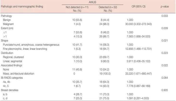
Using AWUS, malignant microcalcifications were more fre- quently visible than were benign microcalcifications (p=
0.003) (Table 3). The detection rate was 86.7% (26/30) for le- sions that were larger than 10 mm and only 46.2% (6/13) for lesions that were smaller than this (p=0.009). Lesions with
Figure 3. A 49-year-old wom- an with fibrocystic disease in the left breast. (A) Spot magni- fication mammogram shows grouped punctate or amor- phous microcalcifications in outer breast (arrows). (B) Auto- mated whole breast ultra- sound (US) shows hyperecho- ic microcalcifications within the hypoechoic area (arrows).
These microcalcifications were not detected by handheld US.
This may be due to the opera- tor dependency of handheld breast US.
A B
B
A C
Figure 4. A 58-year-old woman with ductal carcinoma in situ in the right breast. (A, B) Craniocaudal and mediolateral oblique views of mammogram shows fine pleomorphic microcalcifications in deep central breast (arrows). (C) Axial T1-weighted contrast-enhanced magnetic resonance image shows an irregular enhancing mass in central posterior portion of right breast (arrow). These microcalcifications were not seen by hand held ultra- sound (US) and automated whole breast US.
fine pleomorphic or fine linear shapes (94.7%, 18/19) were more frequently visible than lesions with round, amorphous, or coarse heterogeneous shapes (58.3%, 14/24) (p=0.021).
AWUS detected all 19 microcalcifications associated with masses or architectural distortions but only 13 of 24 (54.2%) lesions without associated abnormalities (p=0.022). In all 17 cases with invasive carcinoma, microcalcifications were ob- served within hypoechoic masses on AWUS. In six of the cases with DCIS, microcalcifications were observed within hy- poechoic masses, abnormal parenchyma, or hypoechoic duct- like structures (Figure 5).
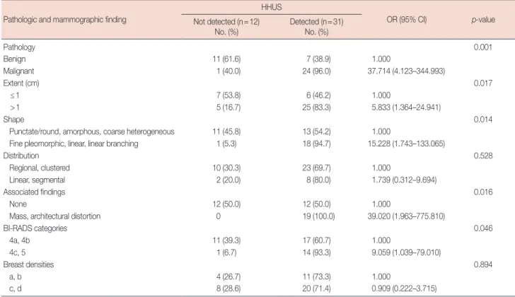
HHUS showed similar results to AWUS in the detection of mammographically suspicious microcalcifications (Table 4).
Using HHUS, malignant microcalcifications were more fre- quently visible than benign microcalcifications (p=0.001).
Table 3. Factors affecting the detectability of AWUS for mammographically suspicious microcalcifications Pathologic and mammographic finding
AWUS
OR (95% CI) p-value
Not detected (n=11)
No. (%) Detected (n=32) No. (%)
Pathology 0.003
Benign 10 (55.6) 8 (44.4) 1.000
Malignant 1 (4.0) 24 (96.0) 30.000 (3.302–272.343)
Extent (cm) 0.009
≤1 7 (53.8) 6 (46.2) 1.000
>1 4 (13.3) 26 (86.7) 7.583 (1.666–34.520)
Shape 0.021
Punctate/round, amorphous, coarse heterogeneous 10 (41.7) 14 (58.3) 1.000
Fine pleomorphic, linear, linear branching 1 (5.3) 18 (94.7) 12.856 (1.466–112.701)
Distribution 0.223
Regional, clustered 10 (30.3) 23 (69.7) 1.000
Linear, segmental 1 (10.0) 9 (90.0) 3.911 (0.436–35.122)
Associated findings 0.022
None 11 (45.8) 13 (54.2) 1.000
Mass, architectural distortion 0 19 (100.0) 33.220 (1.671–660.447)
BI-RADS categories 0.064
4a, 4b 10 (35.7) 18 (64.3) 1.000
4c, 5 1 (6.7) 14 (93.3) 7.778 (0.887–68.189)
Breast densities 0.905
a, b 4 (26.7) 11 (73.3) 1.000
c, d 7 (25.0) 21 (75.0) 1.091 (0.261–4.553)
AWUS=automated whole breast ultrasound; OR=odds ratio; CI=confidence interval; BI-RADS=Breast Imaging Reporting and Data System.
Figure 5. A 43-year-old woman with ductal carcinoma in situ in the left breast. (A) Craniocaudal view of mammogram shows segmental fine pleomor- phic microcalcifications in central (arrowhead) and peripheral outer breast (arrow). (B, C) Handheld ultrasound (US) shows an intraductal hypoechoic mass with microcalcifications (arrowheads), which matched with central microcalcifications on mammogram. (D) Automated whole breast US shows segmental ductal dilatation with intraductal microcalcifications (arrowhead), which matched with central microcalcifications on mammogram. Grouped microcalcifications (arrow) in peripheral breast are found, which matched with peripheral microcalcifications on mammogram.
A C
B
D
The detection rate was higher for lesions that were larger than 10 mm (83.3%) than for lesions that were smaller (46.2%, p=0.017). The detection rate was also higher for lesions with fine pleomorphic or fine linear shapes (94.7%) than for lesions with round, amorphous, or coarse heterogeneous shapes (54.2%, p=0.014). Likewise, the detection rate was higher for lesions associated with masses or architectural distortions (100%) than it was for lesions without associated abnormal- ities (50.0%, p=0.022). Finally, lesions categorized as BI-RADS 4c or 5 (93.3%) were more frequently detected than lesions categorized as BI-RADS 4a or 4b by mammography (60.7%, p=0.046).
DISCUSSION
The objective of this study was to examine the ability of AWUS to detect suspicious microcalcifications initially identi- fied by mammography and to characterize the lesion variables that affect the detectability of AWUS as compared to HHUS.
In this prospective evaluation of 43 cases, we showed that the detectability of AWUS was comparable to that of HHUS. Us- ing AWUS, 74.4% of mammographically suspicious microcal- cifications were identified (96% of malignant microcalcifica- tions and 44.4% of benign microcalcifications). Using HHUS,
Table 4. Factors affecting the detectability of HHUS for mammographically suspicious microcalcifications Pathologic and mammographic finding
HHUS
OR (95% CI) p-value
Not detected (n=12)
No. (%) Detected (n=31)
No. (%)
Pathology 0.001
Benign 11 (61.6) 7 (38.9) 1.000
Malignant 1 (40.0) 24 (96.0) 37.714 (4.123–344.993)
Extent (cm) 0.017
≤1 7 (53.8) 6 (46.2) 1.000
>1 5 (16.7) 25 (83.3) 5.833 (1.364–24.941)
Shape 0.014
Punctate/round, amorphous, coarse heterogeneous 11 (45.8) 13 (54.2) 1.000
Fine pleomorphic, linear, linear branching 1 (5.3) 18 (94.7) 15.228 (1.743–133.065)
Distribution 0.528
Regional, clustered 10 (30.3) 23 (69.7) 1.000
Linear, segmental 2 (20.0) 8 (80.0) 1.739 (0.312–9.694)
Associated findings 0.016
None 12 (50.0) 12 (50.0) 1.000
Mass, architectural distortion 0 19 (100.0) 39.020 (1.963–775.810)
BI-RADS categories 0.046
4a, 4b 11 (39.3) 17 (60.7) 1.000
4c, 5 1 (6.7) 14 (93.3) 9.059 (1.039–79.010)
Breast densities 0.894
a, b 4 (26.7) 11 (73.3) 1.000
c, d 8 (28.6) 20 (71.4) 0.909 (0.222–3.715)
HHUS=handheld breast ultrasound; OR=odds ratio; CI=confidence interval; BI-RADS=Breast Imaging Reporting and Data System.
72.1% of the microcalcifications were identified (96.0% of ma- lignant microcalcifications and 38.9% of benign microcalcifi- cations). These findings are consistent with previous studies evaluating HHUS, in which a 69% to 100% detection rate for malignant microcalcifications and a 23% to 66% detection rate for benign microcalcifications was described [7,8,12].
Both AWUS and HHUS identified malignant microcalcifi- cations more frequently than benign microcalcifications. It is likely that the hypoechoic backgrounds of the masses or duct- like structures increased the conspicuity and detectability of the echogenic microcalcifications. These US findings are simi- lar to those described previously [7, 8,11,12]. In our study, one DCIS lesion located deep in the central portion of the breast was not detected by either AWUS or HHUS. Sonographic posterior acoustic shadowing beneath the nipple may have in- fluenced the detectability of the lesion. In contrast to malig- nant lesions, microcalcifications associated with benign his- tology were visible in only eight of 18 cases. Microcalcifica- tions were detected within hypoechoic masses, anechoic cysts, and normal parenchyma using AWUS, similar to lesions de- scribed in a previous study [7]. The microcalcifications that were not visible on AWUS did not have associated masses or cysts. This discrepancy is believed to be due to a lack of con- trast between normal parenchyma with hyperechoic fibrous
ACKNOWLEDGMENTS
We thank the Clinical Research Coordinating Center at the Catholic University of Korea for statistical analyses.
REFERENCES
1. Sickles EA. Breast calcifications: mammographic evaluation. Radiology 1986;160:289-93.
2. Sickles EA. Mammographic features of 300 consecutive nonpalpable breast cancers. AJR Am J Roentgenol 1986;146:661-3.
3. Ciatto S, Cataliotti L, Distante V. Nonpalpable lesions detected with mammography: review of 512 consecutive cases. Radiology 1987;165:
99-102.
4. Meyer JE, Kopans DB, Stomper PC, Lindfors KK. Occult breast abnor- malities: percutaneous preoperative needle localization. Radiology 1984;
150:335-7.
5. Meyer JE, Eberlein TJ, Stomper PC, Sonnenfeld MR. Biopsy of occult breast lesions: analysis of 1261 abnormalities. JAMA 1990;263:2341-3.
6. Orel SG, Kay N, Reynolds C, Sullivan DC. BI-RADS categorization as a predictor of malignancy. Radiology 1999;211:845-50.
7. Moon WK, Im JG, Koh YH, Noh DY, Park IA. US of mammographi- cally detected clustered microcalcifications. Radiology 2000;217:849- 54.
8. Gufler H, Buitrago-Téllez CH, Madjar H, Allmann KH, Uhl M, Rohr- Reyes A. Ultrasound demonstration of mammographically detected microcalcifications. Acta Radiol 2000;41:217-21.
9. Hashimoto BE, Kramer DJ, Picozzi VJ. High detection rate of breast ductal carcinoma in situ calcifications on mammographically directed high-resolution sonography. J Ultrasound Med 2001;20:501-8.
10. Cheung YC, Wan YL, Chen SC, Lui KW, Ng SH, Yeow KM, et al. Sono- graphic evaluation of mammographically detected microcalcifications without a mass prior to stereotactic core needle biopsy. J Clin Ultra- sound 2002;30:323-31.
11. Soo MS, Baker JA, Rosen EL, Vo TT. Sonographically guided biopsy of suspicious microcalcifications of the breast: a pilot study. AJR Am J Roentgenol 2002;178:1007-15.
12. Soo MS, Baker JA, Rosen EL. Sonographic detection and sonographi- cally guided biopsy of breast microcalcifications. AJR Am J Roentgenol 2003;180:941-8.
13. Shipley JA, Duck FA, Goddard DA, Hillman MR, Halliwell M, Jones MG, et al. Automated quantitative volumetric breast ultrasound data- acquisition system. Ultrasound Med Biol 2005;31:905-17.
14. Tozaki M, Fukuma E. Accuracy of determining preoperative cancer ex- tent measured by automated breast ultrasonography. Jpn J Radiol 2010;
28:771-3.
15. Kim SH, Kang BJ, Choi BG, Choi JJ, Lee JH, Song BJ, et al. Radiologists’
performance for detecting lesions and the interobserver variability of automated whole breast ultrasound. Korean J Radiol 2013;14:154-63.
16. Shin HJ, Kim HH, Cha JH, Park JH, Lee KE, Kim JH. Automated ultra- sound of the breast for diagnosis: interobserver agreement on lesion de- tection and characterization. AJR Am J Roentgenol 2011;197:747-54.
17. Lin X, Wang J, Han F, Fu J, Li A. Analysis of eighty-one cases with breast structures and the microcalcifications.
In our study population, a case with a benign lesion, which displayed discrete hyperechoic dots within normal parenchyma on HHUS, was not detected on AWUS. This may be due to the lower resolution of AWUS resulting from the use of a lower frequency transducer and wider focal zone. In contrast, two be- nign lesions detected by AWUS were not visible using HHUS.
This may be due to the operator dependency of HHUS.
In our study, the mammographic features of the microcalci- fications that predicted their detectability by AWUS were a larger size (>10 mm) and a fine pleomorphic or linear shape.
The factors affecting the detectability of AWUS were similar to those affecting the detectability of HHUS for mammo- graphically suspicious microcalcifications. These results are consistent those of a previous study that found that the visibil- ity of clustered microcalcifications on HHUS was much high- er in cases with malignant microcalcifications, particularly for those larger than 10 mm [7].
Our study had some limitations. First, only a small number of lesions were evaluated, and these had been previously iden- tified by mammography; this is not representative of the gen- eral population. Further studies that include larger numbers of patients undergoing routine screening for breast cancer are required. Second, for lesions that were not visible by US and were instead confirmed by stereotactic biopsy, we considered their size, clock-wise location, and depth of the lesions and made an effort to correlate the findings between US and mammography. Finally, we did not consider the possibility of underestimation of pathologic results by US-guided 14-gauge core needle biopsy and stereotactic 11-gauge vacuum-assisted biopsy. However, 15 lesions diagnosed as benign by US-guid- ed 14-gauge core needle biopsy and stereotactic 11-gauge vac- uum-assisted biopsy showed no significant changes at the 24 to 30 months follow-up visits.
In conclusion, the detectability of AWUS was comparable to that of HHUS for mammographically suspicious microcal- cifications. The detectability of AWUS is much higher for ma- lignant microcalcifications, particularly those larger than 10 mm, having a suspicious morphology, and associated with masses or architectural distortion on mammography. There- fore, for highly suspicious microcalcifications, AWUS might be helpful to predict the feasibility of US-guided percutaneous procedures.
CONFLICT OF INTEREST
The authors declare that they have no competing interests.
lesions using automated breast volume scanner and comparison with handheld ultrasound. Eur J Radiol 2012;81:873-8.
18. Wang HY, Jiang YX, Zhu QL, Zhang J, Dai Q, Liu H, et al. Differentia- tion of benign and malignant breast lesions: a comparison between au- tomatically generated breast volume scans and handheld ultrasound examinations. Eur J Radiol 2012;81:3190-200.
19. Kotsianos-Hermle D, Hiltawsky KM, Wirth S, Fischer T, Friese K, Reiser M. Analysis of 107 breast lesions with automated 3D ultrasound and
comparison with mammography and manual ultrasound. Eur J Radiol 2009;71:109-15.
20. Kotsianos-Hermle D, Wirth S, Fischer T, Hiltawsky KM, Reiser M. First clinical use of a standardized three-dimensional ultrasound for breast imaging. Eur J Radiol 2009;71:102-8.
21. An YY, Kim SH, Kang BJ. The image quality and lesion characterization of breast using automated whole-breast ultrasound: a comparison with handheld ultrasound. Eur J Radiol 2015;84:1232-5.