302
ROOT CANAL TREATMENT OF A MANDIBULAR SECOND PREMOLAR WITH THREE SEPARATE ROOT CANALS
Seok-Ryun Lee, Seol-Hee Shin, Sung-Ok Hong, Chang-Kyu Song, Hoon-Sang Chang, Kyung-San Min*
Department of Conservative Dentistry, School of Dentistry, Wonkwang University, Iksan, Korea
Mandibular premolars show a wide variety of root canal anatomy. Especially, the occurrence of three canals with three separate foramina in mandibular second premolars is very rare. This case report describes the root canal treatment of an unusual morphological configuration of the root canal system and supplements previous reports of the existence of such configuration in mandibular second premolar. [J Kor Acad Cons Dent 35(4):302-305, 2010]
Key words:Mandibular second premolar, Root canal anatomy, Three separate canals
-Received 2010.5.29., revised 2010.6.20., accepted 2010.6.25.-
Ⅰ. Introduction
Successful root canal treatment requires an under- standing of root canal morphology and anatomy.
Accurate diagnosis of root canal morphology and anatomy is essential for thorough shaping and clean- ing of the entire root canal system and consequent successful root canal treatment.1)Mandibular premo- lars show a wide variety of root canal anatomy.2) There seems to be a racial predisposition for the presence of two or more canals in maxillary and mandibular premolars, as well as their bilateral occurrence.2,3) Especially, the occurrence of three canals with three separate foramina in mandibular premolars is very rare. Vertucci4) and Zillich et al5) reported the occurrence of three canals in mandibular first premolars at 0.5% and 0.4% respectively. Their studies in second premolars showed these percent- ages at 0.0% and 0.4%, respectively. To our knowl- edge, however, there is rare case report about treat-
ment of mandibular second premolar with three sep- arate canals divided at the apical level of the root whereas most previous reports showed that the ori- fices were found in the mid root section.6)The present case report describes the root canal treatment of a mandibular second premolar having three indepen- dent root canals with separate foramina.
Ⅱ. Case Report
A 27-year-old man with a noncontributory medical history was referred because of occasionally slight spontaneous pain in the mandibular left second pre- molar. The tooth was diagnosed as irreversible pulpi- tis and initiated to have root canal treatment by gen- eral dentist 2 days ago, but could not negotiate all of the root canals. After this treatment, the initial acute symptom had been relieved, but the tooth was still symptomatic and tender to percussion. A radiograph- ic image of the tooth showed an unusual anatomy of these teeth (Figure 1). According to clinical and radi- ographic examination, a diagnosis of pulp necrosis with apical periodontitis was made.
The tooth was anaesthetized, isolated with rubber dam and temporary filling was removed. The apical third was explored to locate the canal orifices using ABSTRACT
*Corresponding Author: Kyung-San Min Department of Conservative Dentistry, School of Dentistry, Wonkwang University 344-2 Shinyong, Iksan, 570-749, Korea
Tel: +82-63-850-6930 Fax: +82-63-859-2932 E-mail: mksdd@wonkwang.ac.kr
Case Report
Lee SR et al. JKACD Volume 35, Number 4, 2010
303 the operating microscope (OPMI Pico Dental
Microscope; Carl Zeiss, Oberkochen, Germany).
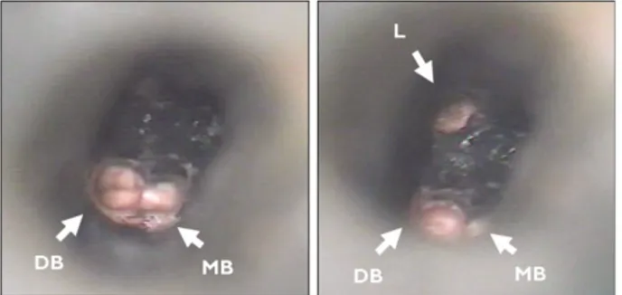
Three separated root canal orifices were found on the same level of the pulp chamber floor at a depth of 14.0 mm: one lingual, one distobuccal, and one mesiobuccal. Each canal had separate portal of exit.
The working lengths of root canals were determined using an electronic apex locator (Root ZX; J Morita Inc., Kyoto, Japan) and then verified by a radiograph (Figure 2). The working lengths of three canals were same as 19.0 mm. In addition, endodontic ultrasonic tips (CPR tips; Spartan, Fenton, MO, USA) were
used to achieve better straight-line access. Small, slightly pre-curved K-files were used to debride the canals and to establish a glide path to the working length. The canals were cleaned and shaped using nickel titanium rotary instrument (Protaper Universal; Dentsply-Mailiefer, Ballaigues, Switzerland). After drying the canals with paper points, all canals were obturated with warm gutta- percha technique with AH 26 (Dentsply, Konstanz, Germany) root canal sealer (Figure 3). A post-opera- tive radiograph showed separate three root canals (Figure 4).
Figure 1. Preoperative periapical radiograph of mandibular left second premolar.
Figure 2. The radiograph of working length measurement demonstrating three root canals. The H-file was inserted in the lingual canal.
Figure 3. Microscopic views of the furcation area and the root canal orifices following obturation.
Figure 4. Post-operative radiograph showing obturation of the root canal system.
Case Report
Root canal treatment of a mandibular second premolar with three separate root canals JKACD Volume 35, Number 4, 2010
304
Ⅲ. Discussion
In this case, the mandibular second premolar with three root canals could be treated successfully using a non-surgical method. The possibility of variations in root canal morphology must be considered before root canal treatment is undertaken.7) Careful inter- pretation of the root outline and the periodontal liga- ment space may suggest the presence of an extra root or canal.8)During the initial placement of scout- ing files in the assumptive site as canal orifice, clini- cians may encounter an obstruction and the files may deflect to the buccal, lingual, mesial or distal before it travels any further. This may indicate a canal divi- sion. Therefore, it is important to develop a sense of tactile feeling and direction with appropriate pre- curved scouting files to detect the multiple canal ori- fices.
When working under the microscope, clinicians can see the sodium hypochlorite bubbling in the extra canal, marking its presence. Furthermore, once it has been established that there are three canals, it is important to obtain straight-line access to all canals.
This may be achieved by ultrasonic tips, which reduce stress on the files used subsequently to shape the canals and minimize the risk of instrument sepa- ration and canal transportation.
In this case, tooth had the C-shaped canal shape as category II9)of the mesiobuccal and ligual distinct
canal. Extravagant use of small files and sodium hypochlorite is a key to thorough debridement of nar- row canal isthmus. Despite the existence of compli- cated dental anatomy, shaping outcomes with nickel- titanium rotary files prepares the canals to a prede- termined shape. Furthermore, warm gutta-percha filling technique has shown to allow the creation of excellent adaptation of filling material to root canals.
References
1. England MC, Hartwell GR, Lance JK. Detection and treatment of multiple canals in mandibular premolars.
J Endod 17:174-178, 1991.
2. Cleghorn BM, Christie WH, Dong CC. The root and root canal morphology of the human mandibular second premolar: a literature review. J Endod 33:1031-1037, 2007.
3. Trope M, Elfenbein L, Tronstad L. Mandibular premo- lars with more than one root canal in different race groups. J Endod 12:343-345, 1986.
4. Vertucci FJ. Root canal morphology of mandibular pre- molars. J Am Dent Assoc 97:47-50, 1978.
5. Zillich R and Dowson J. Root canal morphology of mandibular first and second premolars. Oral Surg Oral Med Oral Pathol 36:738-744, 1973.
6. Nallapati S. Three canal mandibular first and second premolars: a treatment approach. J Endod 31:474- 476, 2005.
7. Slowy RR. Root canal anatomy. Road map to successful endodontics. Dent Clin North Am 23:555-573, 1979.
8. Slowy RR. Radiographic aids in the detection of extra root canal. Oral Surg Oral Med Oral Pathol 37:762- 772, 1974.
9. Melton DC, Krell KV, Fuller MW. Anatomical and his- tological features of C-shaped canals in mandibular second molars. J Endod 17:384-388, 1991.
Case Report
Lee SR et al. JKACD Volume 35, Number 4, 2010
305 국문초록
세 개의 분리된 근관을 갖는 하악 제2소구치의 치험례
이석련∙신설희∙홍성옥∙송창규∙장훈상∙민경산*
원광대학교 치과대학 치과보존학교실
하악소구치의 근관형태는 매우 다양하다. 특히, 근단공까지 분리주행하는 세 개의 근관을 갖는 하악 제2소구치는 매우 드물 다. 본 증례는 하악 제2소구치의 드문 근관형태를 기술하고, 상기 치아들에서 본 증례와 같이 세 개의 근관이 존재한다는 이전 의 연구들을 뒷받침하고 있다.
주요단어: 하악 제2소구치, 근관형태, 분리주행
Case Report
Root canal treatment of a mandibular second premolar with three separate root canals JKACD Volume 35, Number 4, 2010