Background and PurposezzThis study aimed to estimate the changes in gray matter vol- ume (GMV) and their hemispheric difference in patients with mesial temporal lobe epilepsy (MTLE) using a voxel-based morphometry (VBM) methodology, and to determine whether GMV changes are correlated with clinical features.
MethodszzVBM analysis of brain MRI using statistical parametric mapping 8 (SPM8) was performed for 30 left MTLE (LMTLE) and 30 right MTLE (RMTLE) patients and 30 age- and sex-matched healthy controls. We also analyzed the correlations between GMV changes and clinical features of MTLE patients.
ResultszzIn SPM8-based analyses, MTLE patients showed significant GMV reductions in the hippocampus ipsilateral to the epileptic focus, bilateral thalamus, and contralateral puta- men in LMTLE patients. The GMV reductions were more extensive in the ipsilateral hippo- campus, thalamus, caudate, putamen, uncus, insula, inferior temporal gyrus, middle occipital gyrus, cerebellum, and paracentral lobule in RMTLE patients. These patients also exhibited notable reductions of GMV in the contralateral hippocampus, thalamus, caudate, putamen, and inferior frontal gyrus. We observed that GMV reduction was positively correlated with several clinical features (epilepsy duration and seizure frequency in RMTLE, and history of febrile seizure in LMTLE) and negatively correlated with seizure onset age in both the RM- TLE and LMTLE groups.
ConclusionszzOur study revealed GMV decreases in the hippocampus and extrahippocampal regions. Furthermore, the GMV reduction was more extensive in the RMTLE group than in the LMTLE group, since it included the contralateral hemisphere in the former. This difference in the GMV reduction patterns between LMTLE and RMTLE may be related to a longer epilepsy duration and higher seizure frequency in the latter.
Key Wordszz mesial temporal lobe epilepsy, gray matter volume, voxel-based morphometry, hippocampal sclerosis, magnetic resonance imaging.
Asymmetric Gray Matter Volume Changes Associated with Epilepsy Duration and Seizure Frequency
in Temporal-Lobe-Epilepsy Patients with Favorable Surgical Outcome
INTRODUCTION
Mesial temporal lobe epilepsy (MTLE) is the most common type of refractory partial epi- lepsy, and is commonly associated with hippocampal sclerosis (HS).1,2 Voxel-based mor- phometry (VBM) has emerged from recent developments in neuroimaging analysis tech- niques to become a popular technique for whole-brain magnetic resonance imaging (MRI) that allows focal differences between patients and matched healthy controls to be investi- gated.3
Jeong Sik Kima,b* Dae Lim Kooc* Eun Yeon Jooa,b Sung Tae Kimd Dae Won Seoa,b Seung Bong Honga,b
a Department of Neurology, Neuroscience Center, Samsung Medical Center and Samsung Advanced Institute for Health Sciences & Technology (SAIHST), Sungkyunkwan University School of Medicine, Seoul, Korea
b Samsung Biomedical Research Institute (SBRI), Seoul, Korea
c Department of Neurology, Seoul National University Boramae Hospital,
Seoul, Korea
d Department of Radiology, Samsung Medical Center, Sungkyunwan University School of Medicine, Seoul, Korea
pISSN 1738-6586 / eISSN 2005-5013 / J Clin Neurol 2016;12(3):323-331 / http://dx.doi.org/10.3988/jcn.2016.12.3.323
Received November 19, 2015 Revised December 25, 2015 Accepted December 28, 2015 Correspondence Seung Bong Hong, MD, PhD Department of Neurology, Samsung Medical Center, Sungkyunkwan University School of Medicine, 81 Irwon-ro, Gangnam-gu, Seoul 06351 Korea Tel +82-2-3410-3592 Fax +82-2-3410-0052 E-mail sbhong@skku.edu
*These authors contributed equally to this work.
cc This is an Open Access article distributed under the terms of the Creative Commons Attribution Non-Com- mercial License (http://creativecommons.org/licenses/by-nc/3.0) which permits unrestricted non-commercial use, distribution, and reproduction in any medium, provided the original work is properly cited.
JCN
Open Access ORIGINAL ARTICLEAsymmetric GMV Changes in Temporal Lobe Epilepsy Patients
JCN
Previous VBM studies have consistently demonstrated de- creases in the gray matter volume (GMV) or gray matter concentration in the mesiotemporal structures ipsilateral to the epileptic focus, as well as the bilateral extratemporal re- gions, in unilateral MTLE patients.3-7 Gray matter abnormal- ities have been reported most frequently in the hippocam- pus,5,8-11 followed by the thalamus, parietal lobe, and cingulate gyrus. Changes have also been described in the parahippo- campal gyrus, middle temporal gyrus, superior temporal gy- rus, inferior temporal gyrus, fusiform gyrus, temporal pole, entorhinal cortex, amygdala, and perirhinal cortex.3-7,9,10,12-16
However, some of these studies have yielded discrepant re- sults, presumably due to the different methodologies em- ployed. For example, studies have differed with regard to the types of VBM methods applied (e.g., standard vs. optimized VBM), VBM-related parameters (the smoothing kernel, nui- sance variables, and normalization scheme), template choice [statistical parametric mapping (SPM) templates vs. user- created templates], sample size (patients and controls), and characteristics of study subjects.
In the current VBM study, we aimed to identify the distri- bution of focal GMV reductions and hemispheric differences in unilateral MTLE patients who had undergone epilepsy surgery, had histological HS, and who showed a good post- surgical outcome for more than 2 years using SPM8 plus a Diffeomorphic Anatomical Registration using Exponentiat- ed Lie Algebra (DARTEL)17 algorithm and a new segmenta- tion toolbox.
We compared the brain GMV between MTLE patients and controls. Furthermore, we sought to determine the clini- cal implications of GMV changes by investigating their rela- tionship with clinical features of the patients.
METHODS
Subjects and clinical information
This study included 30 left MTLE patients [LMTLE; age 34.2±11.1 years (mean±SD), age range 11–56 years, 16 fe- males and 14 males] and 30 right MTLE patients (RMTLE;
age 36.9±9.5 years, age range 19–57 years of age, 13 females and 17 males). The MTLE patients were enrolled based on the following inclusion criteria: 1) unilateral HS with no oth- er lesion on brain MRI, 2) ictal electroencephalography (EEG) pattern arising from the ipsilateral temporal lobe to HS, 3) an anterior temporal lobectomy with amygdalohippo- campectomy, and 4) seizure-free outcome after the surgery.
All patients underwent a comprehensive presurgical evalua- tion including a combination of seizure semiology, ictal and interictal EEG results, neuropsychology, and neuroimaging findings. They underwent surgery (anterior temporal lobec-
tomy with amygdalohippocampectomy) and showed a good surgical outcome (Engle Class I) for at least 2 years postoper- atively. In addition, HS was the only lesion in all patients pathologically verified by histological analysis. We excluded patients who had bilateral hippocampal atrophy, normal MRI findings, or multiple pathologies. Thirty age- and sex- matched healthy controls (age 39.0±7.9 years, age range 30–
60 years, 15 females and 15 males) with no neurologic or psychiatric disorders were also included. Informed consent was obtained from all participants, and the institutional re- view board of the Samsung Medical Center approved the study protocol (IRB No. 2011-02-049).
MRIAll of the included patients underwent high-resolution brain MRI in a 1.5-tesla scanner (GE Medical Systems, Milwaukee, WI, USA) equipped with an eight-channel head coil, yield- ing T1-weighted three-dimensional spoiled-gradient-recalled (SPGR), T2-weighted, and fluid-attenuated inversion recov- ery (FLAIR) imaging scans. Coronal SPGR MRI images were obtained with the following parameters: 1.6 mm slice thick- ness, no gap, 124 slices, repetition time (TR)=30 ms, echo time (TE)=7 ms, flip angle (FA)=45 degrees, number of exci- tations (NEX)=1, matrix size=256×192, and field of view (FOV)=22×22 cm. The voxel dimensions of the SPGR imag- es were 0.86×0.86×1.6 mm. Oblique coronal FLAIR MRI was performed with the following settings: 4.0 mm slice thickness, 1.0 mm gap, 32 slices, TR/TE=10,002/127.5 ms, 1 NEX, matrix size=256×192, and FOV=20×20 cm. Oblique coronal T2-weighted MRI images were obtained using the following parameters: 3.0 mm slice thickness, 0.3 mm gap, 56 slices, TR/TE=5,300/99 ms, FA=90 degrees, 3 NEX, ma- trix size=256×192, and FOV=20×20 cm.
Data preprocessing
For all subjects, Digital Imaging and Communications in Medicine format images were transformed into the ANA- LYZE format specified by the Mayo Clinic using ITK-SNAP freeware (Paul Yushkevich; http://www.itksnap.org). A recent VBM approach, including the new segmentation toolbox (http://www.fil.ion.ucl.ac.uk/spm/software/spm8) and DARTEL17 algorithm in SPM8, was applied to anatomical T1-weighted MRI data to detect GMV differences between unilateral MTLE patients and healthy controls. In the pre- processing stage, all raw images were segmented and spatial- ly normalized using the new segmentation toolbox that is an extension of the default unified method in SPM8,18 but uses a novel segmentation approach involving an adaptive maxi- mum-a-posterior technique without tissue priori, and addi- tionally uses partial volume estimation with a simplified
Kim JS et al.
JCN
mixed model. Furthermore, two denoising methods (spa- tially adaptive nonlocal-means denoising filter and a classical Markov random-field approach) were applied to the seg- mentation process. Another important extension to the SPM8 segmentation is integration of the DARTEL normal- ization in the toolbox, and thus no additional Montreal Neu- rological Institute (MNI) normalization is necessary. The segmented and normalized images were then modulated and smoothed with a 10-mm full-width-half-maximum isotropic Gaussian kernel to allow for high variability in the intersub- ject gyral anatomy, while also preserving the total gray mat- ter signal in the normalized tissue images.19 We further per- formed manual hippocampal volume (HV) measurements using ANALYZE software (version 10.0, Mayo Clinic) to in- vestigate how changes in GMV were associated with the HV in all patients and healthy controls, according to a previously described anatomical protocol.20 The entire HV was mea- sured from the anterior head to the posterior tail, including the cornu ammonis, gyrus dentatus, hippocampus, and su- biculum. The anterior boundary of the hippocampus was identified as the alveus. The lateral border of the hippocam- pus was delineated against the entorhinal cortex based on the upper margin of the white matter of the subiculum. The pos- terior end of the hippocampus was taken as the point at which the tail of the hippocampus disappeared. The rater manually traced the alveus according to the defined hippo- campal boundary criteria. In order to calculate the intrarater correlation coefficients (ICCs), the brain images of each sub- ject were manually traced twice. The ICCs were 0.96 and 0.95 for the left and right HVs, respectively.
Statistical analysis
Prior to statistical analyses, differences in sex, age, and whole- brain volume between patients and control groups were ex- amined using the independent-samples t-test, with p values of <0.05 being considered indicative of statistical significance.
Spatially processed images from the LMTLE and RMTLE
patients and healthy controls were statistically compared on a voxel-by-voxel basis using the general linear model, which can be used to detect increases or decreases in gray matter in specific brain regions. Whole-brain voxel analyses were cor- rected for multiple comparisons using a topological false dis- covery rate (FDR) statistical threshold of pFDR <0.05 with an extent threshold for clusters with at least 100 contiguous voxels (each voxel had a size of 1×1×1 mm). We confirmed a set of MNI stereotaxic coordinates (x, y, z) provided in the SPM results by visual analyses and further converted them into anatomical names using xjView8.4 (Xu Cui, www.alive- learn.net/xjview8), which is compatible with SPM8. For all voxel-wise analyses, we applied analysis of covariance with age and sex as nuisance covariates to minimize the influence of potential confounding factors, and we corrected for the intracranial volume by globally correcting the individual head sizes between the RMTLE, LMTLE, and healthy con- trol groups. No grand-mean scaling and non-sphericity cor- rection were performed or set. Implicit masking was used to restrict the analysis to one tissue type (gray matter). The in- tracranial volume was manually measured on sagittal T1- weighted MRI images of each subject using ITK-SNAP soft- ware. For MTLE patients, the correlations of changes in GMV with the duration of epilepsy, seizure frequency, age at seizure onset, history of febrile seizure, and number of anti- epileptic drugs (AEDs) were assessed by applying an uncor- rected probability (pUNC) significance value of <0.001. The extent threshold for the whole brain was set to KE >100.
RESULTS
Demographic and clinical characteristics
All patients and healthy controls were right-handed and pre- dominantly middle-aged (mean age 33.4 years). We inspect- ed T1-weighted, T2-weighted, and FLAIR images to exclude subjects with gross structural abnormalities observable in brain MRI scans. The detailed clinical findings for the pa-
Table 1. Demographic and clinical information for patients with left mesial temporal lobe epilepsy (LMTLE), patients with right mesial temporal lobe epilepsy (RMTLE), and healthy controls (HC)
Clinical parameter LMTLE RMTLE HC p
Number of subjects (males/females) 30 (14/16) 30 (17/13) 30 (15/15) NS
Age, years; mean±SD (range) 34.2±11.1 (11–56) 36.9±9.5 (19–57) 39.0±7.9 (30–60) NS
Duration of epilepsy, years 17.3±9.1 (6–40) 18.7±6.2 (8–41) - NS
Number of seizures per month 3.0±3.3 (0.2–18) 3.8±7.9 (0.2–45) - NS
Seizure onset age, years 17.0±8.6 (3–32) 15.5±9.2 (2–37) - NS
History of febrile seizure 20 15 - NS
Number of current AEDs 2.7±0.7 (1–4) 2.8±0.9 (1–5) - NS
Data are mean±SD (range) values or n values.
AED: antiepileptic drug, NS: not significant in independent-samples t-test.
Asymmetric GMV Changes in Temporal Lobe Epilepsy Patients
JCN
tients with MTLE and control subjects are summarized in Table 1. No significant differences in any of the demographic data (age or sex) were observed between the control, LMTLE, and RMTLE groups. The seizure frequency, average duration of epilepsy, seizure onset age, history of febrile seizure, and number of AEDs did not differ significantly between the LMTLE and RMTLE groups.
Reduced GMV in MTLE
Relative to healthy controls, patients with LMTLE showed significant GMV reductions in the left hippocampus, left parahippocampal gyrus, bilateral thalamus, and right puta- men (pFDR<0.05) (Fig. 1, Table 2). In patients with RMTLE, the GMV was significantly decreased in the bilateral hippo- campus, right parahippocampal gyrus, right uncus, right in- sula, right middle temporal gyrus, right inferior temporal gyrus, bilateral thalamus, bilateral putamen, bilateral caudate, left inferior frontal gyrus, right paracentral lobule, right mid- dle occipital gyrus, and right cerebellum (pFDR<0.05) (Fig. 2, Table 2). We observed that the whole-brain GMVs in pa- tients with RMTLE were not only negatively correlated with the duration of epilepsy and seizure frequency, but also posi- tively correlated with seizure onset age (pUNC<0.001). More-
over, the whole-brain GMVs in patients with LMTLE were positively correlated with seizure onset age and negatively correlated with a history of febrile seizure (pUNC<0.001) (Fig.
3, Table 3).
DISCUSSION
We applied VBM analysis to patients with MTLE and ob- served GMV reductions in the hippocampus and extrahip- pocampal regions. The main findings of this study were as follows:
1. Patients with LMTLE or RMTLE exhibited a significant GMV reduction in the ipsilateral mesial temporal lobe (in- cluding in the hippocampus and the parahippocampal gy- rus) on the side of the seizure focus.
2. In patients with RMTLE, widespread reductions of GMV were observed in both cortical and subcortical struc- tures.
3. GMV was negatively correlated with epilepsy duration and seizure frequency in patients with RMTLE, and positively correlated with seizure onset age in both LMTLE and RMTLE groups and with febrile seizure in patients with LMTLE.
Previous VBM studies have drawn conflicting conclusions
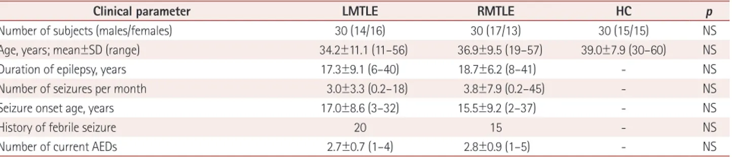
Fig. 1. Statistical brain map revealing gray matter volume (GMV) reductions in the brains of patients with left mesial temporal lobe epilepsy (LM- TLE) compared with controls. Decreased GMV in patients with LMTLE in the ipsilateral (left) hippocampus, bilateral thalamus, and contralateral (right) putamen. These findings were significant at a corrected false discovery rate (FDR) statistical threshold of pFDR<0.05. The left side of the brain images corresponds to the right hemisphere.
R L
8 6 4 2 0
Kim JS et al.
JCN
regarding GMV reduction patterns in patients with temporal lobe epilepsy (TLE), and their association with unilateral HS.
Some studies found that the loss of gray matter was more exten- sive in patients with LMTLE than in those with RMTLE.6,21,22 Another study found that the bilateral frontal lobe and the right cingulate gyrus are more susceptible to injury in pa- tients with left TLE than in those with right TLE, and that these regions are involved in cognitive disorders and im- paired executive functions.23 In contrast, we observed that the GMV reduction was more extensive in patients with RMTLE than in those with LMTLE. A widespread pattern of GMV reduction in RMTLE is consistent with the results obtained in the VBM-MRI study11 for the ipsilateral/contralateral in- sula, especially the contralateral thalamus, in RMTLE com- pared with LMTLE. Those authors concluded that the mag- nitude and extent of right mesiotemporal connectivity to the extrahippocampal structures—especially to the contralateral thalamus—are greater in RMTLE than in LMTLE. Another study also found that the GMV reductions in the contralateral
hippocampus (p=0.065) and thalamus (p=0.052) were more pronounced in RMTLE, even though distinct patterns of structural and metabolic changes within both hippocampal and thalamic regions were observed in two subgroups.24 That study did not explain these differences based on the clinical factors of patients in each group, such as epilepsy duration, seizure frequency, age at seizure onset, or interictal EEG pat- tern, instead explaining these differences by noting that sig- nificant correlations between neuroanatomical and func- tional changes were found only in the RMTLE patients.
Some VBM studies have noted that the degree of atrophy and regional distribution differ between LMTLE and RMTLE groups. Some studies have revealed a more widespread hemispheric GMV reduction in LMTLE6,9,22 or an identical pattern of GMV decrease in LMTLE and RMTLE patients.5 These discrepancies among the studies—in terms of varia- tions in GMV decreases—probably reflect variations in methodology, such as the absence of customized templates,5 the types of correlations compared between patients and Table 2. Brain regions with significant gray matter volume reduction in patients with unilateral mesial temporal lobe epilepsy (MTLE) compared with control subjects
Location Side Peak level MNI coordinates (mm)
T KE ZE PFDR x y z
LMTLE patients
Hippocampus L 8.83 5,251 7.00 <0.001 -27 -30 -6
Parahippocampal gyrus L 8.83 1,548 7.00 <0.001 -24 -32 -8
Thalamus L 7.65 512 6.34 <0.001 -9 -15 11
R 5.19 153 4.69 <0.001 11 -15 11
Putamen R 3.51 128 3.33 0.030 27 9 -5
RMTLE patients
Inferior frontal gyrus L 3.55 132 3.37 0.016 -50 30 -3
Paracentral lobule R 3.38 140 3.23 0.023 26 -33 -6
Hippocampus L 4.99 443 4.53 <0.001 -18 -30 -6
R 11.20 10,095 Inf. <0.001 30 -18 -18
Parahippocampal gyrus R 11.20 2,946 Inf. <0.001 28 -37 -8
Uncus R 11.20 254 Inf. <0.001 8 11 4
Insula R 11.20 1,935 Inf. <0.001 38 -2 3
Superior temporal gyrus R 3.35 127 3.19 0.024 52 10 -12
Middle temporal gyrus R 3.35 121 3.19 0.024 59 4 -14
Inferior temporal gyrus R 3.35 145 3.19 0.024 60 -4 -20
Thalamus L 4.79 174 4.38 0.001 -6 -21 9
R 4.99 685 4.53 0.046 3 -15 9
Putamen L 3.41 180 3.24 0.022 42 -59 11
R 5.29 513 4.76 0.049 -28 -33 -17
Caudate L 4.03 447 3.77 0.005 -15 20 0
R 4.75 463 4.35 0.001 17 18 0
Middle occipital gyrus R 4.22 239 3.92 0.003 33 -75 21
Cerebellum R 3.67 156 3.46 0.012 9 -71 -54
FDR: false discovery rate, Inf: infinity (z-score >7.0), KE: cluster extent threshold (=100 voxels), L: left, MNI: Montreal Neurological Institute, R: right, T:
test statistics, ZE: z-score (the number of standard deviations away from the mean).
Asymmetric GMV Changes in Temporal Lobe Epilepsy Patients
JCN
controls,6 the cortical thickness measurements, or the t-test ap- plied to the adjusted local GMV in the present study. Some results were based on only small samples.22 It is significant that all of our patients had been seizure-free for 24 h, given that seizures very close to the time of scanning might influ- ence the VBM analysis.
The extensive GMV reduction observed in RMTLE in this study is supported by the following findings: first, the GMV reduction was negatively correlated with several clinical fac- tors, including epilepsy duration and seizure frequency, in the RMTLE group, but not in the LMTLE group. Although the correlations detailed in Table 3 (at the level of a cluster extent threshold of KE ≥100) do not cover all of the regions in which the RMTLE and LMTLE patients differed, we iden- tified that, at borderline significance, GMV reductions in the middle frontal gyrus [KE=91, coordinates=(39,48,12), pFDR=0.020]
and temporal subgyral [KE=55, coordinates=(47,-4,-18), pFDR= 0.027] areas were negatively correlated with the duration of epilepsy and seizure frequency, respectively. Second, the av- erage ipsilateral and contralateral HVs were slightly smaller in patients with RMTLE than in patients with LMTLE (ipsilat- eral: RMTLE=1898±559 mm3, LMTLE=1973±462 mm3, p= 0.32;
contralateral: RMTLE= 3110±453 mm3, LMTLE=3287±449
mm3, p=0.094). Moreover, HVs in RMTLE were negatively cor- related with the epilepsy duration (p=0.004, r=-0.505). This conclusion is supported by a recent study25 that examined patterns of changes in GMV and white-matter connectivity in TLE patients with or without HS. That study found that the damage to the white matter was more extensive in TLE patients with HS than in those without HS, despite wide- spread extrahippocampal GMV reduction being observed in both groups. Those authors suggested that the positive corre- lation between ipsilateral HV loss and fractional anisotropy (FA) reduction of the corpus callosum and ipsilateral cingu- lum in the group of patients with TLE and HS supports the hypothesis that neuronal dysfunction and white-matter ab- normality are due to the excitotoxic effects of spreading epilep- togenic activity in the hippocampus, and in extrahippocam- pal brain regions that are directly or indirectly connected to it.
We observed GMV reductions in the subcortical region and bilateral thalamus in both MTLE groups, with the GMV reduction being greater in the ipsilateral thalamus than in the contralateral thalamus. These results are consistent with those of previous studies.12,26 The thalamus has been widely considered to be part of a mesial temporal-limbic network, and to exhibit widespread reciprocal connections with sub-
10 8 6 4 2 0
10 8 6 4 2
B 0
A
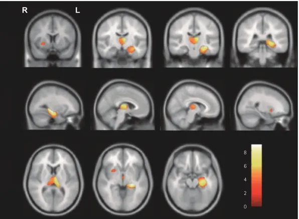
Fig. 2. Statistical brain map revealing GMV reductions in the brains of patients with right mesial temporal lobe epilepsy (RMTLE) compared with control subjects. A: Decreased GMV in patients with RMTLE in the bilateral hippocampus, right parahippocampal gyrus, right uncus, right insula, bilateral thalamus, bilateral putamen, bilateral caudate, right inferior temporal gyrus, right superior/middle/inferior temporal gyrus, right middle occipital gyrus, right cerebellum, and left inferior frontal gyrus. These findings were significant at pFDR<0.05. B: Brain regions with decreased GMV are shown on a rendered three-dimensional brain surface. The left side of the brain images corresponds to the right hemisphere. GMV: gray mat- ter volume.
Kim JS et al.
JCN
cortical structures and other cortical regions. The thalamus might play an important role in determining the extent of seizure propagation to other brain regions.13,17,20-24,26,27 The findings of other studies also support the idea that structural abnormalities in the thalamus influence the pathogenesis of TLE seizures.13,28,29 These studies have suggested that the ipsi- lateral thalamic GMV reduction could be associated with HS, and that contralateral thalamic atrophy could be caused by
the secondary generalization of seizure activities via the in- terhemispheric pathway. Similarly, we found in the present study that the GMV reductions in the caudate and putamen were greater in the RMTLE group than in the LMTLE group.
These results are similar to those obtained in previous VBM studies.9 We hypothesize that the atrophic pattern of basal ganglia could be influenced by clinical factors including the degree of HS, epilepsy duration, seizure onset age, and sei-
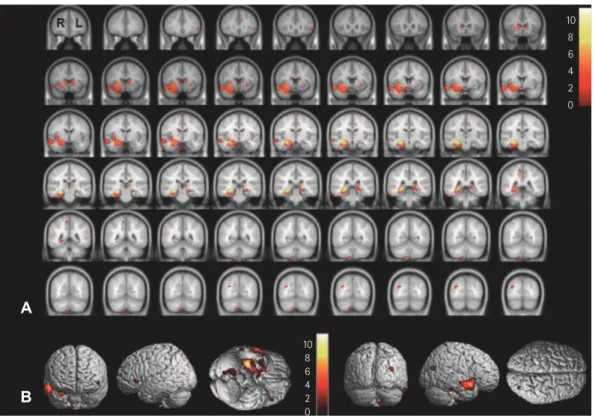
Fig. 3. Brain regions exhibiting significant correlations between changes in GMV and clinical parameters. A and B: Seizure onset age was positive- ly correlated with the GMVs of the right middle temporal gyrus (A) and left parahippocampal gyrus (B) in patients with RMTLE. C and D: In LMTLE patients, seizure onset age was positively correlated with GMV changes in the right extranuclear (C) and right occipital (D) lobes. E and F: Febrile seizures were negatively correlated with GMV changes in the left middle frontal gyrus (E) and right superior temporal gyrus (F) in patients with LMTLE. These findings were significant at an uncorrected p value of <0.001. GMV: gray matter volume, LMTLE: left mesial temporal lobe epilepsy, RMTLE: right mesial temporal lobe epilepsy.
4 3 2 1
E F 0
C D
A
L R L R
B
Table 3. Brain regions with significant correlations between GMV and clinical parameters in patients with unilateral MTLE
Group Location Peak level MNI coordinates (mm)
T ZE PUNC x y z
RMTLE
Duration of epilepsy (-) Middle frontal gyrus R 4.03 3.55 <0.001 29 -1 59
Precentral gyrus R 3.62 3.25 0.001 48 15 9
Parietal lobe (subgyral) L 3.49 3.16 0.001 -32 -45 49
Seizure frequency (-) Temporal lobe (subgyral) R 3.77 3.36 <0.001 46 -33 -5
Seizure onset age (+) Superior frontal gyrus L 3.69 3.30 <0.001 -9 -7 67
Middle temporal gyrus R 4.16 3.64 <0.001 37 -2 -35
Parahippocampal gyrus L 3.99 3.52 <0.001 -23 -2 -27
Medial globus pallidus L 3.85 3.42 <0.001 -17 -7 -6
Cuneus L 3.84 3.41 <0.001 -23 -88 33
LMTLE
Seizure onset age (+) Occipital lobe R 4.18 3.65 <0.001 2 -71 0
Extranuclear R 3.82 3.40 <0.001 34 -2 3
Insula L 3.56 3.20 0.001 -35 2 -5
Febrile seizure (-) Middle frontal gyrus L 3.77 3.36 <0.001 -38 41 21
Superior temporal gyrus R 3.47 3.14 0.001 48 -33 9
+: positive correlation, -: negative correlation, GMV: gray matter volume, L: left, MNI: Montreal neurological institute, MTLE: mesial temporal lobe epi- lepsy, T: test statistics, R: right, UNC: uncorrected, ZE: z-score (the number of standard deviations away from the mean).
Asymmetric GMV Changes in Temporal Lobe Epilepsy Patients
JCN
zure frequency in patients with MTLE.30-32
GMV reduction in the left uncus was previously reported in patients with left TLE.33 Those authors suggested that the uncus damage could be associated with the febrile seizures and status epilepticus being more intense in left TLE than in right TLE. However, we found significant GMV reductions in the right uncus in the RMTLE group. The GMV reduction in the right uncus, which lies anatomically at the anterior and most-medial portion of the parahippocampal gyrus, may be related to the more-frequent seizures propagated from the ipsilateral hippocampus in the patients with RMTLE. We also observed GMV reductions in the bilateral inferior fron- tal gyrus. This finding may be explained by structural or functional deficits caused by the propagation of epileptic dis- charges from the temporal epileptic focus to the frontal lobes that have been observed in MTLE (e.g., damage to fronto- subcortical circuits, volume loss, and metabolic changes in the extratemporal regions).34-36
In conclusion, our study has revealed the extent and asym- metric pattern of brain GMV reductions in patients with RMTLE and LMTLE using the VBM approach in SPM8 plus the DARTEL algorithm and a novel segmentation toolbox.
We detected that the structural abnormalities in the bilateral subcortical and ipsilateral extrahippocampal areas were more extensive in the RMTLE group. Furthermore, we found that the GMV reductions were positively correlated with multiple clinical factors (duration of epilepsy and seizure frequency in RMTLE, and history of febrile seizure in LMTLE) and nega- tively correlated with seizure onset age in both groups. Our study indicates that the SPM8-based VBM method may be useful for obtaining a deeper understanding of the patho- physiology and epileptic networks involved in MTLE.
Conflicts of Interest
The authors have no financial conflicts of interest.
Acknowledgements
This study was funded by a grant of the Korean Health Technology R&D Project, Ministry for Health and Welfare, Republic of Korea (No. A110097) and by Basic Science Research Program through the National Research Foundation of Korea of the Ministry of Science, ICT & Future Planning, Republic of Korea (No. 2014R1A1A3049510).
REFERENCES
1. Bae EK, Jung KH, Chu K, Lee ST, Kim JH, Park KI, et al. Neuro- pathologic and clinical features of human medial temporal lobe epi- lepsy. J Clin Neurol 2010;6:73-80.
2. Regesta G, Tanganelli P. Clinical aspects and biological bases of drug-resistant epilepsies. Epilepsy Res 1999;34:109-122.
3. Keller SS, Roberts N. Voxel-based morphometry of temporal lobe epilepsy: an introduction and review of the literature. Epilepsia 2008;
49:741-757.
4. Bernasconi N, Bernasconi A, Andermann F, Dubeau F, Feindel W, Re- utens DC. Entorhinal cortex in temporal lobe epilepsy: a quantitative
MRI study. Neurology 1999;52:1870-1876.
5. Bernasconi N, Duchesne S, Janke A, Lerch J, Collins DL, Bernasconi A. Whole-brain voxel-based statistical analysis of gray matter and white matter in temporal lobe epilepsy. Neuroimage 2004;23:717-723.
6. Bonilha L, Rorden C, Halford JJ, Eckert M, Appenzeller S, Cendes F, et al. Asymmetrical extra-hippocampal grey matter loss related to hippocampal atrophy in patients with medial temporal lobe epilepsy.
J Neurol Neurosurg Psychiatry 2007;78:286-294.
7. Keller SS, Wilke M, Wieshmann UC, Sluming VA, Roberts N. Com- parison of standard and optimized voxel-based morphometry for analysis of brain changes associated with temporal lobe epilepsy. Neu- roimage 2004;23:860-868.
8. Bonilha L, Edwards JC, Kinsman SL, Morgan PS, Fridriksson J, Ror- den C, et al. Extrahippocampal gray matter loss and hippocampal de- afferentation in patients with temporal lobe epilepsy. Epilepsia 2010;
51:519-528.
9. Bonilha L, Rorden C, Castellano G, Pereira F, Rio PA, Cendes F, et al.
Voxel-based morphometry reveals gray matter network atrophy in re- fractory medial temporal lobe epilepsy. Arch Neurol 2004;61:1379-1384.
10. Keller SS, Mackay CE, Barrick TR, Wieshmann UC, Howard MA, Roberts N. Voxel-based morphometric comparison of hippocampal and extrahippocampal abnormalities in patients with left and right hippocampal atrophy. Neuroimage 2002;16:23-31.
11. Pail M, Brázdil M, Marecek R, Mikl M. An optimized voxel-based morphometric study of gray matter changes in patients with left-sided and right-sided mesial temporal lobe epilepsy and hippocampal scle- rosis (MTLE/HS). Epilepsia 2010;51:511-518.
12. Bonilha L, Rorden C, Castellano G, Cendes F, Li LM. Voxel-based morphometry of the thalamus in patients with refractory medial tem- poral lobe epilepsy. Neuroimage 2005;25:1016-1021.
13. Guye M, Régis J, Tamura M, Wendling F, McGonigal A, Chauvel P, et al. The role of corticothalamic coupling in human temporal lobe epi- lepsy. Brain 2006;129:1917-1928.
14. Keller SS, Cresswell P, Denby C, Wieshmann U, Eldridge P, Baker G, et al. Persistent seizures following left temporal lobe surgery are as- sociated with posterior and bilateral structural and functional brain abnormalities. Epilepsy Res 2007;74:131-139.
15. Mueller SG, Laxer KD, Cashdollar N, Buckley S, Paul C, Weiner MW.
Voxel-based optimized morphometry (VBM) of gray and white mat- ter in temporal lobe epilepsy (TLE) with and without mesial tempo- ral sclerosis. Epilepsia 2006;47:900-907.
16. Nelissen N, Van Paesschen W, Baete K, Van Laere K, Palmini A, Van Billoen H, et al. Correlations of interictal FDG-PET metabolism and ictal SPECT perfusion changes in human temporal lobe epilepsy with hippocampal sclerosis. Neuroimage 2006;32:684-695.
17. Ashburner J. A fast diffeomorphic image registration algorithm. Neuro- image 2007;38:95-113.
18. Ashburner J, Friston KJ. Unified segmentation. Neuroimage 2005;26:
839-851.
19. Ashburner J, Friston KJ. Voxel-based morphometry--the methods.
Neuroimage 2000;11:805-821.
20. Jack CR Jr, Theodore WH, Cook M, McCarthy G. MRI-based hippo- campal volumetrics: data acquisition, normal ranges, and optimal protocol. Magn Reson Imaging 1995;13:1057-1064.
21. Coan AC, Appenzeller S, Bonilha L, Li LM, Cendes F. Seizure fre- quency and lateralization affect progression of atrophy in temporal lobe epilepsy. Neurology 2009;73:834-842.
22. Riederer F, Lanzenberger R, Kaya M, Prayer D, Serles W, Baumgartner C. Network atrophy in temporal lobe epilepsy: a voxel-based mor- phometry study. Neurology 2008;71:419-425.
23. Li J, Zhang Z, Shang H. A meta-analysis of voxel-based morphome- try studies on unilateral refractory temporal lobe epilepsy. Epilepsy Res 2012;98:97-103.
24. Brázdil M, Marecek R, Fojtíková D, Mikl M, Kuba R, Krupa P, et al.
Correlation study of optimized voxel-based morphometry and (1)H
Kim JS et al.
JCN
MRS in patients with mesial temporal lobe epilepsy and hippocam- pal sclerosis. Hum Brain Mapp 2009;30:1226-1235.
25. Scanlon C, Mueller SG, Cheong I, Hartig M, Weiner MW, Laxer KD.
Grey and white matter abnormalities in temporal lobe epilepsy with and without mesial temporal sclerosis. J Neurol 2013;260:2320-2329.
26. Hetherington HP, Kuzniecky RI, Vives K, Devinsky O, Pacia S, Luci- ano D, et al. A subcortical network of dysfunction in TLE measured by magnetic resonance spectroscopy. Neurology 2007;69:2256-2265.
27. Mueller SG, Laxer KD, Barakos J, Cheong I, Finlay D, Garcia P, et al.
Involvement of the thalamocortical network in TLE with and with- out mesiotemporal sclerosis. Epilepsia 2010;51:1436-1445.
28. DeCarli C, Hatta J, Fazilat S, Fazilat S, Gaillard WD, Theodore WH.
Extratemporal atrophy in patients with complex partial seizures of left temporal origin. Ann Neurol 1998;43:41-45.
29. Labate A, Cerasa A, Gambardella A, Aguglia U, Quattrone A. Hip- pocampal and thalamic atrophy in mild temporal lobe epilepsy: a VBM study. Neurology 2008;71:1094-1101.
30. Bonilha L, Rorden C, Appenzeller S, Coan AC, Cendes F, Li LM. Gray matter atrophy associated with duration of temporal lobe epilepsy.
Neuroimage 2006;32:1070-1079.
31. Dabbs K, Becker T, Jones J, Rutecki P, Seidenberg M, Hermann B.
Brain structure and aging in chronic temporal lobe epilepsy. Epilep- sia 2012;53:1033-1043.
32. Faeth WH, Walker AE, Andy OJ. The propagation of cortical and subcortical epileptic discharge. Epilepsia 1954;3:37-48.
33. Santana MT, Jackowski AP, da Silva HH, Caboclo LO, Centeno RS, Bressan RA, et al. Auras and clinical features in temporal lobe epi- lepsy: a new approach on the basis of voxel-based morphometry. Ep- ilepsy Res 2010;89:327-338.
34. Barbas H. Connections underlying the synthesis of cognition, mem- ory, and emotion in primate prefrontal cortices. Brain Res Bull 2000;52:
319-330.
35. Hermann B, Seidenberg M. Executive system dysfunction in tempo- ral lobe epilepsy: effects of nociferous cortex versus hippocampal pa- thology. J Clin Exp Neuropsychol 1995;17:809-819.
36. Lieb JP, Dasheiff RM, Engel J Jr. Role of the frontal lobes in the prop- agation of mesial temporal lobe seizures. Epilepsia 1991;32:822-837.