다발성 경화증의 진단에 자기공명(MR)영상 이 도입된 이래 (1), MR영상은 중요한 비침습적 진단방법이 되었으며 전산화 단층촬영(CT), 유발전위(evoked potential) 및 뇌척수액 검사 보다 우월한 것으로 알려져 있다 (2).
다발성 경화증에서 뇌간은 빈번한 침범부위이며 (3, 4), 다 른 부위를 침범할 때 보다 심한 신경학적 증상을 야기할 수 있 다 (5). 또한, 다발성 경화증이 뇌간을 침범하는 경우 여러 질 환과의 감별이 필요하여 이 질환의 영상소견의 이해는 진단과 치료방침의 결정에 있어 중요하다 (6).
다발성 경화증의 MR영상소견은 국내외의 여러 연구자들에 의해 다수 보고되었으나 (2, 5, 7-11), 뇌간부위를 침범한 다 발성 경화증의 MR영상소견에 대한 기술은 많지 않다 (3. 12,
13). 이에 저자들은 뇌간부위를 침범한 다발성 경화증에서 진 단에 도움이 될 수 있는 소견이 있는지 알아보고자 하였다.
대상과 방법
다발성 경화증의 진단은 임상소견, 신경학적 검사, 유발전위 검사, 뇌척수액 검사 등에 의한 Poser등(14)의 기준 (Table 1)에 따랐으며, 뇌간을 침범하면서 임상적으로 확정적인(clin- ically definite) 다발성 경화증으로 진단된 20예를 대상으로 MR영상소견을 후향적으로 분석하였다. MR을 시행한 다발성 경화증 환자 35명 중 뇌간의 침범 없이 대뇌반구에 특징적인 소견이 있었던 예와 시신경과 척수만을 침범한 예는 대상에서 제외하였고, Poser 등 (14)의 기준에 따라 59세 이상의 환자 도 제외하였다. 대상환자는 남자가 8명, 여자가 12명이었고 연
뇌간을 침범한 다발성 경화증의 자기공명영상 소견
1박정훈・정해웅・김현진・조재굉・김창수
목적: 뇌간을 침범한 다발성 경화증의 MR소견을 분석하여 진단에 도움이 될 수 있는 소견이 있는지 알아보고자 하였다.
대상과 방법: MR을 시행한 다발성 경화증 환자 35예 중에서 뇌간을 침범한 20예(57%)를 대
상으로 뇌 MR영상소견을 후향적으로 분석하였다. 대상환자는 남자가 8명, 여자가 12명이었고, 연령분포는 25세에서 59세로 평균 37.5세였다. MR영상에서 뇌간 및 뇌간외 병변의 침범부위, 신호강도, 다발성유무, 모양, 조영증강유무 등을 알아보았고, 뇌간 병변의 뇌조(cisternal) 혹은 뇌실(ventricular) 뇌척수액 공간과의 근접성(contiguity) 유무를 분석하였다.
결과: 20예 중 5예(25%)에서 뇌간만 침범되었으며 나머지 15예(75%)에서는 뇌간외 다른 부 위의 침범이 있었다. 뇌간 중 중뇌 혹은 연수만을 침범한 예는 없었으며, 중뇌, 뇌교와 연수를 동시에 침범한 경우가 12예(60%)로 가장 많았다. 뇌간 각 부위의 침범 빈도는 연수 18예 (90%), 뇌교 17예(85%), 중뇌 16예(80%)이었다. 신호강도는 전예에서 T1강조영상에서 저신 호 강도를, T2, 양자농도 및 FLAIR영상에서 고신호 강도를 보였다. 뇌간 병변은 17예(85%) 에서 다발성으로 관찰되었다. 병변의 모양은 원형, 난원형, 타원형, 반점형(patchy), 반달형 (crescentic), 융합형(confluent), 무정형(amorphous)으로 다양하였으며, 관상면이나 시상면 영 상을 얻었던 17예중 14예(82%)에서 상하로 긴 막대형(tubular) 병변이 관찰되었다. 뇌간 병 변과 뇌조(cisternal) 혹은 뇌실(ventricular) 뇌척수액 공간과의 근접성은 중뇌와 연수 병변의 경우 전예(100%)에서 관찰되었고, 뇌교 병변의 경우 15예(88%)에서 관찰되었다. 조영증강을 시행한 12예중 7예(58%)에서 뇌간 병변의 조영증강이 관찰되었다.
결론: 뇌간을 침범한 다발성 경화증은 MR영상에서 다양한 모양을 보이는 다발성 병변의 전부 혹은 일부가 뇌조 혹은 뇌실 뇌척수액공간과 접하거나 상하로 긴 병변이 시상면 혹은 관상면 에서 관찰된다.
1메리놀병원 진단방사선과
이 논문은 2001년 5월 4일 접수하여 2001년 8월 27일에 채택되었음.
령분포는 25세에서 59세로 평균 37.5세였다. 주요 증상은 현 훈 또는 어지럼증이 9명, 복시 또는 안구진탕이 6명, 편측 또 는 양측 상하지 마비가 5명, 시력장애가 3명, 운동실조가 2명, 의식소실이 2명, 구음장애가 2명, 경련이 2명, 편측 안면마비 가 1명 등이었다 (Table 2).
1.0T 초전도 MR기기(Shimadzu, Kyoto, Japan)를 이용하여, 전 예에서 고식적 스핀에코 T1강조영상(TR/TE= 500/20
msec), T2강조영상(TR/TE=2500/90 msec), 양자밀도 강조 영 상 (TR/TE=2500/30 msec) 혹 은 FLAIR (TR/TE/TI=6000/110/1700 msec) 축상면 영상을 얻었으며, 17예에서는 T2 강조영상 관상면 또는 시상면영상을 얻었다.
20예 중 12예에서는 Gd-DTPA(0.1 mmol/kg) 조영증강을 시 행하였다.
MR영상에서 분석한 내용은 뇌간과 뇌간외 병변의 침범부위, 신호강도, 다발성 유무, 모양, 조영증강 등이었고, 뇌간 병변의 뇌조 혹은 뇌실 뇌척수액 공간과의 근접성 유무를 함께 분석 하였다. 이 분석을 위하여 두 명의 신경방사선과 전문의와 한 명의 전공의가 참여하였다.
결 과
뇌간을 침범한 임상적으로 확정적인 다발성 경화증의 임상 증상 및 MR영상에서의 침범부위를 요약하였다 (Table 2). 20 예중 5예(25%)에서 뇌간부위만 침범되었고, 5예중 뇌교만 침 범된 경우가 1예, 중뇌와 연수가 동시에 침범된 경우가 2예, 전체 뇌간부위가 침범된 경우가 2예에서 있었으며, 중뇌 혹은 연수만을 침범한 경우는 없었다. 15예(75%)에서는 뇌간외 다 른 부위의 침범이 함께 있었으며, 이중 중뇌와 뇌교가 동시에 침범된 경우가 1예, 중뇌와 연수가 동시에 침범된 경우가 1예, 뇌교와 연수가 동시에 침범된 경우가 3예, 전체 뇌간부위가 침 범된 경우가 10예에서 있었으며, 중뇌, 뇌교 혹은 연수만을 침 범한 경우는 없었다 (Table 3). 뇌간 각 부위의 침범 빈도는 연수 18예(90%), 뇌교 17예(85%), 중뇌 16예(80%)의 순이 었다 (Table 4). 뇌간외 침범부위는 뇌실 주변 백질 10예, 경 부 척수 7예, 뇌량 6예, 시상 2예, 소뇌 백질 1예였다.
Table 1. Diagnostic Criteria for Multiple Sclerosis Category : Diagnostic criteria
Clinically definite MS:
Two attacks at least 1 month apart with clinical evidence of two separate lesions
Two attacks at least 1 month apart with clinical evidence of one lesion and paraclinical evidence of another lesion
Laboratory-supported definite MS:
Two attacks at least 1 month apart with clinical or paraclinical evidence of one lesion and a positive laboratory test on CSF One attack with clinical evidence of two separate lesions and a
positive laboratory test on the CSF
One attack with clinical evidence of one lesion, paraclinical evi- dence of another lesion and a positive laboratory test on the CSF
Clinical probable MS:
Two attacks with clinical evidence of one lesion One attack with clinical evidence of two separate lesions One attack with clinical evidence of one lesion and paraclinical
evidence of another lesion Laboratory-supported probable MS:
Two attacks and a positive laboratory test on the CSF CSF: cerebrospinal fluid
Table 2. Summary of Clinical Symptoms and Location of the Lesions in Multiple Sclerosis of Total 20 Cases
Case Number Sex/ Age Symptoms Brainstem Locations Other Locations
1 F/36 dysarthria, paraplegia MB, pons, MO
2 F/42 Lt. upper limb weakness Lt. pons. MO cervical cord
3 M/26 Rt. quadraopsia, dizziness pons, Rt. MCP, MO PVWM
4 F/32 vertigo, dizziness MB, pons, both MCP, MO PVWM, CC, Rt. thalamus
5 M/29 vertigo, dizziness MB, pons, MO PVWM, CC, cbll
6 F/36 vertigo, ataxia, nystagmus MB, Lt. pons, Lt. MCP, MO PVWM
7 F/45 Rt. hemiparesis, gait disturbance MB, pons PVWM
8 M/46 diplopia, Rt. facial paresthesia MB, Rt. MO
9 F/42 Lt. nystagmus, dysarthria Rt. pons, MO cervical cord
10 F/25 paraplegia MB, pons, MO PVWM, CC, cervical cord
11 M/28 vertigo, dizziness, nystagmus MB, Rt. MO
12 F/59 visual loss MB, pons, MCP, MO CC
13 F/30 Lt. hemiparesis MB, pons, MO PVMW, cervical cord
14 M/57 vertigo, diplopia MB, Rt. pons, Rt. MO cervical cord
15 M/39 seizure, mental loss MB, pons, Lt. MCP, MO PVWM, CC, cervical cord
16 F/39 seizure, dizziness, nystagmus MB, pons, MO PVWM, CC, cervical cord
17 M/33 dizziness, vertigo MB, Lt. pons, MO
18 M/25 visual loss Rt. pons
19 F/40 mental loss Lt. MCP, MO Lt. thalamus, cervical cord
20 F/40 vertigo, diplopia MB, Lt. MCP, MO PVWM
MB: midbrain, MCP: middle cerebellar peduncle, MO: medulla oblongata, cbll.: cerebellum, PVWM: periventricular white matter, CC: corpus callosum
뇌간 병변의 신호강도는 전 예에서 정상 뇌백질과 비교하여 T1강조영상에서 미약하거나 뚜렷한 저신호 강도, T2, 양자농 도 및 FLAIR영상에서 고신호 강도를 보였다 (Fig. 2).
뇌간 병변은 17예(85%)에서 다발성으로 관찰되었고, 뇌간 병변의 모양은 축상면 영상에서 원형, 난원형, 타원형, 반점형, 반달형, 융합형, 무정형 등으로 다양하였으며 관상면이나 시상 면 영상을 얻었던 17예중 14예(82%)에서 상하로 긴 막대형 의 병변이 관찰되었다 (Fig. 3, 4).
뇌간 병변과 뇌조 혹은 뇌실 뇌척수액 공간과의 근접성은 중 뇌와 연수 병변의 경우 전예(100%)에서 관찰되었고, 뇌교 병 변의 경우 88%(15/17)에서 관찰되었다 (Fig. 1) (Table 4).
조영증강을 시행한 12예중 7예(58%)에서 뇌간 병변의 조 영증강이 관찰되었는데 이중 5예에서는 균일한 조영증강을 보 였고, 2예에서는 반점상 혹은 미약한 조영 증강을 보였다 (Fig.
2).
고 찰
다발성 경화증은 재발과 완화의 양상이 있고 중추신경계의 백질에 다발성 병변을 일으키는 성인에 있어서 가장 흔한 탈 수초성 병변이다 (15). 20-40세의 젊은 성인에서 주로 발생
하며, 여자에서 2-3배 높은 빈도로 발생한다. 원인은 확실치 않으나 환경적 요인과 유전적 요인이 작용할 것으로 생각되며
Table 3. The Distribution of Brainstem Lesions in Total 20 Cases of Multiple Sclerosis
Involvement site Brainstem only Brainstem+Other sites Total
Midbrain only 0 0 0
Pons only 1 0 1
MO only 0 0 0
Midbrain+Pons 0 1 1
Midbrain+MO 2 1 3
Pons+MO 0 3 3
Midbrain+Pons+MO 2 10 12
Total 5 15 20
MO: Medulla oblongata
Table 4. The Frequency and Contiguity with Cisternal or Ventri- cular CSF Space in Total 20 Brainstem Lesions
Location No. of Patients(%) No. of Contiguity to CSF space(%)
Midbrain 16(80) 16(100)
Pons 17(85) 150(88)
Medulla oblongata 18(90) 18(100)
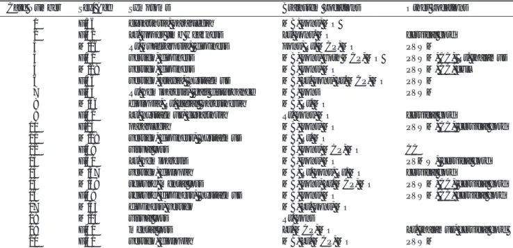
Fig. 1. Schematic drawing of brain- stem MR lesions in 20 patients with multiple sclerosis (A: midbrain, B:
middle pons, C: medulla oblongata, D:
mid-sagittal section of brainstem).
A B
D C
자가면역질환으로 추정된다 (16). 임상적으로 대부분의 환자 는 재발과 완화를 반복하면서 만성적 진행기로 이행되는데, 초 기 급성기 환자보다는 만성적 진행기 환자에서 천막하 병변이 더 호발하는 것으로 보고되고 있고 (9), 뇌간이 침범되면 다른 부위를 침범할 때 보다 심한 신경학적 증상을 야기한다 (5).
뇌간 침범시의 임상증상은 중심성 현훈, 복시, 안구진탕, 안면 신경마비, 청력장애에서부터 편측 또는 양측 상하지 마비, 의 식소실까지 다양하다 (16).
다발성 경화증으로 확진하기 위한 확정적인 검사방법은 없 는 것으로 알려져 있다 (5). 따라서 진단의 확실성 정도를 정 하기 위하여 여러 가지 기준이 있으며, 저자들의 연구에서는 Poser 등(14)의 기준을 사용하였다. 저자들의 연구에서는 뇌 간을 침범하는 다발성 경화증의 보다 정확한 MR 소견을 얻기 위하여 임상적으로 확정적인 다발성 경화증만을 대상으로 하 였다. 다발성 경화증에서 MR은 가장 유용한 부가적인 진단방 법으로 85-97%의 민감도를 보인다 (5, 9).
다발성 경화증은 뇌실 주위 백질, 시신경, 척수, 뇌간 등에 호발하는 것으로 알려져 있으며, Sheldon 등(5)에 의하면 대
뇌 반구 병변은 주로 뇌실주위, 피질하 백질, 내포(internal cap- sule) 등에 위치하며, 후두와(posterior fossa)에서는 주로 뇌 간(특히 뇌교)에 발생하였다고 하였다.
Uhlenbrock 등(10)은 뇌간 병변은 성인 환자의 13%에서 관 찰되었다고 하였으며, Ormerod 등(4)은 68%의 환자에서 뇌 간 병변이 관찰되었다고 하였다. 저자들의 연구에서는 35명의 다발성 경화증 환자에서 20명(57%)에서 뇌간의 병변이 관찰 되었다.
Brainin 등(12)에 의하면 뇌간에 발생한 다발성 경화증의 분 포는 뇌교(71%), 연수(50%), 중뇌(25%) 순으로 뇌교를 침범 한 경우가 가장 많았으며, 저자들의 연구에서는 연수, 뇌교, 중 뇌를 침범한 경우가 각각 18예(90%), 17예(85%), 16예(80%) 순으로 빈도의 차이는 없었다. 전체 뇌간을 침범한 경우가 12 예(60%)에서 관찰되어 뇌간이 침범되는 경우에는 뇌간의 일 부보다는 전체 뇌간이 동시에 침범되는 경우가 많은 것을 알 수 있었다.
다발성 경화증의 MR에서 신호강도는 T1강조영상에서 저신 호 강도를, T2강조영상에서 고신호 강도를 보이는 것으로 알
A B
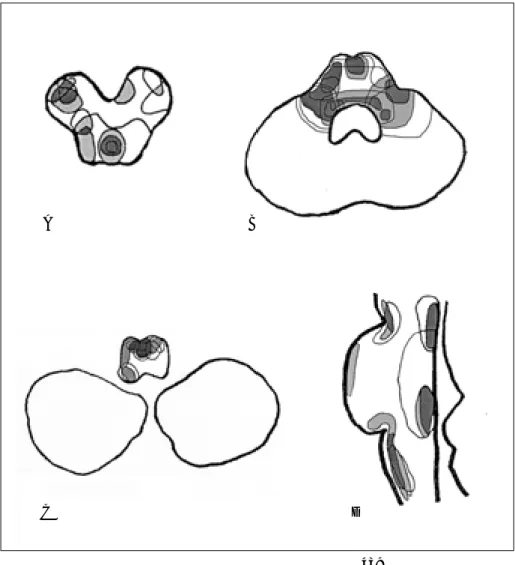
Fig. 2. A 39-year-old female with seizure, dizziness and nystagmus (Case 16).
A. Axial T2 weighted images show high signal intensity lesion in pons and this lesion is contiguous with ventricular CSF space.
B. Axial T1 weighted image shows sub- tle low signal intensity lesions on dorsal aspect of pons.
C. Contrast-enhanced axial T1 weight- ed image shows multiple patch en- hanced lesions on dorsal aspect of pons.
D. Sagittal T2 weighted image shows patchy high signal intensity lesions in corpus callosum, dorsal pons, medulla oblongata, and high cervical cord, and these lesions are contiguous with cis- ternal or ventricular CSF space.
C D
려져 있다 (2, 17). 저자들의 연구에서도 모든 병변은 T1강조 영상에서 미약하거나 뚜렷한 저신호 강도로 보이고, T2강조영 상에서 고신호 강도로 보였다. 다발성 경화증의 MR 촬영방법 으로서 Wilms 등(11)은 뇌간 병변의 경우 주로 뇌척수액 근 처에 위치하므로 시상면 영상보다 축상면 영상이 우수하다고 하였다. 그러나, Brainin 등(12)은 뇌간 병변이 축상면 혹은 시 상면 T2강조영상에서 잘 관찰되었다고 하였다. 저자들의 연구 에서는 뇌간 병변의 인지에 있어서 축상면 T2강조영상이 우 수하였으나, 상하로 긴 막대형 병변의 발견에는 시상면 혹은 관상면 T2강조영상이 우수하였다. 따라서 뇌간 병변의 발견에 있어서 시상면 혹은 관상면 T2강조영상이 도움이 될 것으로 생각된다.
Brainin 등(12)은 뇌간부위를 침범한 다발성 경화증의 MR 소견의 위치적 특성을 보고하였는데, 대부분의 뇌간 병변은 뇌 조 혹은 뇌실 뇌척수액 공간과 인접하면서 연속적인 모양을 보 인다고 하였다. Sheldon 등(5)에 의하면 대뇌반구 병변은 주 로 뇌실 뇌척수액공간과 접하였다고 하였으며, Offenbacher 등
(17)은 소뇌각, 중뇌, 제4뇌실에 인접하여 발생한 병변은 99%
의 특이도를 보인다고 하였다. 본 연구에서도 뇌간 부위중 중 뇌와 연수 병변은 전예에서, 뇌교 병변은 15예(88%)에서 병 변의 전부 혹은 일부가 뇌조 혹은 뇌실 뇌척수액 공간과 접하 여 다른 저자들의 보고와 일치하였다. 뇌간 병변이 뇌조 혹은 뇌실 뇌척수액 공간과 인접해서 발생하는 원인은 밝혀져 있지 않으나, 대뇌에서 뇌척수액주위에 발생하는 측뇌실주위의 침범 과 뇌간부위에서의 뇌실주위 침범의 병리해부학적 연관성을 생 각해 볼 수 있다.
Comi 등(13)은 뇌간에서 발견된 병변의 49%는 단일 병변 이며, 51%는 다발성이라고 보고하였고, Runge 등(18)은 뇌간 에서 발견된 병변의 모양은 원형이거나 막대모양이었다고 기 술하였다. 저자들의 연구에서는 뇌간 병변이 17예(85%)에서 다발성이었으며, 모양은 원형, 난원형, 타원형, 반점형, 융합형, 무정형 및 상하로 긴 막대형 등으로 다양하였다. 관상면이나 시상면 영상에서 상하로 긴 막대형 모양의 병변은 뇌간 병변 의 82%(14/20)에서 관찰되어 높은 빈도를 보였고 이는 뇌조
A B
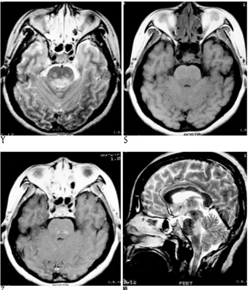
Fig. 3. A 40-year-old woman with verti- go and diplopia (Case 20).
A, B, C. Axial T2 weighted images show high signal intensity lesions in midbrain, pons and medulla oblongata and these lesions are contiguous with ventricular CSF space.
D. FLAIR coronal images show long tubular high signal intensity lesions in midbrain and medulla oblongata.
C D
혹은 뇌척수액 공간과 인접하였다.
다발성 경화증 병변의 조영증강은 활동성 염증부위에 파괴 된 혈관뇌장벽(blood brain barrier)을 조영제가 통과하여 나 타난다. 따라서 조영증강 MR 영상이 재발과 완화를 반복하는 다발성 경화증의 특징적인 임상경과 중에서 병변의 활동성을 인지하는데 유용한 보조적 진단방법이다 (9, 12, 17). 저자들 의 연구에서 조영증강을 시행한 예중 58%에서 병변이 조영증 강되었다.
다발성 경화증의 감별진단으로서 급성 파종성 뇌척수염 (acute disseminated encephalomyelitis), 진행성 다병소성 백 질뇌증(progressive multifocal leukoencephalopathy), 베체트 증후군(Behcet’s syndrome), 혈관염, 소혈관 허혈성질환, 전 신성 홍반성 루푸스, 종양(뇌교 교종, 임파종 등), 라임병(Lyme disease) 등이 뇌간에서 뇌척수액공간과 인접하여 병변을 나 타낼 수 있으므로 감별을 요한다 (6). 소혈관 허혈성질환의 경 우는 젊은 성인에서는 드물고, 하부뇌간이나 척수는 잘 침범하
지 않으며, 뇌간에 발생할 때에도 경계가 불분명하고 뇌간 실 질중심부 근처에 잘 발생한다는 점이 감별진단에 도움이 된다 (12, 19).
결론적으로, 뇌간을 침범한 다발성 경화증은 MR에서 다양 한 모양을 보이는 다발성 병변의 전부 혹은 일부가 뇌조 혹은 뇌실 뇌척수액공간과 접하거나 상하로 긴 병변이 시상면 혹은 관상면에서 관찰된다.
참 고 문 헌
1. Young IR, Hall AS, Pallis CA, Legg NJ, Bydder GM, Steiner RE.
Nuclear magnetic resonance imaging of the brain in multiple scle- rosis. Lancet 1981;2:1063-1066
2. Scotti G, Scialfa G, Biondi A, Landoni L, Caputo D, Cazzullo CL.
Magenetic resonance in multiple sclerosis. Neuroradiology 1986;28:
319-323
3. Nakashima I, Fujihara K, Okita N, Takase S, Itoyama Y. Clinical and MRI study of brain stem and cerebellar involvement in
A B
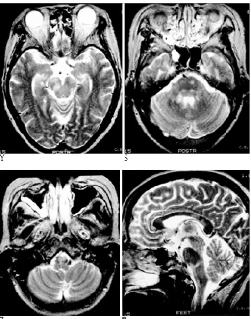
Fig. 4. A 59-year-old woman with visu- al loss (Case 12).
A, B, C. Axial T2 weighted images show multiple high signal intensity le- sions of various size and shape in mid- brain, pons, both middle cerebellar pe- duncles and medulla oblongata, and most of these lesions are contiguous with cisternal or ventricular CSF space.
D. Sagittal T2-weighted image shows patchy high signal intensity lesions in corpus callosum, and long tubular shaped high signal intensity lesion in pons and medulla oblongata.
C D
Japanese patients with multiple sclerosis. J Neurol Neurosurg Psychiatry 1999;67:153-157
4. Ormerod IEC, Miller DH, McDonald WI, et al. The role of NMR imaging in the assessment of multiple sclerosis and isolated neuro- logical lesions. A quantitative study. Brain 1987;110:1579-616 5. Sheldon JJ, Siddharthan R, Tobias J, Sheremata WA, Soila K,
Viamonte M. Jr. MR imaging of multiple sclerosis: comparison with clinical and CT examinations in 74 patients. AJR Am J Roentgenol 1985;145:957-964
6. Gass A, Filippi M, Grossman RI. The contribution of MRI in the differential diagnosis of posterior fossa damage. J Neurol Sci 2000;
172:S43-S49
7. 최민연, 설창효, 정춘필, 김병수, 박병호. 다발성 경화증의 자기공명 영상 소견. 대한방사선의학회지 1993;29:627-633
8. 이상건, 노재규, 김재우 등. 국내 다발성 경화증에서의 핵자기 공명 영상소견. 임상양상에 따른 고찰. 대한신경과학회지 1991;9:18-24 9. Wallace CJ, Seland TP, Fong TC. Multiple sclerosis: the impact of
MR imaging. AJR Am J Roentgenol 1992;158:849-857
10. Uhlenbrock D, Siedel D, Gehlen W, et al. MR imaging in multiple sclerosis: Comparison with clinical, CSF, and visual evocated po- tential findings. AJNR Am J Neuroradiol 1988;9:59-67
11. Wilms G, Marchal G, Kersschot E, et al. Axial vs sagittal T2- weighted brain MR images in the evaluation of multiple sclerosis. J Comput Assist Tomogr 1991;15:359-364
12. Brainin M, Reisner T, Neuhold A, Omasits M, Wicke L.
Topological characteristics of brainstem lesions in clinically defi- nite and clinically probable cases of multiple sclerosis: an MRI- study. Neuroradiology 1987;29:530-534
13. Comi G, Filippi M, Martinelli V, et al. Brain stem magnetic reso- nance imaging and evoked potential studies of symptomatic multi- ple sclerosis patients. Eur Neurol 1993;33:232-237
14. Poser CM, Party DW, Scheinberg L, et al. New diagnostic criteria for multiple sclerosis: guidelines for research protocols. Ann Neurol 1983;13:227-231
15. Glasier CM, Robbins MB, Davis PC, Ceballos E, Bates SR. Clinical neurodiagnostic, and MR findings in children with spinal and brain stem multiple sclerosis. AJNR Am J Neuroradiol 1995;16:87- 95
16. Victor M, Ropper AH. Adams and Victor’s priciples of neurology, 7th edition. New york: McGraw-Hill, 2001:954-975
17. Offenbacher H, Fazekas F, Schmidt R, et al. Assessment of MRI criteria for a diagnosis of multiple sclerosis. Neurology 1993;43:905- 909
18. Runge VM, Price AC, Kirshner HS, Allen JH, Partain CL, James AE. Magnetic resonance imaging of multiple sclerosis: a study of pulse-technique efficacy. AJR Am J Roentgenol 1984;143:1015-1026 19. Palmer S, Bradley WG, Chen DY, Patel S. Subcallosal striations:
early findings of multiple sclerosis on sagittal, thin-section, fast FLAIR MR images. Radiology 1999;210:149-153
J Korean Radiol Soc 2001;45:437-444
Address reprint requests to : Hae Woong Jeong, M.D., Department of Diagnostic Radiology, Maryknoll Hospital, 4-12, Daechung-dong, Chung-gu, Pusan 600-094, Korea.
Tel. 82-51-461-2282 Fax. 82-51-467-6744 E-mail: hwjeong2000@lycos.co.kr
MRI Findings of Multiple Sclerosis Involving the Brainstem
1Jeong Hoon Park, M.D., Hae Woong Jeong, M.D., Hyun Jin Kim, M.D., Jae Kwoeng Cho, M.D., Chang Soo Kim, M.D.
1Department of Diagnostic Radiology, Maryknoll Hospital
Purpose: To describe MRI findings of multiple sclerosis involving the brainstem.
Materials and Methods: Among 35 cases of clinically definite multiple sclerosis, the authors retrospectively analysed 20 in which the brainstem was involved. MR images were analysed with regard to involvement sites in the brainstem or other locations, signal intensity, multiplicity, shape, enhancement pattern, and contiguity of brainstem lesions with cisternal or ventricular CSF space.
Results: The brainstem was the only site of involvement in five cases (25%), while simultaneous involvement of the brainstem and other sites was observed in 15 cases (75%). No case involved only the midbrain or medul- la oblongata, and simultaneous involvement of the midbrain, pons and medulla oblongata was noted in 12 cas- es (60%). The most frequently involved region of the brainstem was the medulla oblongata (n=18; 90%), fol- lowed by the pons (n=17; 85%) and the midbrain (n=16; 80%). Compared with normal white matter, brain- stem lesions showed low signal intensity on T1 weighted images, and high signal intensity on T2 weighted, proton density weighted, and FLAIR images. In 17 cases (85%), multiple intensity was observed, and the shape of lesions varied: oval, round, elliptical, patchy, crescentic, confluent or amorphous areas were seen on axial MR images, and in 14 cases (82%), coronal or sagittal scanning showed that lesions were long and tubu- lar. Contiguity between brainstem lesions and cisternal or ventricular CSF space was seen in all cases (100%) involving midbrain (16/16) and medulla oblongata (18/18) and in 15 of 17 (88%) involving the pons. Contrast enhancement was apparent in 7 of 12 cases (58%).
Conclusion: In the brainstem, MRI demonstrated partial or total contiguity between lesions and cisternal or ventricular CSF space, and coronal or sagittal images showed that lesions were long and tubuler.
Index words :Brainstem, MR Sclerosis, multiple