간경색은 비교적 드문 간병변으로 대개는 간이식이나 복강 경을 이용한 담낭절제술 등을 포함하는 각종 수술 및 중재적 시술의 합병증으로 발생하며 드물게는 동맥경화, 쇽, 패혈증, 전색증, 혈관염, 자간증, 경구피임제의 복용 등과 관련된 간동 맥의 폐색으로 발생하는 것으로 알려져 있다 (1). 하지만 급 성 췌장염과 관련된 간경색은 보고된 바 없어서 저자들은 급 성 췌장염의 합병증으로 발생한 간경색 1 예를 보고한다.
증례 보고
31세 남자 환자가 심한 심와부 동통을 주소로 내원하였다.
과거력상 환자는 평소에 술을 자주 많이 마시는 편이었고 내 원 2일 전에도 술을 많이 마신 후 상기 증상이 발생하였으며 외상병력은 없었다. 급성 췌장염 진단하에 금식과 수액요법을 시행받았지만 증상이 호전되지 않고 오히려 동통이 더 심해지 며 복부팽만이 발생하여 본원 응급실로 내원하였다. 입원 당 시 주요 검사소견은 다음과 같았다 (Amylase, 624 U/L; WBC, 14,170/mm3; Glucose, 241 mg%; aspartate aminotrans- ferase[AST], 3,565 IU/L; lactate dehydrogenase[LDH], 5,825 IU/L).
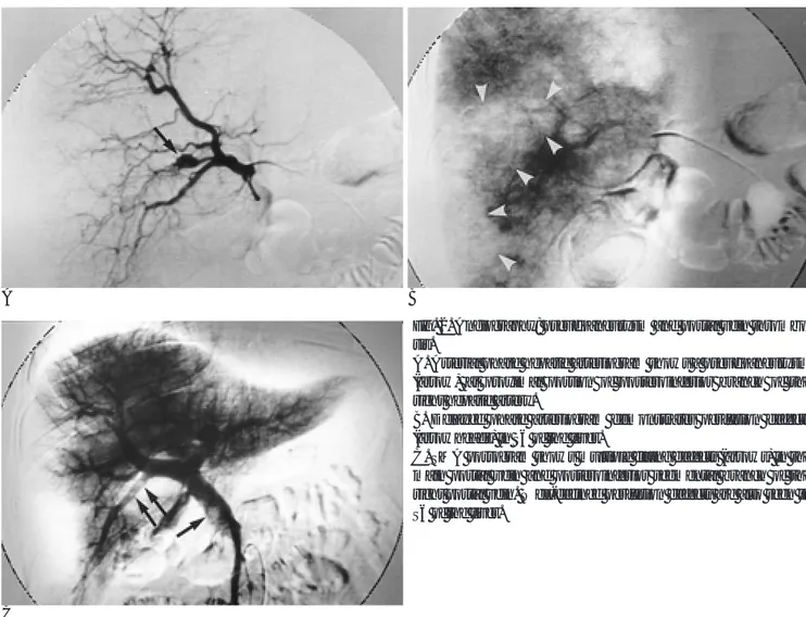
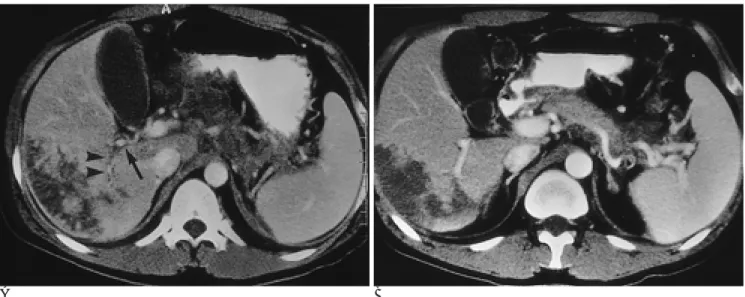
내원당시 CT에서 췌장이 미만적으로 커져 있고 췌장 내부 및 주변에 심한 수액저류가 있었으며 우문맥의 후하분절에 저 감쇄의 혈전과 함께 간우엽 후하분엽에 국한된 경색이 관찰되 었다 (Fig. 1). 간동맥조영술의 동맥기에서는 우간동맥 후하분 절 근위부에 가성동맥류가 보였고 (Fig. 2A), 지연기에서는 간 우엽 후하분엽부위와 일치하는 관류결손이 증명되었다 (Fig.
2B). 그러나 간동맥 자체가 막히거나 좁아진 소견은 없었다.
상장간막동맥을 통한 문맥조영술에서는 주문맥과 우문맥 후하 분절에 충만결손이 관찰되었으며 (Fig. 2C), 이 병변의 위치는 간동맥의 가성동맥류가 있는 위치의 원위부에 해당하였다.
추적 CT에서 우문맥 후하분절 혈전 근위부에 간동맥의 가 성동맥류가 위치하는 것을 볼 수 있었다 (Fig. 3A). 환자의 증 상이 호전되면서 췌장주위 수액저류는 그 양이 감소하였다. 경 색은 범위가 점차 줄어들고 그 전과 비교하여 주변 정상 간조 직과의 경계가 더욱 명확해지는 변화를 보였으며 문맥의 혈전 은 소실되었다 (Fig. 3B). 경피적 흡인생검에 의해 조직학적으 로 간경색을 확진하였다.
고 찰
간의 혈류공급은 간동맥과 문맥으로부터 이중으로 이루어지 고 간세포는 비교적 낮은 산소환경에 대하여 잘 견디기 때문 에 간경색은 매우 드물게 발생한다 (1, 2). 그러나 최근 말기 간질환에 대해 간이식을 시행하는 빈도가 늘고 각종 수술 및 중재적 시술이 광범위하게 시행되면서 보고되는 간경색의 빈 도가 점차 늘고 있는 추세이다 (3). 혈류이상에 의한 간경색 의 일차적인 발생기전이 간동맥 혈류장애인지 혈전등에 의한 문맥폐색인지 또는 이 두 가지 요소가 모두 작용해야 하는지 에 대해서는 여러 가지 견해가 있으나 (2-7), 간문맥만의 문 제로는 발생하지 않는다는 주장도 있다 (8). 이는 예를 들어 간의 우엽절제술에 앞서 우문맥 색전술을 시행한 경우에 경색 이 발생하지 않는 것을 봄으로써도 알 수 있다 (9).
췌장염 환자의 경우 국소적인 염증반응이나 가성낭종에 의 한 외부압박에 의해 문맥의 혈전이 발생한다 (10). 따라서 대 개의 경우에는 췌장염이 있는 부위와 인접한 문맥에 혈전이 형성되는데 이 증례에서는 주간문맥은 물론, 간내의 문맥에도 혈전이 형성된 것을 볼 수 있었다. 이와 같이 췌장염과는 직 대한방사선의학회지 2000;43:73-76
─ 73 ─
급성 췌장염에 병발된 간경색: 1예 보고1
김현석・홍성환・박홍석・이일성・강익원
간경색은 수술이나 중재적 시술과 관련되어 발생하는 경우를 제외하면 드물게 발생하는데 특히 급성 췌장염에 병발하는 증례는 보고된 바가 없다. 이에 저자들은 급성 췌장염 환자에서 발생한 간경색 1예를 보고한다. 복부전산화단층촬영(CT)상 췌장의 미만적 종창과 주위의 수 액저류가 있었고, 문맥의 혈전과 함께 간우엽 후하분엽에 경색이 관찰되었다. 혈관조영술에서 는 우간동맥의 가성동맥류 및 우문맥의 혈전이 확인되었고 간우엽 후하분엽에 국한된 관류결 손이 증명되었다.
1한림대학교 의과대학 방사선과학교실
이 논문은 1999년 11월 22일 접수하여 2000년 5월 12일에 채택되었음.
김현석 외 : 급성 췌장염에 병발된 간경색
─ 74 ─
A B
C
Fig. 2. Angiography: pseudoaneurysm and portal vein thrombo- sis.
A. Arterial phase hepatic arteriogram shows a pseudoaneurysm (arrow) at proximal portion of posteroinferior branch of the right hepatic artery.
B. Delayed phase arteriogram demonstrates perfusion defects (arrowheads) in S6 of the liver.
C. SMA portogram shows multiple filling defects (arrows) in the main portal vein and posteroinferior segmental branch of the right portal vein. Well-defined perfusion defects are also seen in S6 of the liver.
A B
Fig. 1. Initial CT scans.
A. Contrast-enhanced CT scan shows a segmental infarction in S6 of the liver along with thrombus (arrows) in the posteroinferior branch of the right portal vein.
B. The pancreas is poorly defined and reveals heterogeneous low attenuation.
접적인 관련이 없을 것으로 보이는 간내 문맥에까지 혈전이 형성된 것은 질환의 초기에 이 부위에까지 염증이 파급되었거 나, 주문맥내의 혈전이 일부 떨어져 나가서 간내 문맥에 색전 을 초래하였기 때문으로 생각하였다.
췌장염 환자에서 발생하는 가성동맥류는 췌장효소의 단백분 해효과에 의해 동맥벽에 미란을 일으킴으로써 형성되는 것으 로 알려져 있고 따라서 대개는 췌장 내부 혹은 췌장 주변 혈 관에서 발생한다 (11, 12). 흔히 나타나는 혈관은 췌장 주변 의 위십이지장동맥, 비장동맥과 상장간막동맥이다. 이 증례에 서는 췌장과는 떨어져 있는 간내 간동맥에 가성동맥류가 형성 되었는데, 간내 문맥혈전과 더불어 생각할 때 간내의 간동맥 및 문맥 주변으로 염증이 직접 파급되어 가성동맥류와 문맥혈 전을 형성하였을 가능성이 높다. 그리고 비록 간동맥조영술의 동맥기에서 우측 간동맥과 분지에서 폐색이나 협착은 보이지 않았으나 지연기에서 관류결손이 있어 간동맥 혈류장애가 있 다고 볼 수 있다. 따라서 이 증례는 급성 췌장염의 합병증으 로 간의 동맥 및 문맥 혈류장애가 병합되어 간경색이 발생한 것으로 생각한다.
참 고 문 헌
1. Heiken JP, Liver, In Lee JKT, Sagel SS, Sanley RJ, Heiken JP.
Computed body tomography with MRI correlation. 3rd ed. Philadel- phia, Lippincott-Raven 1998:701-777
2. Seige M, Schweigart U, Moessmer G, Schneider KTM, Classen M.
Extensive hepatic infarction caused by thrombosis of right portal
vein branches and arterial vasospasm in HELLP syndrome associ- ated with homozygous factor V Leiden. Am J Gatroenterol 1998;93:
473-474
3. Holbert BL, Baron RL, Dodd Ⅲ GD. Hepatic infarction caused by arterial insufficiency: spectrum and evolution of CT findings. AJR Am J Roentgenol 1996;166:815-820
4. Belli AM, Jennings CM, Nakielny RA. Splenic and portal venous thrombosis: a vascular complication of pancreatic disease demon- strated on computed tomography. Clin Radiol 1990;41:13-16 5. Yamashita K, Tsukuda H, Mizukami Y, et al. Hepatic infarction
with portal thrombosis. J Gastroenterol 1997;32:684-688
6. Mathieu D, Vasile N, Grenier P. Portal thrombosis: dynamic CT features and course. Radiology 1985;154:737-741
7. 박기순, 옥인돈, 강진화. 간경색의 방사선학적 소견: 1예 보고. 대한 방사선의학회지 1991;27:105-107
8. Smith GS, Birnbaum BA, Jacobs JE. Hepatic infarction secondary to arterial insufficiency in native livers: CT findings in 10 patients.
Radiology 1998;208:223-229
9. 방선우, 성규보, 송호영, 조경식, 이승규, 권태원. 광범위 간절제술 에서 수술전 간문맥 색전술의 역할. 대한방사선의학회지 1995;32:
769-774
10. Zalcman M, Gansbeke DV, Matos C, Engelholm L, Struyven J.
Sonographic demonstration of portal venous system thromboses secondary to inflammatory diseases of the pancreas. Gastrointest Radiol 1987;12:114-116
11. Sawlani V, Phadke RV, Baijal SS, et al. Arterial complications of pancreatitis and their radiological management. Australas Radiol 1996;40:381-386
12. Perez C, Llauger J, Pallardo Y, Sanchis E, Sabate JM. Radiologic di- agnosis of pseudoaneurysms complicating pancreatitis. Eur Radiol 1993;16:102-106
대한방사선의학회지 2000;43:73-76
─ 75 ─
A B
Fig. 3. Follow-up CT scans.
A. CT scan obtained 7 days after initial CT shows a partial recanalization (arrowheads) of the posteroinferior branch of the right portal vein and hepatic pseudoaneurysm (arrow) nearby. The extent of the hepatic infarction is decreased and some enhancing portions appear in the periphery of the lesion.
B. CT scan 19 days later shows a patent portal vein. The hepatic infarction is sharply demarcated with surrounding normal parenchyma. The pancreas has smooth contour and reveals homogeneous enhancement.
김현석 외 : 급성 췌장염에 병발된 간경색
─ 76 ─
J Korean Radiol Soc 2000;43:73-76
Address reprint requests to : Sung Hwan Hong, M.D., Department of Radiology, Hangang Sacred Heart Hospital 94-195 Yongdungpo-dong, Yongdungpo-gu, Seoul 150-030, Korea.
Tel. 82-2-2639-5204 Fax. 82-2-679-0121
Hepatic Infarction Complicating Acute Pancreatitis: A Case Report1
Hyun Suk Kim, M.D., Sung Hwan Hong, M.D., Hong Suk Park, M.D., Eil Seong Lee, M.D., Ik Won Kang, M.D.
1Department of Radiology, College of Medicine, Hallym University
Hepatic infarction is relatively uncommon and is usually related to surgery or interventional procedures.
Pancreatitis-associated hepatic infarction has not been reported in the literature, and we now describe a case of hepatic infarction in a 31-year-old man with acute pancreatitis. Initial CT scanning demonstrated an enlarged pancreas with multifocal fluid collection, and a large wedge-shaped low attenuation lesion was seen in the right lobe of the liver along with thrombi in the posteroinferior branch of the right portal vein. Hepatic arteri- ography and SMA portography revealed a pseudoaneurysm in the right hepatic artery, thrombi in the main portal vein and its posteroinferior branch, and perfusion defects confined to S6 of the liver.
Index words :Liver, infarction Pancreatitis
Portal vein, thrombosis Hepatic arteries, injuries