www.krspine.org
A Thoracolumbar Pure Spinal Epidural Cavernous Hemangioma - A Case Report -
Byeong Sam Choi, M.D., Ju Yeon Kim, M.D.*, Sungjoon Lee, M.D.
J Korean Soc Spine Surg 2018 Dec;25(4):169-174.
Originally published online December 31, 2018;
https://doi.org/10.4184/jkss.2018.25.4.169
Korean Society of Spine Surgery
Asan Medical Center 88, Olympic-ro 43 Gil, Songpa-gu, Seoul, 05505, Korea Tel: +82-2-483-3413 Fax: +82-2-483-3414
©Copyright 2017 Korean Society of Spine Surgery pISSN 2093-4378 eISSN 2093-4386
The online version of this article, along with updated information and services, is located on the World Wide Web at:
http://www.krspine.org/DOIx.php?id=10.4184/jkss.2018.25.4.169
This is an Open Access article distributed under the terms of the Creative Commons Attribution Non-Commercial License (http://
creativecommons.org/licenses/by-nc/4.0) which permits unrestricted non-commercial use, distribution, and reproduction in any medium, provided the original work is properly cited.
Journal of Korean Society of
Spine Surgery
A Thoracolumbar Pure Spinal Epidural Cavernous Hemangioma - A Case Report -
Byeong Sam Choi, M.D., Ju Yeon Kim, M.D.
*, Sungjoon Lee, M.D.
Department of Neurosurgery, Inje University Haeundae Paik Hospital, Busan, Korea
*Department of Pathology, Inje University Haeundae Paik Hospital, Busan, Korea
Study Design: Case report.
Objectives: We report a case of pure epidural cavernous hemangioma located at the thoracolumbar spine in a 53-year-old woman that mimicked a neurogenic tumor on magnetic resonance imaging (MRI).
Summary of Literature Review: A pure spinal epidural cavernous hemangioma without bony involvement is a very rare lesion about which limited information is available in the literature.
Materials and Methods: A 53-year-old woman visited our clinic for hypoesthesia with a tingling sensation in the left anterolateral thigh that had begun a month ago. No other neurologic symptoms or signs were present upon a neurologic examination. MRI from an outside hospital showed a 2.0×0.5 cm elongated mass at the T11-12 left neural foramen. The tumor was completely removed in piecemeal fashion.
Results: The histopathologic examination revealed a cavernous hemangioma, which was the final diagnosis. The outcome was favorable in that only operation-related mild back pain remained, without any neurologic deficits, after a postoperative follow-up of 2 years and 3 months. No recurrence was observed on MRI at 2 years postoperatively.
Conclusion: Pure epidural spinal cavernous hemangioma is very rare, and it is very difficult to differentiate from other epidural lesions.
However, we believe that it should be included in the differential diagnosis of spinal epidural tumors due to its favorable prognosis.
Key Words: Cavernous hemangioma, Epidural, Thoracic vertebrae, Spine
Received: April 24, 2018 Revised: August 22, 2018 Accepted: October 8, 2018
Published Online: December 31, 2018 Corresponding author: Sungjoon Lee, M.D.
ORCID ID: Byung Sam Choi: http://orcid.org/0000-0002-8760-0959 Ju Yeon Kim: http://orcid.org/0000-0002-4259-667x Sungjoon Lee: https://orcid.org/0000-0002-1675-0506 Department of Neurosurgery, Inje University Haeundae Paik Hospital 875 Haeundae-ro, Haeundae-gu, Busan, 48108, Korea
TEL: +82-51-797-0241, FAX: +82-51-797-0841 E-mail: potata98@naver.com
Hemangiomas are hamartomatous malformations showing vascular endothelial proliferations.1) According to the predominant type of vascular channel, they are classified into four histopathologic types: cavernous, capillary, arteriovenous, and venous.1) Hemangiomas can be found in any part of the body. In the spine, they are most frequently found in the vertebral bodies,1-3) and they are very common lesions that we easily encounter in our daily practices. However, pure spinal epidural cavernous hemangiomas (PSECHs) without bony involvement are very rare lesions. The authors intend to present a case of an PSECH in the thoracic spine. We will also discuss the clinical presentations, radiologic findings and treatment outcomes with a review of the relevant literature.
Case report
A 53-year-old woman visited our clinic for hypoesthesia with a tingling sensation in left lateral thigh, which had begun
a month ago. No other neurologic symptoms or signs were presented upon neurologic examination. Four months before the visit, an MRI was taken at a local hospital for evaluation of a tingling sensation in the right arm, which had begun 7 months prior. An epidural mass at the T11-12 level of the left neural foramen was incidentally found. Initially, a benign, non-symptomatic neurogenic tumor was suspected, and it was put to observation. Three months later, the sensory
Byeong Sam Choi et al Volume 25 • Number 4 • December 31 2018
www.krspine.org 170
change in the left antero-lateral thigh appeared. It appeared to aggravate during walking and relieve in resting. Because the symptom duration and intensity were gradually aggravating, she consulted the other hospital. They recommended her for a surgical excision of the mass and referred her to our hospital.
On MRI, a 2.0×0.5 cm elongated mass was observed at the T11-12 left neural foramen along the right T11 nerve root (Fig.
1A-E). It showed iso-signal intensity on T1-weighted images (Fig. 1B) and high signal intensity on T2-weighted images (Fig.
1C). It was homogenously well enhanced after gadolinium injection (Fig. 1D). The mass was extended to outside the left T11-12 neural foramen and to the left T12-L1 foramen along the medial side of the T12 pedicle (Fig. 1E). It was a pure epidural mass without involvement of vertebral bodies or adjacent bony structures. In addition, the mass stretched to the dorsal side of the dura, encircling the dura with a small mass effect (Fig. 1B-D). No neural foramen widening or bony erosion was observed.
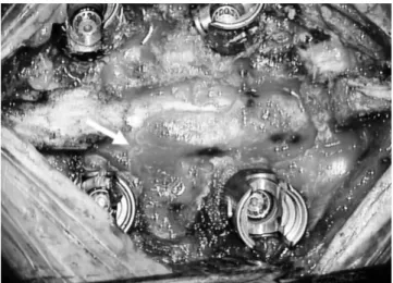
Under general anesthesia, a T11-12 total laminectomy and left facetectomy were performed, and a red-colored mass was found at the epidural space (Fig. 2). It was a vascular mass with soft consistency, and it easily bled even with gentle touch. However, there was no massive bleeding, and it could be controlled easily with a bipolar coagulator. Clear dissection between the mass and the dura was possible. The mass was removed in piecemeal fashion. After complete removal, T11- 12 posterolateral fusion with pedicle screws was performed.
On frozen biopsy, a vascular tumor such as a hemangioma was reported.
Histopathological examination revealed a well-demarcated vascular tumor, measuring 2.0 cm in diameter. The tumor was composed of dilated blood-filled vessels in various sizes.
The vessels were arranged in a diffuse, haphazard pattern or
Fig. 1. Preoperative magnetic resonance images. (A) T1-weighted sagit- tal image after gadolinium injection. A well-enhanced mass located at the left T11-12 neural foramen (white arrows) was identified. The mass showed iso-signal intensity on a T1-weighted image (B), high signal in- tensity on a T2-weighted image (C), and homogenous enhancement (D).
The mass showed plastic growth features encircling the dorsal side of the dura sac and creeping out of the neural foramen with lobulated con- tours (white arrow heads, D). Note that there was almost no widening of the T11-12 neural foramen. The mass also extended caudally, encircling the left T12 pedicle (white arrow heads, E).
A
B
C
D
E
Fig. 2. Intraoperative photo. It was a reddish, highly-vascular epidural mass (white arrow).
in partly lobular arrangement. The vessel wall was focally thickened by fibrosis and lined by flattened endothelial cells without obvious cytologic atypia and mitotic activity.
Microscopically, the mass was diagnosed as a cavernous hemangioma (Fig. 3).
The sensory change in the left leg did not improve immediately after the surgery. However, it gradually disappeared in 4 months. Other than mild back pain, the patient was symptom-free through 2-years follow-up period.
MRI taken at the last follow-up showed no evidence of residual or new lesions.
This report had been reviewed after gaining the institutional review board approval (2018-07-017).
Discussion
PSECHs are very rare. Since the first report of one in 1929 by Globus and Doshay,4) approximately 130 cases have been reported in the literature.3,5) It has generally been understood that PSECHs have a female predominance, as reported in previous literature.4) However, several recently reported case series have presented more male patients, making it unclear whether PSECHs have any sexual predominancy.2,3,6,7) They frequently occur at 30 to 60 years of age, with a peak at approximately 40 years.2,6,7) The most common location is thoracic (approximately 60%), followed by cervical (30%) and lumbar (10%).6) They are usually located at dorsal or dorso- lateral epidural spaces of the spinal canal, where venous plexus is abundant.3,5,7)
The clinical presentation of PSECH depends on its location, growth rate, and biological behavior.2) Considering its slow growth and predilection for thoracic and cervical location, the most common clinical symptom is progressive myelopathy.2,3,5-8) Spinal pain or radiculopathy could be combined or presented as a sole clinical symptom. Usually, its clinical course is benign.
However, it can show perilous behavior characterized by acute paraplegia caused by extradural hemorrhage or thrombotic occlusion within the lesion, which are reported quite often in the literature.2,3,5-7,9)
MRI is the most recommended diagnostic modality. There is no pathognomonic imaging finding for PSECHs, and some cases are impossible to distinguish from neurogenic tumors of the spinal epidural space.10) In addition, other epidural pathologies such as lymphoma, meningioma, angiolipoma, eosinophilic granuloma, disc herniation, pure epidural hematoma, abscess, extramedullary hematopoiesis, metastasis, and multiple myeloma should be considered for the differential diagnosis since they share many common radiologic findings.3,10)
However, they may have some characteristic MRI features that should be noted. They usually appear as solid oval, hypervascular lesions with lobular contours.3,10) Most of them appear isointense on T1-weighted images, hyperintense on T2-weighted images, and with strong enhancement after gadolinium injection.2,3,5-7,10) Because of the soft texture of the tumor, they show creeping, plastic growth and form a characteristic lobulated spindle shape with two tapered ends.6) Fig. 3. Microscopic findings. (A, B) The mass was characterized by a
large number of vessels in various sizes. Predominantly, large dilated vessels (arrows) were revealed (hematoxylin and eosin, ×100). (C) The vessels were lined by flat endothelial cells (arrows) and filled with blood or transudate (arrow head) (hematoxylin and eosin, ×200).
A
B
C
Byeong Sam Choi et al Volume 25 • Number 4 • December 31 2018
www.krspine.org 172
In addition, bony erosion is rare, and the neural foramen widening is minimal compared to that in a neurogenic tumor of similar size.3) The range of the tumor is often long, such that involvement of multiple vertebral segments is quite common.6) Heterogenous signal intensities on MRI images indicate intralesional hemorrhage or thrombus.2,3,6) Unlike cavernous hemangiomas in the central nervous system, a low peripheral signal intensity on T2-weighted images due to hemosiderin deposits is typically not present in PSECH.2,3,6) Some authors think that the blood-brain barrier might be the cause of this difference since it would be easier for blood products to be removed in highly vascularized spinal epidural space than in the central nervous system with the blood-brain barrier.3,6)
Complete surgical removal is currently the treatment of choice for PSECH.2,3,5-7,9) Many authors emphasized total en bloc excision of the mass to minimize the risk of massive intraoperative bleeding.2,3,5) This could be achieved since most of the lesion could be easily dissected from the dura, and its bulk shrinks well by bipolar coagulation on the surface. If en bloc resection of the mass is not possible due to ventral or extraforaminal extension, piecemeal resection should be considered as an alternative. Although concerns of massive intraoperative bleeding are present in piecemeal resection, most of the bleeding could be kept under control with microsurgical resection as shown in our case.7) In cases of incomplete resection, radiosurgery has shown benefits in tumor control.11) However, its therapeutic effect is unclear because of the limited number of cases and unclear natural history of PSECH.
The clinical outcome is mostly good if total resection of the mass is achieved.3-7,9) Prognosis usually depends on the preoperative neurologic status.3,9) Incomplete resection may also lead to poor outcome due to bleeding from the residual mass and recurrence.2,9) To achieve total removal of the lesion during the initial attempt, careful preoperative surgical planning and adequate exposure are mandatory. Considering that PSECHs carry a relatively high risk of acute spinal cord compression by intralesional bleeding or rapid growth after thrombotic occlusion, early surgical excision is recommended.2) For the reasons stated above, making the correct preoperative diagnosis of PSECH is important. It is generally believed that digital subtraction spinal angiography is not useful for diagnosis PSECH because it is an angiographically silent lesion.3,4,7,9) In patients with acute paraplegia with epidural hemorrhage,
it may help to distinguish other vascular pathologies from PSECH. However, as urgent exploration and decompression of the spinal canal are more important, many authors are opposed to performing the angiography.3,5)
In conclusion, PSECHs are rare benign lesions that may present with various clinical symptoms and radiographic findings. Because they are dynamic lesions that often cause acute paraplegia, early total surgical removal is recommended.
Therefore, an exact preoperative diagnosis of PSECH is important. Although there are no known pathognomonic MRI findings of PSECH, it should be included in the differential diagnosis for spinal epidural lesions if they show certain MRI features such as solid hypervascularity, lobular contours with plastic growth, minimal bony erosion and multi-segment involvement.
REFERENCES
1. Murphey MD, Fairbairn KJ, Parman LM, et al. From the archives of the AFIP. Musculoskeletal angiomatous le- sions: radiologic-pathologic correlation. Radiographics.
1995 Jul;15(4):893-917. DOI: 10.1148/radiograph- ics.15.4.7569134.
2. Khalatbari MR, Abbassioun K, Amirjmshidi A. Solitary spinal epidural cavernous angioma: report of nine surgically treated cases and review of the literature. Eur Spine J. 2013 Mar;22(3):542-7. DOI: 10.1007/s00586-012-2526-2.
3. Li TY, Xu YL, Yang J, et al. Primary spinal epidural cav- ernous hemangioma: clinical features and surgical outcome in 14 cases. J Neurosurg Spine. 2015 Jan;22(1):39-46.
DOI: 10.3171/2014.9.SPINE13901.
4. Hatiboglu MA, Iplikcioglu AC, Ozcan D. Epidural spinal cavernous hemangioma. Neurol Med Chir (Tokyo). 2006 Sep;46(9):455-8. DOI: 10.2176/nmc.46.455.
5. Esene IN, Ashour AM, Marvin E, et al. Pure spinal epidu- ral cavernous hemangioma: A case series of seven cases. J Craniovertebr Junction Spine. 2016 Jul-Sep;7(3):176-83.
DOI: 10.4103/0974-8237.188419.
6. Feng J, Xu YK, Li L, et al. MRI diagnosis and preopera- tive evaluation for pure epidural cavernous hemangiomas.
Neuroradiology. 2009 Nov;51(11):741-7. DOI: 10.1007/
s00234-009-0555-2.
7. Zhong W, Huang S, Chen H, et al. Pure spinal epidural
cavernous hemangioma. Acta Neurochir (Wien). 2012 Apr;154(4):739-45. DOI: 10.1007/s00701-012-1295-3.
8. Min HJ, Kim KW, Kim YH, et al. Epidural hemangioma: a case report. J Korean Soc Spine Surg. 2001 Sep;8(3):253-8.
DOI: 10.4184/jkss.2001.8.3.253.
9. Aoyagi N, Kojima K, Kasai H. Review of spinal epidural cavernous hemangioma. Neurol Med Chir (Tokyo). 2003 Oct;43(10):471-5. DOI: 10.2176/nmc.43.471.
10. Lee JW, Cho EY, Hong SH, et al. Spinal epidural heman- giomas: various types of MR imaging features with his- topathologic correlation. AJNR Am J Neuroradiol. 2007 Aug;28(7):1242-8. DOI: 10.3174/ajnr.A0563.
11. Sohn MJ, Lee DJ, Jeon SR, et al. Spinal radiosurgical treat- ment for thoracic epidural cavernous hemangioma present- ing as radiculomyelopathy: technical case report. Neu- rosurgery. 2009 Jun;64(6):E1202-3. DOI: 10.1227/01.
NEU.0000345940.21674.AE.
174
J Korean Soc Spine Surg. 2018 Dec;25(4):169-174. https://doi.org/10.4184/jkss.2018.25.4.174
Case Report
© Copyright 2018 Korean Society of Spine Surgery
Journal of Korean Society of Spine Surgery. www.krspine.org. pISSN 2093-4378 eISSN 2093-4386
This is an Open Access article distributed under the terms of the Creative Commons Attribution Non-Commercial License (http://creativecommons.org/licenses/by-nc/4.0/) which permits unrestricted non-commercial use, distribution, and reproduction in any medium, provided the original work is properly cited.
흉요추부에서 발견된 경막외 해면상 혈관종 - 증례 보고 -
최병삼 • 김주연* • 이승준
해운대백병원 신경외과학교실, *해운대백병원 병리과학교실
연구계획: 증례 보고
목적: 신경인성 종양과 유사한 흉요추부의 경막외 해면상 혈관종 환자를 보고하고자 한다.
선행 연구문헌의 요약: 뼈에 침습이 없는 경막외 해면상 혈관종은 매우 드문 질환으로 제한된 문헌 보고만이 존재한다.
대상 및 방법: 53세 여자 환자가 내원 한달여 전부터 시작된 좌측 하지의 감각저하를 동반한 저린 느낌을 주소로 내원하였다. 내원시 시행한 신경학적 검 진에서 기타 다른 신경학적 이상 소견은 관찰되지 않았다. 자기공명영상에서는 흉추 11-12번간 좌측 추간공에 2x0.5 cm 크기의 종괴가 관찰되었다. 수 술을 시행하여 이를 완전 적출하였다.
결과: 병리 검사에서 해면상 혈관종이 확진되었다. 수술 부위의 약한 통증을 제외하고, 추적 관찰 2년동안 추가적인 신경학적인 증상 발생 없이 좋은 결 과를 보였다. 수술 후 2년째에 촬영한 MRI에서 종양의 재발 소견은 관찰되지 않았다.
결론: 척추의 경막외 해면상 혈관종은 매우 드문 질환이며, 다른 경막외 병변들과 감별하기는 쉽지 않다. 그러나, 수술적 치료의 좋은 예후를 보이는 점을 감안하면, 경막외 병변의 감별진단시 이를 반드시 고려해야 할 것으로 생각한다.
색인 단어: 해면상 혈관종, 경막외, 흉추, 척추 약칭 제목: 흉추의 경막외 해면상 혈관종
접수일: 2018년 4월 24일 수정일: 2018년 8월 22일 게재확정일: 2018년 10월 8일 교신저자: 이승준
부산광역시 해운대구 해운대로 875 해운대백병원 신경외과학교실
TEL: 051-797-0241 FAX: 051-797-0841 E-mail: potata98@naver.com
