83
발효유제품에서
Lactobacillus rhamnosus
GG의 생육 특성방미선1․정안나1․박동준3․임광세2*․오세종1*
1전남대학교 동물자원학부, 2㈜매일유업 중앙연구소, 3한국식품연구원
Acid Stress Response of Lactobacillus rhamnosus GG in Commercial Yogurt
Miseon Bang1, Anna Jeong1, Dong-June Park3, Kwang-Sei Lim2* and Sejong Oh1*
1Division of Animal Science, Chonnam National University, Gwangju 500-757, Korea
2R & D Center, Maeil Dairy Co. Ltd, Pyeongtaek 451-861, Korea
3Korea Food Research Institute, Seongnam 463-746, Korea
Abstract
Yogurt is a product of the acidic fermentation of milk, which affects the survival of lactic acid bacteria (LAB). The aim of this present study was to examine the survival and acid stress response of Lactobacillus rhamnosus GG to low pH environment. The survival of LAB in commercial yogurt was measured during long-term storage. The enumeration of viable cells of LAB was determined at 15-day intervals over 52-weeks at 5℃. L. acidophilus, L. casei, and Bifidobacterium spp.
showed low viability. However, L. rhamnosus GG exhibited excellent survival throughout the refrigerated storage period.
At the end of 52-weeks, L. rhamnosus GG survived 7.0 log10 CFU/mL. F0F1 ATPase activity in L. rhamnosus GG at pH 4.5 was also evaluated. The ATPase activities of the membranes were higher when exposed at pH 4.5 for 24 h. The survival of L. rhamnosus GG was attributable to the induction in F0F1 ATPase activity. In addition, the mRNA expression levels of acid stress-inducible genes at low pH were investigated by qRT-PCR. clpC and clpE genes were up-regulated after 1 h, and atpA and dnaK genes were up-regulated after 24 h of incubation at pH 4.5. These genes could enhance the survival of L. rhamnosus GG in the acidic condition. Thus, the modulation of the enzymes or genes to assist the viability of LAB in the low pH environment is thought to be important.
Keywords: Lactobacillus rhamnosus GG, stress respond, probiotics, yogurt
* Corresponding authors: Kwang-Sei Lim, R & D Center, Maeil Dairy Co. Ltd, Pyeongtaek 451-861, Korea. E-mail: kwangslim
@hotmail.com. Sejong Oh, Division of Animal Science, Chonnam National University, Gwangju 500-757, Korea. Tel: +82-62-530- 2116, Fax: +82-62-530-2129, E-mail: [email protected]
서 론
사람의 위장관(gastrointestinal tract)은 수백 종 이상의 다 양한 미생물들이 서식하는 복잡한 생태계이며, 이 안에는 인체를 구성하는 모든 세포의 수보다 10배나 많은 미생물 로 구성되어 있는 것으로 알려져 있다. 또한 이들 장내 미 생물의 대부분은 미생물과 미생물 또는 미생물과 숙주 사
이의 상호작용을 통하여 건강에 기여한다.
Probiotics는 살아있는 미생물로 적절한 양으로 투여되었 을 때 숙주에게 건장 증진 작용을 하는 미생물이다(FAO/
WHO, 2001). Probiotics는 정상적으로 내재하는 미생물군 으로 적응하여 병원성 세균의 부착을 억제시킬 수 있을 뿐 아니라, 면역기능을 조절하는 것으로 밝혀졌다(Wolvers et
al., 2010). 또한 장내의 산도를 낮추어 장 점막을 손상시킬
수 있는 세균의 증식을 억제하고, 손상된 장 점막의 재생을 촉진시키며, 또한 독성이 있는 세균에 대한 항체의 생성을 유도하거나, 장 점막을 보호하는 점액 생성을 조절하는 등 의 여러 작용을 통하여 장염을 치료하거나 예방할 수 있다.
Probiotics로 가장 널리 이용되는 미생물로 Lactobacillus 속, Bifidobacterium 속 그리고 비병원성 효모인 Saccharomyces boulardii(Kligler and Cohrssen, 2008)를 들 수 있으며, 이들 미생물들은 국내에서 정장작용을 하는 대표적인 건강기능 식품으로 인식되어 왔다.
Probiotics로 사용되는 미생물들이 효과가 있으려면 위장 관내에서 생존할 수 있어야 하고, 장내에서 분화를 할 수 있 어야 한다. 종류가 다른 Probiotics는 그 효과도 다른 것으로 알려져 있다. 내산성이 높고 장내 정착성이 우수한 것으로 알려 져 있는 Lactobacillus rahmnosus GG(LGG)는 1985년 Gorbach 교수와 Goldin 교수에 의해 성인의 분변에서 처음 분리되었 다. 오늘날 이 미생물은 전 세계에서 가장 연구가 많이 된 probiotics이다. 700편이 넘는 학술논문에서 LGG균의 유익한 건강작용들을 보고하였으며, 1990년 핀란드에서 처음 제품화 된 이후로 단일 probiotics로써 전 세계적으로 가장 많은 제품 에 이용된 유일한 균주이다(Salminen et al., 2002; 2004).
LGG는 인체에서 발견되는 여러 균주들 중 여러 가지 인 체에 유익한 특성(장 상피세포 부착능, 내산성과 내담즙성, 독성 세균에 대한 항균물질의 분비능)이 가장 탁월하다고 알려졌다. 또한 LGG는 장에서 IgA 분비능을 증가시킨다든 지, 소화관에서 장 점막의 보호역할을 하고 있는 점액의 성 분을 바꾸어 장내에서 장벽으로 작용하게 한다는 등 장 질 환 관련 유용성에 대한 실험적 가설이 제시되어 있으며, 동 물실험 및 임상실험으로 꾸준히 연구되고 있다.
LGG 균을 포함한 다양한 유산균들은 발효유와 음료에
주로 사용되고 있고, 이들의 건강 기능성 관련 연구는 점점 늘어가는 추세이며, 이를 토대로 유산균의 건강 기능성 식 품에서의 활용 범위를 넓힐 수 있는 계기가 될 것이다.
본 연구는 상업적으로 시판되는 발효유제품과 LGG를 함 유한 호상농후발효유의 저장 중 유산균의 생존성을 조사하 여 probiotics로서의 이용성을 증진시키는 기초적 자료를 제 공하고자 실시하였다.
재료 및 방법
1. 제품 준비 및 LGG 생균수 측정
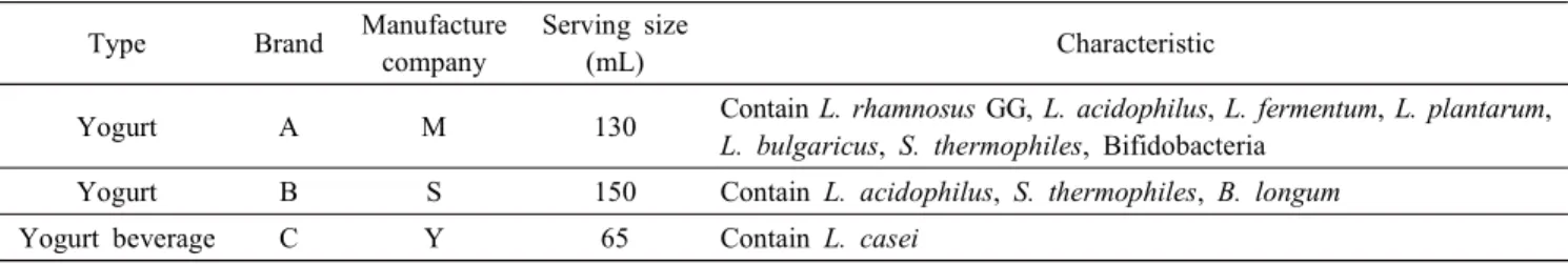
Table 1. Tested commercial yogurt and yogurt beverage in this study
Type Brand Manufacture company
Serving size
(mL) Characteristic
Yogurt A M 130 Contain L. rhamnosus GG, L. acidophilus, L. fermentum, L. plantarum, L. bulgaricus, S. thermophiles, Bifidobacteria
Yogurt B S 150 Contain L. acidophilus, S. thermophiles, B. longum
Yogurt beverage C Y 65 Contain L. casei
샘플은 지역 마트에서 구입할 수 있는 3가지 브랜드의 요거트를 준비했다(Table 1). 샘플은 냉장 온도(5℃)에 저장 하며, 52주 동안 15일 간격으로 생균수 계수와 pH 변화를 측정하였다.
제품 내 LGG 생균수 측정에 배지는 vancomycin을 첨가한 BL(glucose-blood-liver) medium을 사용하여 pour plating 법 으로 도말하고, 혐기상태에서 37℃, 48시간 동안 배양한 다 음 콜로니 수를 계수하였다. 제품은 4℃에서 저장하며, 15 일 간격으로 6개월 이상 생균수 측정을 진행했다. 균주의 보존을 위해 모든 균주를 MRS 배지에 3회 계대 배양한 후, 원심 분리한 다음 cell pellet에 skim milk 10%, glucose 3%, yeast extract 0.3%의 배지를 제조하여 혼합한 후 -80℃의 냉동고에 보관하면서 사용하였다.
2. Bifidobacteria 및 S. thermophilus 생균수 측정 Bifidobacteria 생균수 측정은 NPNL-BL medium을 사용하 여 pour plating 법으로 도말하고, 혐기상태에서 37℃, 48시 간 동안 배양한 다음 콜로니 수를 계수하고, S. thermophilus 생균수 측정은 M17 medium을 사용하여 pour plating 법으 로 도말하고, 혐기상태에서 43℃, 24시간 동안 배양한 다음 콜로니 수를 계수하였다. 진행 과정 중 LGG 유산균과의 생 균수 변화를 측정하였다.
3. 산성 환경에서의 스트레스 반응
제품 내에서 분리한 LGG 균을 pH 7.0인 MRS 배지에 37℃에서 배양하였다. 그리고 A600에서 0.6 값을 가질 때까 지 배양하였다(mid-log phase, ~3×10⁸ CFU/mL). 배양균을 5,000 g에서 10분간 원심분리 하여 모은다(Micro17TR, Hanil Scientific Co., Gangneung, Korea). Pellet을 pH 4.5인 MRS 배지에 배양한다. 37℃에서 1 h, 12 h, 24 h 그리고 48 h 동 안 배양한 후, cell pellet을 5,000 g에서 10분간 원심분리 하 여 준비하였다. 이 pellet은 glycerol이 포함된 stock 상태로
-70℃에 저장하였다.
4. pH 4.5에서 Lactobacillus spp.의 생균수 측정 준비한 균을 A600에서 0.6에 도달할 때까지 pH 4.5 MRS
배지에서 배양하여 준비하였다. 그 샘플을 at 0 h, 4 h, 8 h, 12 h, 24 h, 48 h 간격으로 두고, 단계적으로 희석한 후 MRS agar 배지에 도말하였다. 배양된 plate를 48 h 동안 37℃
에서 배양하고, 콜로니 수를 계수하였다.
5. F1F0-ATPase activity 측정
1) 세포 파쇄 및 조효소액 제조
Cell membrane에 존재하는 ATPase의 Lactobacillus 종 별 activity를 측정하기 위하여 각 유산균을 37℃ 배양기에 서 MRS broth를 이용하여 24시간 동안 정치 배양하였다.
균액을 10,000 g에서 20분간 원심분리 하여 균체를 회수하 였고, 0.85% NaCl로 3회 세척하였으며, 세척된 균체 0.5 g (wet wt.)를 0.4 mM sucrose와 2 mM MgCl2를 함유한 Tris HCl buffer (pH 7.5, 75 mM)에 현탁하여 beadbeater를 이용 하여 세포를 파쇄하였다. 그 다음, DNase와 RNase를 각각 10 μL/mL 되도록 첨가한 후, 45분간 실온에서 반응시켰다.
반응이 끝난 균체 파쇄액을 15,000 g에서 20분간 원심분리 하여 일부 파쇄 되지 않은 균체와 cell wall skelton을 버리 고, 상등액을 모아 다시 20,000 g에서 20분간 원심분리한 후 상등액을 ATPase 활성 측정용 조효소용액으로 사용하였다.
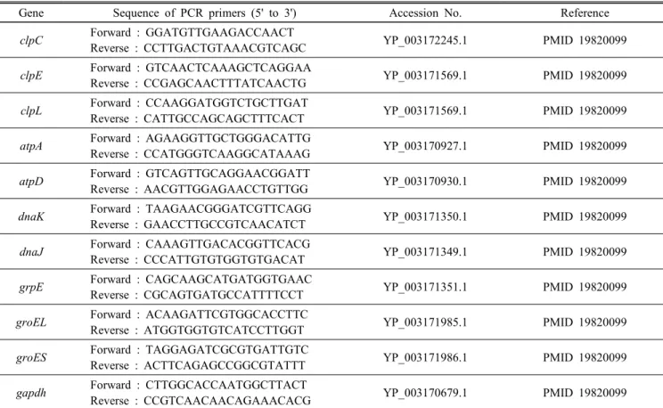
Table 2. Oligonucleotides primers used for qRT-PCR analysis of stress gene expression
Gene Sequence of PCR primers (5' to 3') Accession No. Reference
clpC Forward : GGATGTTGAAGACCAACT
Reverse : CCTTGACTGTAAACGTCAGC YP_003172245.1 PMID 19820099
clpE Forward : GTCAACTCAAAGCTCAGGAA
Reverse : CCGAGCAACTTTATCAACTG YP_003171569.1 PMID 19820099
clpL Forward : CCAAGGATGGTCTGCTTGAT
Reverse : CATTGCCAGCAGCTTTCACT YP_003171569.1 PMID 19820099
atpA Forward : AGAAGGTTGCTGGGACATTG
Reverse : CCATGGGTCAAGGCATAAAG YP_003170927.1 PMID 19820099
atpD Forward : GTCAGTTGCAGGAACGGATT
Reverse : AACGTTGGAGAACCTGTTGG YP_003170930.1 PMID 19820099
dnaK Forward : TAAGAACGGGATCGTTCAGG
Reverse : GAACCTTGCCGTCAACATCT YP_003171350.1 PMID 19820099
dnaJ Forward : CAAAGTTGACACGGTTCACG
Reverse : CCCATTGTGTGGTGTGACAT YP_003171349.1 PMID 19820099
grpE Forward : CAGCAAGCATGATGGTGAAC
Reverse : CGCAGTGATGCCATTTTCCT YP_003171351.1 PMID 19820099
groEL Forward : ACAAGATTCGTGGCACCTTC
Reverse : ATGGTGGTGTCATCCTTGGT YP_003171985.1 PMID 19820099
groES Forward : TAGGAGATCGCGTGATTGTC
Reverse : ACTTCAGAGCCGGCGTATTT YP_003171986.1 PMID 19820099
gapdh Forward : CTTGGCACCAATGGCTTACT
Reverse : CCGTCAACAACAGAAACACG YP_003170679.1 PMID 19820099
2) ATPase activity 측정
Fiske-Subbarow 방법을 이용하여 ATPase 활성을 측정하며, 조효소 용액 100 μL에 10 mM MgCl2와 5 mM ATP(sigma)를 함유한 Tris maleate buffer(pH 7.5, 50 mM) 900 μL를 가하 고, 37℃에서 10분 동안 효소반응 시킨 후, 3 mL 증류수와 1 mL의 3.5N 황산 용액을 가하여 효소 반응을 종결시켰다.
효소 반응이 완료된 용액에 3.5% ammonium molybdate 용액 1 mL를 가한 후 2.1% NaHSO3에 0.7% Developer(Kodak, D-76)를 혼합한 용액 1 mL를 첨가했다. 실온에서 20분간 반응시킨 후 spectrophotometer를 이용하여 660 nm에서 흡광 도를 측정하고, ATPase에 의해 ATP로부터 유리된 inorganic phosphate의 양을 standard curve로부터 환산하였다.
효소 단위는 1분 동안 1 μmole의 inorganic phosphate를 유리시 키는 효소의 양을 1 unit으로 하였으며, specific activity를 산출했다.
Specific activity = enzyme unit / protein (mg)
6. RNA 분석
1) Primer design
Primer는 qRT-PCR 분석을 위해 L. rhamnosus GG에 기
반을 둔 Primer3plus software(http://www. bioinformatics.nl/
cgibbin/primer3plus/primer3plus.cgi)를 사용하여 설계하였다.
이 primer는 작은 amplicon size(200~300 bp)를 포함하고 있으며, 45~55% 사이 범주의 GC 함량을 가지고, 60에서 64℃까지의 Tm 값을 측정하였다. 증폭된 결과물을 1% agrose gel에서 확인하고, 뒤이어 primer 이량체 형성 가능성을 배 제하기 위한 PCR-amplified products의 melt 곡선 분석을 확인했다(Table 2).
2) Total RNA 분리와 cDNA 분석
L. rhamnosus GG의 Total RNA는 MRS broth에서 OD600 0.6에 도달할 때까지 배양하고, pH 7.0 조건이 control이며, pH 4.5 조건에서 1시간, 12시간, 24시간, 48시간 배양한 샘 플을 RNeasy Mini Kit(Qiagen, Valencia, CA)를 사용하여 cell로부터 추출하고, genomic DNA를 제거하기 위해 RNase- free-DNaseⅠ(Qiagen, Valencia, CA)를 사용했다. 역전사를 하는 동안, total RNA 1,000 ng이 qRT-PCR kit(Intron, Seong- nam, Korea)의 RT premix oligo(dt)를 반응 mixture에 첨가 하였다. 역전사효소 반응 조건은 45℃에서 60분, 95℃에서 5분으로 실시하였다.
3) 실시간 RNA 정량 분석(Real-time qPCR)
Value method는 다른 샘플로부터 유전자 발현을 비교하 기 위해 사용했다(Livak and Schmittgen, 2001; Meng et al., 2007). Glyceraldehyde-3-phosphate dehydrogenase(GAPDH) 는 PCR 증폭에서 internal control로 사용했다. qRT-PCR은 1μL cDNA 주형, 각 primer, SYBR Green을 사용하여 수행 했고, 처음 denaturation은 95℃에서 5분간 실행한 후, 95℃
에서 10초(denaturation), 54℃에서 15초(annealing), 72℃에서 15초(extension)를 1 cycle로, CFX96TX(Bio-Rad, California, USA)를 사용하여 35 cycle을 반복 하였다. 각 PCR cycle마 다 melting curves는 비특이적 증폭을 피하기 위해 모니터 링 했다. 증폭 효율의 연구는 primers가 CT 값을 기준으로 비교 분석하기에 적합한 것으로 나타났다.
Relative expression=2-ΔCT with ΔCT= CTgene-CTactin.
7. 통계 분석
이 실험은 3번 반복하여 수행했다. Duncan’s multiple range test를 이용하여 시료간의 유의적 차이(p<0.05)를 검정하였다.
결과 및 고찰
1. 제품의 pH 및 생균수 변화
LGG 유산균이 함유된 요거트 제품을 52주 동안 냉동보 관했을 때, 그 결과 저장 초기 pH 3.6에 4.4의 범위를 보였 다(Fig. 1).
제품의 생균수 결과는 Fig. 2~4에서 보는 바와 같다. 생 균수 측정결과 Lactobacillus spp.는 약 107~108 CFU/mL, S.
thermophilus는 109 CFU/mL를 5주 동안 유지하였다. 비슷 하게 A, B 제품에서 초기 Bifidobacterium spp. 수는 105~107 CFU/mL이었다. 모든 샘플의 저장 기간을 고려했을 때, Bifido- bacterium spp.는 Lactobacillus와 S. thermophilus와 비교했을 때, 매우 급격하게 감소하였다. 저장 말기에는 Bifidobacterium spp.는 모두 사멸하였다. L. casei는 20주의 저장 기간 동안 낮은 생존성을 보였다. 반면에 Lactobacillus rhamnosus GG 는 저장 기간 동안 제품에서 가장 높은 생존성을 나타냈다.
저장 52주 후에도 107 CFU/mL를 유지하며, 매우 높은 생 존성을 보이는 것으로 확인되었다.
Fig. 1. Change of the pH in yogurt over 52-weeks storage at 5℃. A(◆), B(■), C(▲).
Fig. 2. Changes in viable count of lactic acid bacteria in yogurt (A) over 52-weeks storage at 5℃. S. thermophilus (◆), L. rhamnosus GG (■), Bifidobacterium (▲).
Fig. 3. Changes in viable count of lactic acid bacteria in yogurt (B) over 52-weeks storage at 5℃. S. thermophilus (◆), L. rhamnosus GG (■), B. longum (▲).
Fig. 4. Changes in viable cells counts of L. casei (■) in yogurt (C) over 52-weeks storage at 5℃.
발효유의 최종 pH는 lactobacilli, S. thermophilus와 Bifido- bacterium spp.의 성장과 생존성에 영향을 미칠 수 있다(Laroia and Martin, 1991; Hekmat and McMahon, 1992). 유산균에 의한 생산된 젖산은 특히 후산발효(post-acidification) 과정중 에 lactobacilli와 Bifidobacterium spp.의 생존성에 많은 영향 을 미치는 것으로 보고되었다(Ishibashi and Shimamura, 1993;
Shah et al., 1995).
2. pH 4.5에서 Lactobacillus spp.의 생존력
Probiotic Lactobacillus spp.의 pH 4.5 조건에서 생존력은 0시간, 4시간, 8시간, 12시간, 24시간, 48시간의 간격으로 생균수를 측정하였고, 그 결과는 Fig. 5에 나타난 바와 같 다. L. rhamnosus GG가 가장 높은 생존율을 나타냈고, 특히 12시간 이상 산성 조건에 노출되었을 때, 다른 Lactobacillus strains보다 높은 생존율을 보였다. 이 결과는 L. rhamnosus
Fig. 5. The viability of Lactobacillus spp. at pH 4.5. a) L.
rhamnosus GG, b) L. plantarum, c) L. acidophilus, d) L. casei.
GG가 in vitro 산성 조건에서 가장 높은 내산성을 가진다는 것을 의미한다.
3. 산성 조건에서 Lactobacillus spp.의 ATPase 활성 Lactobacillus strain의 세포막 ATPase 활성은 Fig. 6에 나 타난 바와 같으며, 다른 유산균들에 비해 L. rhamnosus GG 의 ATPase 활성이 가장 높게 나타났다.
산성 조건에서 ATPase 활성을 측정한 결과는 Fig. 7에서 보는 바와 같다. 그 결과, pH 7.0에서 보다 더 높은 활성을 나타냈다. 초기 ATPase의 활성은 pH 7.0에서 0.61 nmol/min/
mg이었고, 24시간 산성 조건에서 노출 후 ATPase 활성이 2.09 nmol/min/mg로 계속해서 증가했다.
F0F1 ATPase는 그람 양성균에서 산성 환경으로부터 보 호해주는 역할을 하는 것으로 잘 알려져 있으며 세포 안 pH 조절을 위해 원형질막의 proton 삼투압을 유도하는 것으로 알려져 있다. 몇 가지 mechanism들이 세포 안의 pH 항상성 을 조절한다고 알려져 있으며 그중 Proton-translocating ATPase 는 LAB에서 가장 중요하다(Hutkins and Nannen, 1993). 일 반적인 stress 반응에 포함된 ATPase의 유사성과 낮은 pH 에서의 강한 대응반응 때문에, F0F1 ATPase proton pump는 산성 환경에서 여러 미생물의 생존성에 영향을 미친다(Cotter and Hill, 2003). 이 효소는 L. casei와 L. plantarum에도 존재 하며, pH 5.0~5.5 부근에서 최고 활성을 보이는 것으로 알 려져 있다. 원형질막의 proton 투과성은 세포 안의 pH 유지 능력과 밀접한데, L. casei와 L. plantarum의 막 투과성은
Fig. 6. F1F0 ATPase activity of Lactobacillus strains.
Fig. 7. F1F0 ATPase activity at pH 4.5 in L. rhamnosus GG.
pH 4.0에서 가장 낮은 것으로 보고되었다(Bender and Marquis, 1987; Hong et al., 1999). 또한 adenosine-triphosphate(ATP) 는 세포질의 proton을 F0F1 ATPase에 의해 방출시킬 수 있게 하고, 에너지원 고갈 후에 세포가 좀 더 오래 살아남을 수 있 도록 도움을 줄 수 있다고 밝혀졌다(Stuart et al., 1999; Arena et al., 1999).
선행 연구에서, L. rhamnosus GG의 ATPase 활성은 다른
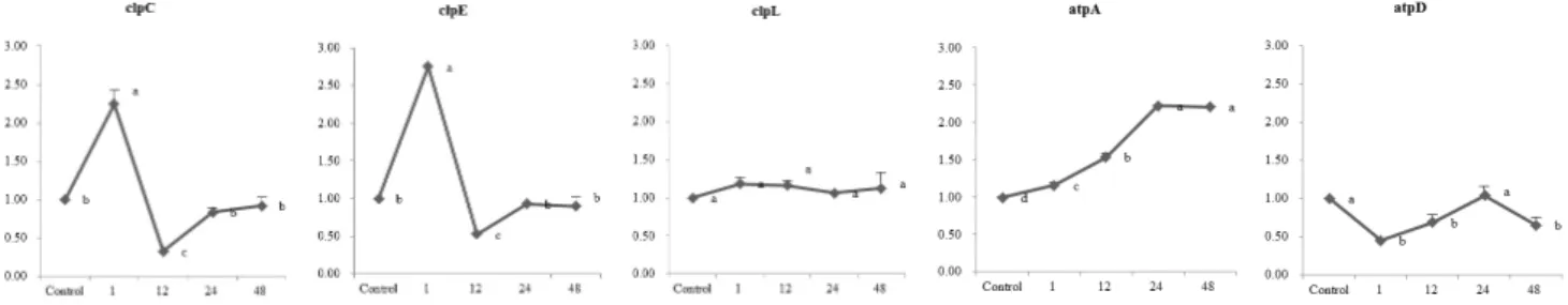
Fig. 8. Expression profile of the clp and atp genes at pH 4.5 for different time in L. rhamnosus GG. The cells exposed at pH 4.5 for different time. qRT-PCR was performed with 1 μL cDNA template, each primer, SYBR Green and subjected initial denaturation at 95℃ for 5 min, followed by 35 cycles of amplification at 95℃ for 10 s, 54℃ for 15 s, 72℃ for 15 s.
Expression of GAPDH was used as an internal control for equal loading. The x axis represents the exposure time at pH 7.0 (control) or pH 4.5 (acid stress), and the y axis represents the normalized fold expression of genes. a~d Means with same superscripts in a same row are not different (P>0.05).
Lactobacillus spp.보다 더 높게 나타났고, pH 4.5에 노출되 는 동안 증가함을 보였는데, 이는 세포막에 연결된 F0F1
ATPase가 산성 조건에 대한 내성에 중요한 역할을 하는 것으로 추정된다. 궁극적으로 L. rhamnosus GG는 산성 환 경에 노출되었을 때, 내산성이 증가되는 것으로 보아, 발 효유와 같은 비교적 낮은 pH에서 적응이 된 유산균은 다 른 조건에서 적응된 경우보다 내산성이 높을 것으로 판단 된다.
4. 유전자 발현 비교
pH에 따른 각 시간대 별 L. rhamnosus GG의 유전자 발 현은 relative expression software tool로 분석하였다. pH 4.5 에서 clpC/E/L, atpA/D, dnaK--dnaJ-grpE, and groES-groEL의 산성 유도성 특성은 qRT-PCR을 이용하여 입증하였다(Fig.
8, 9). pH 4.5에서 clpC/E/L, atpA/D, dnaK--dnaJ-grpE, and groES-groEL의 유전자가 높게 발현되는 것을 확인하였다.
clpC와 clpE gene은 pH 4.5에서 1시간 incubation되었을 때 각각 2.25 fold, 2.75 fold로 control과 비교하여 2~3배 정도 높은 발현량을 나타내었다. 반면, pH 4.5에서 노출시간이 증 가될수록 clpC와 clpE 유전자 발현은 각각 0.01~0.16 fold와 0.53~0.9 fold 감소되었다.
몇몇의 연구에서 일반적으로 clp 유전자의 발현은 스트레 스에 반응한다고 밝혔다. 그람 양성균에서 ClpC는 스트레 스에 의해 손상된 단백질을 분해하는 샤페론 역할을 하는 것으로 알려져 있다. ClpE는 단백질 가수분해 과정에서 기능 을 하고, ClpL은 가수분해보다는 단백질 수선의 기능이 있다 고 알려졌다. 최근 L. reuteri ATCC 55730의 late-logarithmic phase에서 산 자극의 반응에 대한 연구는 esterase 및 pepti- doglycan과 세포막 생합성에 관여하는 phosphatidyl-glycero- phosphatase를 코딩하는 유전자의 유도를 보여주었다. ClpL 및 esterase 돌연변이는 GI관을 통한 운송 중 산 충격에 대한
초기 반응 및 생존 가능성에 대한 중요성을 증명하였다(Wall et al., 2007). 본 연구에서는 산 조건의 반응초기에 CLPC/E 유전자의 발현이 증가했다. CLP 유전자는 배양초기에 발현 되며 산 스트레스 하에서 세포 생존성을 유지하기 위해 기 여하는 것으로 추정된다.
산 조건에서의 L. rhamnosus GG는 배양 24시간에 atpA 가 2.2 fold로 과발현되었다. Kullen과 Klaenhammer(1999) 는 낮은 pH에서 L. acidophilus가 pH-조절 proton 전위 F1F0
ATPase의 mRNA level의 증가를 일으킨다는 것을 diffe- rential display-PCR 장치를 이용하여 증명하였다(Ventura et al., 2004). ATP 오페론은 주로 박테리아 세포질에서 외막으로 proton pumping과 연관되어 있으며, 이러한 이유로 박테리 아 세포질에서 pH를 중성으로 유지시킬 수 있다. 또한 F1F0
ATPase는 proton pump를 유도하고, 세포 내의 ATP 양을 증가시키는 것으로 알려져 있는데, 그로 인해 박테리아의 세포 내 pH를 조절한다. 이 조절 기작은 산성 스트레스에 대 응하여 박테리아의 생육에 도움을 준다(Duary et al., 2010).
L. rhamnosus GG에서는 atpA 유전자 발현이 산성 조건에 노출된 시간이 증가된 후에 F1F0 ATPase의 알파 subunit이 높 은 수준을 나타내었다. 이 결과는 산성 조건에서 F1F0 ATPase 활성이 증가했고, 이것이 장시간 동안 산성 환경에서 다른 유산균과 비교해서 높은 생존율을 가지게 하는 것으로 생 각된다.
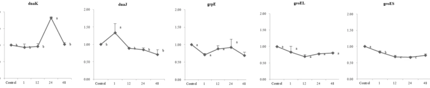
Fig. 9에서 나타난 바와 같이 dnaK는 24시간 배양한 후 1.85 fold 증가하였다. DnaK(Dank-DnaJ-GrpE)와 GroEL(GroEL- GroES) 시스템은 새로 합성된 polypeptides의 folding을 지원 한다고 알려져 있다(Houry et al., 1999; Teter et al., 1999).
이 단백질은 비정상적인 단백질의 분해에 연관되어 있다고 알려져 있으며 게다가 분자적 샤페론은 기질에 결합 cycle로 부터 운반되는 ATP를 사용하는 단백질의 folding을 도울 수 있고, 이 단백질의 refolding을 적극적으로 도울 수 있다(Bukau
Fig. 9. Expression profile of the stress genes at pH 4.5 for different time in L. rhamnosus GG. The cells exposed at pH 4.5 for different time. qRT-PCR was performed with 1 μL cDNA template, each primer, SYBR Green and subjected initial denaturation at 95℃ for 5 min, followed by 35 cycles of amplification at 95℃ for 10 s, 54℃ for 15 s, 72℃ for 15 s.
Expression of GAPDH was used as an internal control for equal loading. The x axis represents the exposure time at pH 7.0 (control) or pH 4.5 (acid stress), and the y axis represents the normalized fold expression of genes. a~c Means with same superscripts in a same row are not different (P>0.05).
and Horwich, 1998). 유산균에 있어서 stress 조건이나 정상 조건에서 단백질의 항상성을 유지하는 것이 매우 중요하 다. 본 연구결과, dnaK 유전자는 pH 4.5에서 과 발현되었 고, L. rhamnosus GG는 산 조건일 때 보호 기능을 지니는 것으로 나타났다.
qRT-PCR 결과로 볼 때, clpC, clpE, atpA, dnaK 유전자 발현이 증가하였는데, 이는 낮은 pH 환경에서 L. rhamnosus GG의 생존율을 향상시키는데 도움을 주며, L. rhamnosus
GG가 다른 유산균 보다 산성 조건에서 잘 적응하도록 기
여하는 것으로 생각된다.
감사의 글
본 연구는 농림수산식품기술기획평가원 고부가가치식품기 술개발사업(A2012-0447)에 의해 이루어졌으며, International Journal of Dairy Technology(67:229-236, 2014)에 게재된 결과를 사용하도록 허가해준 John Wiley and Sons에 감사 드립니다.
참고문헌
1. Alander, M., Satokari, R., Korpela, R., Saxelin, M., Vilpponen-Salmela, T., Mattila-Sandholm, T. and von Wright, A. 1999. Persistence of colonization of human colonic mucosa by a probiotic strain, Lactobacillus rhamnosus GG, after oral consumption. Appl. Environ. Microbiol.
65:351-354.
2. Arena, M. E., Saguir, F. M. and Manca de Nadra, M. C.
1999. Arginine, citrulline and ornithine metabolism by lactic acid bacteria from wine. Food Microbiology 47:
203-209.
3. Bender, G. R. and Marquis, R. E. 1987. Membrane ATPases and acid tolerance of Actinomyces viscosus and Lacto- bacillus casei. Applied and Environmental Microbiology 53:2124-2128.
4. Booth, I. R. 1985. Regulation of cytoplasmic pH in bacteria. Microbiol Rev. 49:359-378.
5. Bukau, B. and Horwich, A. L. 1998. The Hsp70 and Hsp60 chaperone machines. Cell 92:351-366.
6. Bukau, B., Weissman, J. and Horwich, A. 2006. Molecular chaperones and protein quality control. Cell 125:443-451.
7. Casadei, M. A., Ingram, R., Hitchings, E., Archer, J. and Gaze, J. E., 2001. Heat resistance of Bacillus cereus, Salmonella typhimurium and Lactobacillus delbrueckii in relation to pH and ethanol. Int. J. Food Microbiol 63:
125-134.
8. Cotter, P. D. and Hill, C. 2003. Surviving the acid test:
responses of grampositive bacteria to low pH. Micro- biology and Molecular Biology 67:429-453.
9. De Angelis, M. and Gobbetti, M. 2004. Environmental stress responses in Lactobacillus: A review. Proteomics 4:106-122.
10. Desmond, C., Stanton, C., Fitzgerald, G. F., Collins, K.
and Ross, R. P. 2001. Environmental adaptation of pro- biotic lactobacilli towards improvement of performance during spray drying. Int. Dairy J. 11:801-808.
11. Doron, S., Snydman, D. R. and Gorbach, S. L. 2005.
Lactobacillus GG: Bacteriology and clinical applications.
Gastroenterol Clin. North Am. 34:483-498.
12. Duary, R. K., Batish, V. K. and Grover, S. 2010. Ex- pression of the atpD gene in probiotic Lactobacillus plantarum strains under in vitro acidic conditions using RTqRCR. Research in Microbiology 161:399-405.
13. Fillingame, R. H. and Divall, S. 1999. Proton ATPase in bacteria: Comparison to Escherichia coli F1F0 as the prototype. Novartis Foundation Symposium 221:218-229.
14. Fiske, C. H. and Subbarow, Y. 1925. The colorimetric determination of phosphorous. J. Biol. Chem. 66: 375- 389.
15. Glaasker, E., Konings, W. N. and Poolman, B. 1996.
Osmotic regulation of intracellular solute pools in Lacto- bacillus plantarum. J. Bacteriol. 178:575-582.
16. Gorbach, S. L. 2000. Probiotics and gastrointestinal heal- th. Am. J. Gastroenterol 95:S2-S4.
17. Gorbach, S. L., Chang, T. W. and Goldin, B. 1987.
Successful treatment of relapsing Clostridium difficile colitis with Lactobacillus GG. Lancet 2:1519.
18. Hartl, F. U. and Hayer-Hartl, M. 2002. Molecular cha- perones in the cytosol: From nascent chain to folded protein. Science 295:1852-1858.
19. Hekmat, S. and McMahon, D. J. 1992. Survival of Lacto- bacillus acidophilus and Bifidobacterium bifidumin ice cream for use as a probiotic food. Dairy Science 75:
1415-1422.
20. Henriksson, A., Khaled, A. K. D. and Conway, P. L.
1999. Lactobacillus colonization of the gastrointestinal tract of mice after removal of the non-secreting stomach region. Microb. Ecol. Health Dis. 11:96-99.
21. Hong, S. I., Kim, Y. J. and Pyun, Y. R. 1999. Acid tole- rance of Lactobacillus plantarumfrom Kimchi. Food Science and Technology 32:142-148.
22. Houry, W. A., Frishman, D., Eckerskorn, C., Lottspeich, F. and Hartl, F. U. 1999. Identification of in vivo sub- strates of the chaperonin GroEL. Nature 402:147-154.
23. Hutkins, R. W. and Nannen, N. L. 1993. pH Homeostasis in lactic acid bacteria. J. Dairy Science 76: 2354-2365.
24. Ishibashi, N. and Shimamura, S. 1993. Bifidobacteria:
Research and development in Japan. Journal of Food Technology 47:129-134.
25. Isolauri, E., Salminen, S. and Ouwehand, A. C. 2004.
Probiotics. Best Practice & Research Clinical Gastro- enterology 18:299-313.
26. Kailasapathy, K. and Rybka, S. 1997. L. acidophilus and Bifidobacterium spp.: Their therapeutic potential and survival in yogurt. Australian Journal of Dairy Tech- nology 52:47-72.
27. Kaneko, T., Sato. S., Kotani, H., Tanaka, A., Asamizu, E., Nakamura, Y., Miyajima, N., Hirosawa, M., Sugiura, M., Sasamoto, S., Kimura, T., Hosouchi, T., Matsuno, A., Muraki, A., Nakazaki, N., Naruo, K., Okumura, S., Shimpo, S., Takeuchi, C., Wada, T., Watanabe, A., Yamada, M., Yasuda, M. and Tabata, S. 1996. Sequence analysis of the genome of the unicellular Cyanobacterium Syne- chocystis sp. strain PCC6803. II. Sequence determination of the entire genome and assignment of potential protein- coding regions. DNA Res. 3:109-136.
28. Kim, W. S., Perl, L., Park, J. H., Tandianus, J. E. and Dunn, N. W. 2001. Assessment of stress response of the probiotic Lactobacillus acidophilus. Curr. Microbiol.
43:346-350.
29. Kligler, B. and Cohrssen, A. 2008. Probiotics. Am. Fam.
Physician. 78:1073-1078.
30. Koponen, J., Laakso, K., Koskenniemi, K., Kankainen, M., Savijoki, K., Nyman, T. A., De Vos, W. M., Tynkkynen, S., Kalkkinen, N. and Varmanen, P. 2012. Effect of acid stress on protein expression and phosphorylation in Lactobacillus rhamnosus GG. Proteomics 75:1357-1574.
31. Kullen, M. J. and Klaenhammer, T. R. 1999. Identifica- tion of the pH-inducible, proton-translocating F1F0 ATPase (atpBEFHAGDC) operon of Lactobacillus acidophilus by differential display: Gene structure, cloning and cha- racterization. Mol. Microbiol. 33:1152-1161.
32. Laroia, S. and Martin, J. H. 1991. Effect of pH on sur- vival of Bifidobacterium bifidum and Lactobacillus acido- philus in frozen fermented dairy desserts. Cultured Dairy Products 26:13-21.
33. Lemay, M. J., Rodrigue, N., Garièpy, C. and Saucier, L.
2000. Adaptation of Lactobacillus alimentarius to environ- mental stresses. Int. J. Food Microbiol. 55:249-253.
34. Lim, E. M., Ehrlich, S. D. and Maguin, E. 2000. Iden- tification of stress-inducible proteins in Lactobacillus delbrueckii subsp. bulgaricus. Electrophoresis 21:2557-2561.
35. Obembe, O. O., Jacobsen, E., Vincken, J. and Visser, R.
G. F. 2009. Differential expression of cellulose synthase (CesA) gene transcripts in potato as revealed by QRT- PCR. Plant Physiology and Biochemistry 49:1116-1118.
36. Obokata, J., Ohme, M. and Hayashida, N. 1991. Nu- cleotide sequence of a cDNA clone encoding a putative glycine-rich protein of 19.7 kDa in Nicotiana sylvestris.
Plant Mol. Biol. 17:953-955.
37. Olson, E. R. 1993. Influence of pH on bacterial gene expression. Mol. Microbiol. 8:5-14.
38. Ouwehand, A. C., Salminen, S. and Isolauri, E. 2002.
Probiotics: An overview of beneficial effects. Antonie Van Leeuwenhoek. 82:279-289.
39. Rijkers, G. T., Bengmark, S., Enck, P., Haller, D., Herz, U., Kalliomaki, M., Kudo, S., Lenoir-Wijnkoop, I., Mercenier, A., Myllyluoma, E., Rabot, S., Rafter, J., Szajewska, H., Watzl, B., Wells, J., Wolvers, D. and Antoine, J. M.
2010. Guidance for substantiating the evidence for bene- ficial effects of probiotics: Current status and recommen-
dations for future research. J. Nutr. 140:6715-6765.
40. Schmidt, G., Hertel, C. and Hammes, W. P. 1999. Mole- cular characterisation of the dnaK operon of Lactobacillus sakei LTH681. System. Appl. Microbiol. 22:321-328.
41. Seigumfelt, H., Rechinger, K. B. and Jakobsen, M. 2000.
Dynamic changes of intracellular pH in individual lactic acid bacterium cells in response to a rapid drop in extra- cellular pH. Applied and Environmental Microbiology 66:2330-2335.
42. Shah, N. P., Lankaputhra, W. E. V., Britz, M. L. and Kyle, W. S. A. 1995. Survival of Lactobacillus acidophilus and Bifidobacterium bifidum in commercial yoghurt during refrigerated storage. Dairy Journal 5:515-521.
43. Stuart, M. R., Chou, L. S. and Weimer, B. C. 1999. In- fluence of carbohydrate starvation and arginine on cul- turability and amino acid utilization of Lactococcus- lactis subsp. lactis. Applied and Environmental Microbio- logy 65:665-673.
44. Sugimoto, S. and Sonomoto, K. 2011. Quality control of protein structure in lactic acid bacteria. Pages 143-155 in Lactic acid bacteria and bifidobacteria. Sonomoto, K., Yokota, A. Caister Avademic Press.
45. Teter, S. A., Houry, W. A., Ang, D., Tradler, T., Rock- abrand, D., Fischer, G., Blum, P., Georgopoulos, C. and Hartl, F. U. 1999. Polypeptide flux through bacterial Hsp70: DnaK cooperates with trigger factor in chape- roning nascent chains. Cell 97:755-765.
46. Udvardi, M. K., Czechowski, T. and Scheible, W. R. 2008.
Eleven golden rules of quantitative RT-PCR. The Plant Cell 20:1736-1737.
47. Wall, T., Bath, K., Britton, R. A., Jonsson, H., Versalovic, J. and Roos, S. 2007. The early response to acid shock in Lactobacillus reuteri involves the ClpL chaperone and a putative cell wall altering esterase. Applied and Environmental Microbiology 73:3924-3935.
48. Wouters, J. A., Mailhes, M., Rombuts, F. M., de Vos, W. M., Kuipers, O. P. and Abee, T. 2000. Physiologi- cal and regulatory effects of controlled overproduction of five cold shock proteins of Lactococcus lactis MG1363.
Appl. Environ. Microbiol. 66:3756-3763.
(Received 1 February 2015 / Accepted 26 March 2015)