ISSN 2234-3806 • eISSN 2234-3814
http://dx.doi.org/10.3343/alm.2013.33.3.174
Flow Cytometric Human Leukocyte Antigen-B27 Typing with Stored Samples for Batch Testing
Bo Young Seo, M.D. and Dong Il Won, M.D.
Department of Clinical Pathology, Kyungpook National University School of Medicine, Daegu, Korea Background: Flow cytometry (FC) HLA-B27 typing is still used extensively for the diagno- sis of spondyloarthropathies. If patient blood samples are stored for a prolonged duration, this testing can be performed in a batch manner, and in-house cellular controls could eas- ily be procured. In this study, we investigated various methods of storing patient blood samples.
Methods: We compared four storage methods: three methods of analyzing lymphocytes (whole blood stored at room temperature, frozen mononuclear cells, and frozen white blood cells [WBCs] after lysing red blood cells [RBCs]), and one method using frozen platelets (FPLT). We used three ratios associated with mean fluorescence intensities (MFI) for HLA- B27 assignment: the B27 MFI ratio (sample/control) for HLA-B27 fluorescein-5-isothiocya- nate (FITC); the B7 MFI ratio for HLA-B7 phycoerythrin (PE); and the ratio of these two ra- tios, B7/B27 ratio.
Results: Comparing the B27 MFI ratios of each storage method for the HLA-B27+ sam- ples and the B7/B27 ratios for the HLA-B7+ samples revealed that FPLT was the best of the four methods. FPLT had a sensitivity of 100% and a specificity of 99.3% for HLA-B27 assignment in DNA-typed samples (N=164) when the two criteria, namely, B27 MFI ratio
>4.0 and B7/B27 ratio <1.5, were used.
Conclusions: The FPLT method was found to offer a simple, economical, and accurate method of FC HLA-B27 typing by using stored patient samples. If stored samples are used, this method has the potential to replace the standard FC typing method when used in combination with a complementary DNA-based method.
Key Words: HLA-B27 typing, Flow cytometry, Sample storage, Frozen platelets
Received: September 10, 2012 Revision received: November 28, 2012 Accepted: January 24, 2013
Corresponding author: Dong Il Won Department of Laboratory Medicine, Kyungpook National University Hospital, 135 Dongdeok-ro, Jung-gu, Daegu 700-721, Korea
Tel.: +82-53-420-5291 Fax: +82-53-426-3367 E-mail: wondi@knu.ac.kr
© The Korean Society for Laboratory Medicine.
This is an Open Access article distributed under the terms of the Creative Commons Attribution Non-Commercial License (http://creativecom- mons.org/licenses/by-nc/3.0) which permits unrestricted non-commercial use, distribution, and reproduction in any medium, provided the original work is properly cited.
INTRODUCTION
HLA-B27 typing is widely practiced as an aid for diagnosing spondyloarthropathies [1]. Many HLA-B27 typing methods are in use, and several DNA-based methods are increasingly being used in clinical laboratories [2]. However, flow cytometry (FC) using monoclonal antibodies (FC HLA-B27 typing) is still the most extensively used method, because it is economical and relatively simple [3].
However, it has been recommended that FC HLA-B27 typing be performed using blood samples within 24 hr of venipuncture [4], and therefore, “batch testing” of stored samples is not pos- sible. Routine batch testing is usually performed on a set day of the week by using stored blood samples collected during the previous week. Furthermore, the results of HLA-B27 typing are not usually reported immediately, because they are required for
“next” patient visits. However, clinical laboratories serving low- volume hospitals inevitably face inefficiencies in terms of time,
cost, and labor because of the small numbers of samples col- lected in order to comply with the principle of testing “fresh”
blood samples. Whole blood can be stored after being treated with commercially available white blood cell (WBC) stabilization solutions such as TransFix (Cytomark, Buckingham, UK) or Cyto-Chex Reagent (Strek Laboratories, Omaha, NE, USA);
however, these reagents were developed for control materials [5], and are not intended for nor have been evaluated using pa- tient samples.
According to the guidelines issued by the European Federa- tion for Immunogenetics (EFI) [6] and the American Society for Histocompatibility and Immunogenetics (ASHI) [7], cellular con- trols should be run as a part of each FC HLA-B27 typing batch to verify reagent specificity [5, 8]. Fresh blood samples with known HLA-B27 typing results would be ideal, but are logisti- cally difficult to obtain. Cryopreserved and thawed mononuclear cell suspensions with known results can be used, if each labo- ratory validates that thawed cells exhibit reactivity patterns simi- lar to those of the same cells when tested freshly [9]. A few manufacturers provide HLA-B27+ control cells, which are stabi- lized preparations of human cell lines. However, if the prolonged storage of patient blood samples is allowed, FC HLA-B27 typing can be performed in a batch manner, and in-house cellular controls could easily be prepared from known patient samples.
One of the commercial monoclonal antibody reagents for FC HLA-B27 typing, the IOTest HLA-B27-FITC/HLA-B7-PE (Beck- man Coulter, Miami, FL, USA) consists of two kinds of monoclo- nal antibodies: ABC-m3 directed toward HLA-B27/B2708 anti- gens and BB7.1 directed toward HLA-B7 antigens [4]. The for- mer is a fluorescein-5-isothiocyanate (FITC)-conjugated anti- body that targets HLA-B27, but is also weakly reactive to the HLA-B7 antigens, while the latter is a phycoerythrin (PE)-conju-
gated antibody that targets HLA-B7 and blocks the cross-reac- tivity of the former to the HLA-B7 antigens [10].
Batch testing using stored blood samples for FC HLA-B27 typing demands that the antigenicities of HLA Class I antigens on stored cells remain detectable to allow differentiation within the B7 cross-reactive group (CREG), which includes the HLA- B7, 13, 27, 2708, 37, 41, 42, 47, 48, 54, 55, 56, 60, 61, 73, and 81 antigens [11]. In contrast, platelets carry HLA Class I anti- gens and not HLA Class II antigens [12]. Furthermore, the cryo- preservation of platelets is relatively straightforward because of their simple anuclear structures [13], and therefore, we sup- posed that platelets might be adequate for FC typing after cryo- preservation.
Little data are available regarding the reliability of FC HLA-B27 typing using stored samples. In this study, we sought to deter- mine which of the four methods described above is best for storing patient blood samples without using a commercial pre- serving solution; three of these methods involved the analysis of lymphocytes (whole blood stored at room temperature, frozen mononuclear cells, and frozen WBCs after lysing red blood cells [RBCs]) and one involved the use of frozen platelets.
METHODS
We compared six FC HLA-B27 typing methods: two “immedi- ate” methods and four “storage-based” methods (Table 1). We then selected the best storage method and evaluated it using patient samples.
1. Samples
In the present study, we used 164 consecutive EDTA-anticoagu- lated blood samples sent to the histocompatibility laboratory at
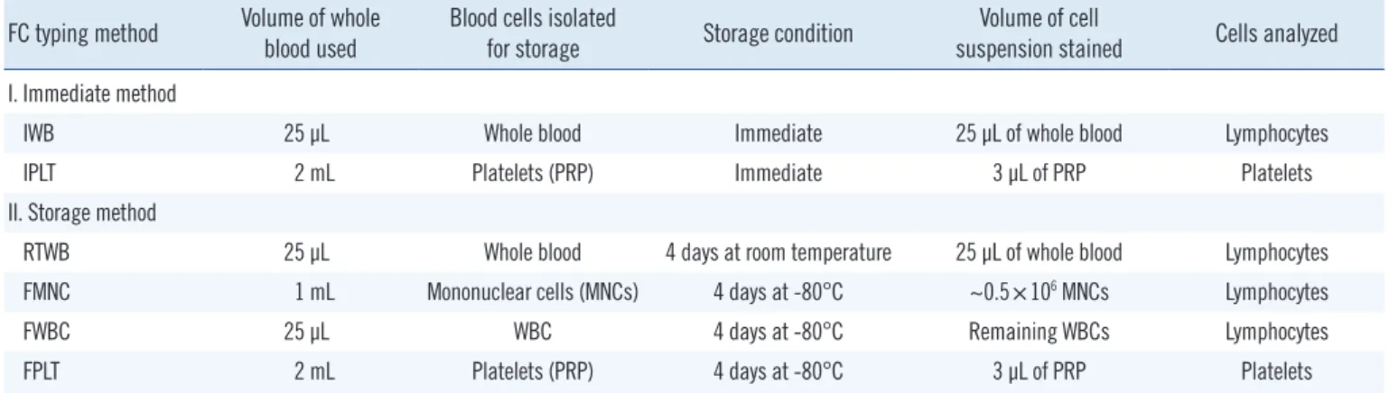
Table 1. Outline of the six different methods (two immediate and four storage) used for FC HLA-B27 typing FC typing method Volume of whole
blood used Blood cells isolated
for storage Storage condition Volume of cell
suspension stained Cells analyzed I. Immediate method
IWB 25 µL Whole blood Immediate 25 µL of whole blood Lymphocytes
IPLT 2 mL Platelets (PRP) Immediate 3 µL of PRP Platelets
II. Storage method
RTWB 25 µL Whole blood 4 days at room temperature 25 µL of whole blood Lymphocytes
FMNC 1 mL Mononuclear cells (MNCs) 4 days at -80°C ~0.5×10⁶ MNCs Lymphocytes
FWBC 25 µL WBC 4 days at -80°C Remaining WBCs Lymphocytes
FPLT 2 mL Platelets (PRP) 4 days at -80°C 3 µL of PRP Platelets
Abbreviations: FC, flow cytometry; IWB, immediate whole blood; IPLT, immediate platelets; RTWB, room temperature-stored whole blood; FMNC, frozen mononuclear cells; FWBC, frozen WBCs after lysing RBCs; FPLT, frozen platelets.
Kyungpook National University Hospital during 2011 for DNA- based HLA-A/B/DR or HLA-B27 typing. Of these, 154 samples were obtained from Korean patients and 10 samples were from HLA proficiency surveys (B27A2011 and B27B2011) conducted by the College of American Pathologists. Informed consent was obtained from all patients prior to the study.
2. FC HLA-B27 typing
Each FC typing method shared identical final steps, namely, flu- orescent staining, data acquisition, and data analysis, as de- scribed later. One IgG2a-FITC/IgG1-PE isotype control (Beck- man Coulter) and one negative control were always run simulta- neously with the test samples. Cells lacking specific and cross- reacting antigens (i.e., having neither HLA-B27 nor the other B7 CREG antigens) were selected as negative controls from known samples. For each FC typing method, we used negative control cells of the same cell type (lymphocytes or platelets) that had been stored under the same conditions as the test samples.
FC typing methods were designated names by using the fol- lowing credentials: “(storage temperature)-(status of stored cells);” for example, “RTWB” stands for “(Room Temperature)- (Whole Blood).” One “wash” in this study means: 1) 3 mL of phosphate-buffered saline (PBS) was added to a tube; 2) the tube was vortexed and centrifuged; 3) the supernatant was de- canted; and 4) the cell pellet was re-suspended in the remain- ing or additional PBS in its tube.
The outline of the six FC typing methods is provided in Table 1. IWB is the “standard” method performed according to the manufacturer’s recommendations [4]. The pre-staining assay steps for each FC typing method (numbered one to six) were as follows:
1) Immediate whole blood (IWB)
Twenty-five microliters of whole blood in EDTA was aliquoted into a microcentrifuge tube. After “fluorescent staining” (de- scribed later), stained whole blood was mixed with 1 mL of RBC lysing solution (BD Biosciences, San Jose, CA, USA), vortexed, and incubated for 10 min at room temperature in the dark. The mixture was then moved to a 5 mL polystyrene tube, centrifuged for 5 min at 400×g, and the supernatant was decanted. The re- maining WBCs were washed once.
2) Immediate platelets (IPLT)
Whole blood collected in a 5 mL EDTA tube was centrifuged for 10 min at 200×g, and the top 3/4 of the supernatant platelet rich plasma (PRP) was moved to another tube [14]. Three mi-
croliters of PRP was then mixed with 22 µL of PBS in a micro- centrifuge tube to achieve the same reaction volume as IWB.
Platelets in this suspension underwent fluorescent staining, dur- ing which one wash was performed with 3 mL of EDTA-PBS (3.36 g EDTA in 1,000 mL PBS) for 10 min at 1,000×g.
3) Room temperature-stored whole blood (RTWB)
Samples were held for four days at room temperature before processing. The remaining processes were as described for IWB.
4) Frozen mononuclear cells (FMNC)
Using the density gradient centrifugation method, mononuclear cells (MNCs) were separated from 1 mL of whole blood and washed (×3) with RPMI. This suspension was then mixed with the same volume of cryosolution (6% hydroxyethyl starch, 4% fetal calf serum, 5% dimethyl sulfoxide in RPMI; final concen- tration) in ice water [15], and stored at -80˚C. Just prior to test- ing, frozen MNCs were immediately thawed in thawing solution (2% fetal calf serum in RPMI). The suspension was then vor- texed and centrifuged, and supernatants were discarded. Re- maining MNCs were distributed in ~0.5×10⁶ cells per tube for fluorescent staining.
5) Frozen WBCs after lysis of the RBCs (FWBC)
One milliliter of RBC lysing solution was added to 25 µL of whole blood in a microcentrifuge tube. After vortexing and incubating for 10 min at room temperature, the mixture was immediately frozen at -80˚C [16]. Just prior to testing, it was briefly (< 5 min) thawed in a 37˚C water bath, and after centrifugation for 5 min at 400×g, the supernatant was decanted. Remaining WBCs were washed once and then fluorescent stained.
6) Frozen platelets (FPLT)
Platelets were frozen as described by Helmerhorst et al. [13].
Briefly, PRP (obtained as described for IPLT) was frozen directly at -80˚C, and just prior to testing, it was thawed at room temper- ature. The remaining processes were as described for IPLT.
7) Fluorescent staining
Five microliters of IOTest HLA-B27-FITC/HLA-B7-PE reagent was added to each immediate or stored sample prepared at its defined volume in a microcentrifuge tube, and incubated for 20 min at room temperature with gentle agitation in the dark. For IWB, RTWB, and FWBC, RBCs were removed using RBC lysing solution. After one wash, 50 µL of PBS was added to achieve the volume required for data acquisition. Samples were stored
at 4˚C in the dark shortly until data acquisition.
8) Data acquisition
FC was performed using a FACSCalibur flow cytometer running CELLQuest pro software (BD Biosciences). The instrument was calibrated daily using CaliBRITE beads and FACSComp software (BD Biosciences). Fluorescence signals were logarithmically amplified using a 4-log decade scale (100 to 104) (Fig. 1).
When analyzing lymphocytes, both fluorescence 1 (FL1) and FL2 photomultipliers were adjusted by running an isotype con- trol prior to a negative control or a sample tube, so that > 90% of the lymphocyte fluorescence signals would be in the first de- cade counter on the FL1 vs. FL2 plot [4]. Scatter signals were acquired using linear amplification on a forward scatter (FSC) vs. side scatter (SSC) plot using a lymphocyte gate. A total of
~10,000 events of gated lymphocytes were acquired per tube.
When analyzing platelets, FL1 and FL2 photomultipliers were adjusted for platelets as described above for the lymphocytes.
However, scatter signals were acquired using logarithmic ampli- fication on a FSC vs. SSC plot using a platelet gate. A total of
~20,000 events of gated platelets were acquired per tube.
9) Data analysis
Logarithmically amplified fluorescence signals were expressed as linear values (not channel values) (Fig. 1). Levels of antibody binding to lymphocytes or platelets were measured as mean flu- orescence intensities (MFI) within markers set equidistantly around the peak maxima. The upper and lower ends of the markers were set to maximally cover the symmetric portion of the peak.
Fig. 1. FC HLA-B27 typing using the two immediate methods. The black and blue peaks denote a test sample (HLA-B27+) and a negative control, respectively. On each FSC vs. SSC plot, a lymphocyte gate was drawn for IWB using linear amplification, and a platelet gate was drawn for IPLT using logarithmic amplification. For IPLT, the FSC and SSC threshold values (the two purple arrows) were set in order to ef- fectively collect the platelet signals by eliminating unwanted events. MFI values were obtained as the geometric means of the peaks on the HLA-B27 FITC or B7 PE histogram of the gated lymphocytes or platelets. An example of the B27 and B7 MFI ratios obtained by analyzing lymphocytes and the corresponding B7/B27 ratio is presented.
Abbreviations: FC, flow cytometry; IWB, immediate whole blood; IPLT, immediate platelets; MFI, mean fluorescence intensity; FITC, fluorescein-5-isocyanate;
PE, phycoerythrin; FSC, forward scatter; SSC, side scatter.
SSC
HLA-B27 FITC IWB
0 200 400 600 800 1,000
100 80 60 40 20 0
100
80
60
40 20
0 104
103
102
101
100 104
103 102 101 100 101
101 101
101 100
100 100
100 103
103 103
103 104
104 104
104 102
102 102
102
IPLT Platelet gate
threshold Lymphocyte gate
R1
M1 M1
M2 M2
linear
HLA-B27 FITC
HLA-B7 PE logarithmic
FSC HLA-B7 PE
Counts B7/B27 ratio
= 1.2 16.2
= 0.1 MFI ratio
= 8.0 6.6
= 1.2 MFI ratio
= 140.7 8.7
= 16.2
In this study, MFI “ratio,” rather than MFI value, was consid- ered for the HLA-B27 assignment. Three ratios were calculated per sample as follows:
- MFI ratios:
B 27 MFI ratio =test sample MFI/negative control MFI for HLA-B27 FITC
B 7 MFI ratio =test sample MFI/negative control MFI for HLA-B7 PE
- B7/B27 ratio=B7 MFI ratio/ B27 MFI ratio
3. DNA-based HLA-B27 typing
Real-time PCR was performed for melting curve analysis by us- ing a Real-Q HLA-B*27 detection kit (BioSewoom, Seoul, Korea) [17] on an ABI 7500 real-time PCR system (Applied Biosystems, Foster city, CA, USA).
4. HLA-A/B/DR typing
Typing was performed using Dynal RELI SSO HLA typing kits (Dynal A.S, Oslo, Norway), which use the PCR-reverse sequence specific oligonucleotide probe (SSOP) method.
RESULTS
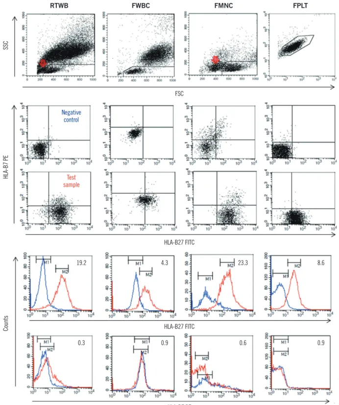
1. Representative examples of the four storage methods Many dead cells were seen in FMNC and in RTWB (Fig. 2).
FWBC induced significant nonspecific staining of the negative controls and test samples. FPLT generally diminished the MFI values for the test samples and negative controls, because of the size difference between the platelets and lymphocytes.
However, for the HLA-B27+ samples, the B27 MFI “ratios” for the platelets were near those for the lymphocytes. For example, the median MFI ratio obtained with FPLT was similar to that for IWB in Experiment A, described below (10.9 and 14.8, respec- tively, N=11).
2. Changes in the B27 MFI ratios for each storage method We investigated whether stored cells A) have patent HLA-B27 antigenicity, and B) do not overexpress HLA antigenicity for B7 CREG. In Experiment A, 11 HLA-B27+ samples were used (Fig.
3A). In Experiment B, 11 samples with ABC-m3-reactive speci- ficities, such as B7 CREG and HLA-B44 [10], were used (Fig.
3B): nine HLA-B13, B37, or B44+ samples and two HLA-B7+ samples.
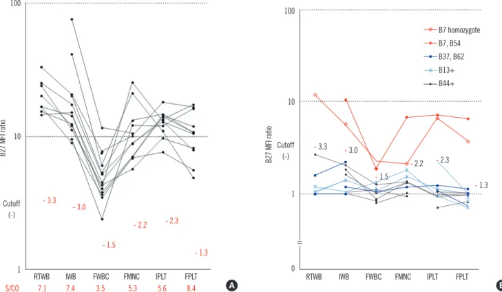
In Experiment A, we compared the discriminatory powers of the six FC typing methods (Fig. 3A); that is, we examined the ability of each FC typing method to discriminate HLA-B27+ from
other B7 CREG samples. For this purpose, we used an index, the sample/cutoff (S/CO) ratio, which was calculated as follows:
S/CO ratio=sample MFI ratio/cutoff MFI ratio
Where the sample MFI ratio is the average B27 MFI ratio of Experiment A samples using a given FC typing method, and the cutoff MFI ratio is (the average B27 MFI ratio of the samples for Experiment B (N=9) after excluding two HLA-B7+ samples) + (3×SD) using the same FC typing method.
In Experiment A, for a given FC typing method, HLA-B27+ samples always gave higher B27 MFI ratios than the cutoff MFI ratio for the same FC typing method (Fig. 3A). Therefore, all FC typing methods had a sensitivity of 100%. Comparing the S/CO ratios of the six FC typing methods, FWBC yielded the lowest value of 3.5. In contrast, relatively satisfactory discriminatory powers were obtained for the other five methods; FPLT had the highest value (8.4), suggesting that it had the greatest discrimi- natory power.
In Experiment B, nine HLA-B13+, B37+, or B44+ samples showed relatively low B27 MFI ratios, resulting in a specificity of 100% for all FC typing methods (Fig. 3B). Two HLA-B7+ sam- ples always showed higher B27 MFI ratios for a given FC typing method than the cutoff of the same FC typing method, except for one case of FMNC. This finding suggests that neither stor- age conditions nor platelet analysis enhanced the cross-reactiv- ity of anti-HLA-B27 FITC antibodies compared with the standard method (IWB).
3. Changes of B7/B27 ratios for each storage method We sought to determine which storage method was best at dis- criminating HLA-B7, B27 from the HLA-B7 homozygote sam- ples based on the B7/B27 ratios (Fig. 4). To be assigned as HLA-B27+, samples should have a relatively low B7/B27 ratio (<1.5) (plotted under the red line on the B27 vs. B7 MFI ratio plot in Fig. 4). In this experiment, both HLA-B27+ donors (HLA- B7, B27 and HLA-B27, B51) had B7/B27 ratios of <1.0 or were plotted under the line y=x for all FC typing methods, and were successfully assigned as HLA-B27+.
On the other hand, in principle, HLA-B7 homozygotes should have a relatively high B7/B27 ratio (>1.5), not for HLA-B27, but rather for B7 assignments. In this experiment with one HLA-B7 homozygote, FPLT yielded the highest value (2.7, note * in Fig.
4), but RTWB and FWBC yielded the lowest values (1.4 and 1.0, respectively). These low values could be mistakenly interpreted as HLA-B27+ reactions. This finding also suggests that FPLT is the method with the highest discriminatory capability.
Fig. 2. Representative examples of the four storage methods. The HLA-B phenotype of the test sample (red line) was HLA-B27, B62. The negative control (blue line) for FMNC was a random donor pool showing a broad peak. MFI ratios are presented in the right upper region of each histogram. Red arrows indicate dead cells detected during or after storage by FMNC or RTWB.
Abbreviations: RTWB, room temperature-stored whole blood; FWBC, frozen WBCs after lysing RBCs; FMNC, frozen mononuclear cells; FPLT, frozen plate- lets; FSC, forward scatter; SSC, side scatter; FITC, fluorescein-5-isocyanate; PE, phycoerythrin; MFI, mean fluorescence intensity.
SSC
FSC
19.2 4.3 23.3 8.6
0.3 0.9 0.6 0.9
HLA-B7 PE
HLA-B27 FITC
HLA-B27 FITC
HLA-B7 PE Negative
control
sampleTest
Counts
RTWB FWBC FMNC FPLT
ples) were further tested by FPLT (Fig. 6).
Regarding the B7/B27 ratio, we supposed that its cutoff value should be at the midpoint between two groups of samples, that is, HLA-B7, B27 and HLA-B7+. The average of the latter group (N =7) was 3.9, and that of the former group (N =1) was 0.8. Thus, the cutoff value for the B7/B27 ratios was defined as 1.5, despite the small sample size. Receiver operating characteris- tics (ROC) curve analysis revealed that the cutoff value for the B27 MFI ratio was 4.0. These findings indicate that a sample can be assigned as HLA-B27+ if it meets the two criteria: 1) B27 MFI ratio >4.0 and 2) B7/B27 ratio <1.5.
Using those criteria, all determinations obtained by FPLT were in agreement with those obtained by DNA typing, except for one case. This case (Fig. 6, black arrow) was found to be false posi- tive using FPLT, with a B27 MFI ratio of 6.4 and a B7/B27 ratio of 0.1. IWB also produced a false-positive result for this sample (B27 MFI ratio 4.3 (cutoff 3.0 in Fig. 3) and B7/B27 ratio 0.22).
Accordingly, using the above-mentioned cutoff values, FPLT achieved a sensitivity of 100% and a specificity of 99.3% for the HLA-B27 assignment.
4. The caveat of using RTWB
RTWB seemed to be vulnerable in terms of false positivity and negativity. We experienced both these problems. As an example of false negativity, an HLA-B27+ sample had an additional higher peak with a low MFI on the fluorescence histogram (Fig. 5), which was probably caused by the accumulation of dead cell debris during prolonged storage at room temperature. As an ex- ample of false positivity, an HLA-B7 homozygote sample gave the following results: B27 MFI ratio 12.0; B7 MFI ratio 17.1; and B7/B27 ratio 1.4. This low B7/B27 ratio could be mistakenly in- terpreted as a HLA-B27+ reaction.
5. Definition of the FPLT cutoff in a large number of known samples
Because FPLT appeared to be the best of the four storage meth- ods, we defined its formal cutoff values in terms of maximizing both assay sensitivity and specificity compared with DNA typing as a reference. A total of 164 samples with known DNA typing results (including 24 HLA-B27+ and 80 other B7 CREG sam-
Fig. 3. Changes in B27 MFI ratios by storage method in HLA-B27+ (N=11, panel A) and B7 CREG or HLA-B44+ (N=11, panel B) sam- ples. The cutoff B27 MFI ratio for each FC typing method was calculated in Experiment B after excluding two HLA-B7+ samples. Sample/
cutoff (S/CO) ratios for each FC typing method are presented in panel A.
Abbreviations: MFI, mean fluorescence intensity; IWB, immediate whole blood; IPLT, immediate platelets; RTWB, room temperature-stored whole blood; FMNC, frozen mononuclear cells; FWBC, frozen WBCs after lysing RBCs; FPLT, frozen platelets.
B27 MFI ratio
100
10
1 RTWB IWB FWBC FMNC IPLT FPLT
Cutoff (-)
S/CO 7.1 7.4 3.5 5.3 5.6 8.4
- 3.3 - 3.0
- 1.5
- 2.2 - 2.3 - 1.3
- 1.3
RTWB IWB FWBC FMNC IPLT FPLT
B27 MFI ratio
100
10
1
0 Cutoff
(-) - 3.3 - 3.0
- 2.2 - 1.5
- 2.3 B7 homozygote B7, B54 B37, B62 B13+
B44+
A B
DISCUSSION
Sample storage is a prerequisite for batch testing. Our study provides an evaluation of various simple storage methods for FC HLA-B27 typing, and shows that FPLT is the best of the four methods examined, with a sensitivity of 100% and a specificity of 99.3% in 164 samples compared with DNA typing as the ref- erence. In “standard” FC HLA-B27 typing, the cells used for analysis are restricted to lymphocytes. However, in the present
study, we found that platelets have sufficient HLA molecules for testing.
Out of the 164 samples, FPLT produced a false-positive result in a case with the HLA- B61, B67 phenotype. McKenna and Takemoto [18] classified HLA-B61 and B67 antigens as the same CREG (B07C CREG); however, Rodey et al. [11] classified them as different CREGs (B7 and B8 CREG, respectively). IWB
B7, 7
Fig. 6. B27 vs. B7 MFI ratio plot for FC HLA-B27 typing by FPLT (N=
164). The results of DNA typing are indicated as red (HLA-B27+) or black (HLA-B27-) dots, or are written alongside the dots. The cutoff line (red) is composed of two straight lines: 1) x =4; and 2) y=1.5x. Dots under this broken border were assigned HLA-B27+
by FPLT. FPLT was concordant with DNA typing in all cases, except for a single false-positive case indicated by the black arrow (pheno- type HLA-B61, B67). Cases in the indeterminate region (purple, shaded area) required confirmation by DNA typing for a robust de- termination to be reached. Refer to the DISCUSSION for a definition of the indeterminate region. There were two HLA-B27- samples in the indeterminate region, but not in the HLA-B27+ region, whose phenotypes were determined as HLA-B55+ by DNA typing.
Abbreviations: MFI, mean fluorescence intensity; FPLT, frozen platelets.
B7 MFI ratio
20
16
12
8
4
0
0 4 8 12 16 20 B27 MFI ratio
HLA-B27+region B7, 27 cutoff
B7, 7 B7, 75
B7, 37
B7, 35
B7, 54
B7, 54 B7, 48
B61, 67 Indeterminate
region B55+
y=x
Fig. 4. Changes in B7/B27 ratios on the B27 vs. B7 MFI ratio plot according to storage method in three donors: 1) HLA-B7 homozy- gote (red ■), 2) HLA-B7, B27 (purple ◆), and 3) HLA-B27, B51 (blue ●). The six results obtained for a given donor are connected by thin colored lines. The three results obtained for a given FC typ- ing method are connected by thick colored lines. The slope of the virtual straight line, which passes through the origin of the coordi- nate, corresponds to its B7/B27 ratio. One asterisk (*) indicates the highest B7/B27 ratio (2.7) of the six FC typing methods for a HLA- B7 homozygous donor. This value was obtained using FPLT.
Abbreviationds: MFI, mean fluorescence intensity; IWB, immediate whole blood; IPLT, immediate platelets; RTWB, room temperature-stored whole blood; FMNC, frozen mononuclear cells; FWBC, frozen WBCs after lysing RBCs; FPLT, frozen platelets.
B7 MFI ratio
20
15
10
5
00 10 20 30 40 B27 MFI ratio
HLA-B7, B27 RTWB
FWBCFMNC FPLT
IPLTIWB HLA-B7 homozygote
HLA-B27, B51 y=1.5 x y=X
*
Fig. 5. An example of a caveat of using RTWB. An HLA-B27+ sample, which was held for seven days at room temperature, showed many particles with weak fluorescence (red arrow) on the FL1 vs. FL2 plot of gated lymphocytes. These particles constituted a main peak (2 blue arrows) on each fluorescence histogram, which could have been mistakenly interpreted as a negative reaction.
Abbreviations: SSC, side scatter; FSC, forward scatter; PE, phycoerythrin; FITC, fluorescein-5-isocynate; M1, marker 1.
SSC Counts
HLA-B7 PE
HLA-B27 FITC HLA-B27 FITC HLA-B7 PE
FSC
also produced a false-positive result for this sample. Particular combinations of non-B27 HLA-B antigens are known to cause false-positive reactions [19]. In contrast, the HLA-B27+ sample with the lowest B27 MFI ratio (5.3) achieved by FPLT was ob- tained from a patient with aplastic anemia who had undergone hematopoietic stem cell transplantation; this sample also re- sulted in a relatively low B27 MFI ratio (14.0) by IWB.
It has been reported that FC HLA-B27 typing can be performed on blood samples stored for up to 16 days at room temperature.
In that study, no erroneous HLA-B27 assignment was made for 39 blood samples held for 11 to 16 days [19]. In the present study, blood samples could be held for four days as RTWB also showed no apparent changes in the MFI ratios. However, there is little guarantee of accuracy in samples containing high levels of dead cell debris, as occurred in the case depicted in Fig. 5, or when contaminating bacteria proliferate during storage. To ana- lyze pure viable lymphocytes in such cases, a T-cell marker such as anti-CD3 should be incorporated into the fluorescent staining protocol [19].
Although FMNC had a high discriminatory power, it required the most complex procedure, involving MNC isolation, freezing, thawing, and the adjustment of cell counts, to the extent that the method would be too cumbersome for routine clinical labo- ratory application. FWBC is much simpler than FMNC, but the non-specific binding of anti-B27 and anti-B7 antibodies was pronounced even in negative controls (Fig. 2, 3A), which mark- edly diminished its discriminatory power; therefore, this method cannot be recommended.
The processing required for FPLT is much simpler than that required for FMNC: 1) platelets can be simply isolated by differ- ential centrifugation (neither RBC lysing solution nor density- gradient centrifugation is required), 2) platelets can be frozen and thawed in plasma (requiring neither cryosolution nor thaw- ing solution), and 3) FPLT does not require the platelet counts to be adjusted. The present study shows that platelets procured using the FPLT protocol retain patent HLA antigenicity, and that FPLT had the greatest discriminatory power. Therefore, we sug- gest FPLT as the method of choice for stored samples.
If FC HLA-B27 typing were performed on Friday, a sample re- ferred on Monday would be stored for four days. This is why
“four days” was adopted as the storage period in the present study. However, the same conclusions for FPLT would have been reached had the PRP storage period been extended, be- cause freezing and thawing steps more substantially affect cell viability than storage period when a deep freezer is used. It has already been reported that the platelet surface glycoproteins
such as HLA and the human platelet antigen (HPA)-1a are sta- ble and are clearly detected by their corresponding antibodies when stored for 12 months at -80˚C [19].
Although a minimum of two HLA-B27 monoclonal antibody reagents is required to guarantee consistently reliable HLA-B27 typing [20], we used only a single reagent because our aim was to search for the best storage method, and because only two HLA-B27 alleles (HLA-B*27:04 and B*27:05) are found in the Korean patients with spondyloarthropathies [21, 22]. Neverthe- less, we suppose that the diagnostic efficiency of FPLT would have been improved had other reagents been incorporated.
Although absolute MFI values have been directly compared for the HLA-B27 assignment in the majority of studies [20], we utilized the MFI “ratio” in the present study. This normalization of test samples versus a negative control allows comparisons of measured values between different cell types (lymphocytes and platelets) and between different batches. However, we suggest that each laboratory defines its own cutoff values, because even MFI ratios are subject to variation between laboratories.
The cutoff B27 MFI ratio should be defined by a statistical analysis of different known samples, including B7 CREG and non-B7 CREG samples. In the present study, the cutoff was de- fined as 4.0 based on the results of a ROC curve analysis of the 164 samples. Furthermore, when a sample had a B7/B27 ratio of <1.5, a B27 MFI ratio ranging from 2.4 to 6.6 was regarded
“indeterminate.” This range was defined based on the following statistics: 1) the lower limit, 2.4, was calculated as (the average of 78 HLA-B27- samples with a B27 MFI ratio of <4.0)+(3×SD);
and 2) the upper limit, 6.6, was calculated as (the average of 21 HLA-B27+ samples excluding the four outliers)-(2×SD). There- fore, on the B27 vs. B7 MFI ratio plot (Fig. 6), an “indetermi- nate” region was defined as that defined by 1) 2.4 <x(B27 MFI ratio) <6.6 and 2) below the line y=1.5x. Samples plotted within this region cannot be accurately determined for HLA-B27 as- signment. In the present study, these samples accounted for 3.7% (6/164), and thus, the HLA-B27 assignment would have required complementary DNA typing [23], which could have been performed using previously frozen blood samples without re-sampling, if the blood cells remaining after removing PRP had been frozen for possible further testing.
It is difficult to discriminate HLA-B7, B27 from other HLA-B7+ samples. When a B7 MFI value of >4 was obtained on a Coul- ter Epics XL-MCL flow cytometer (Beckman Coulter), Coates and Darke [9] applied a cutoff B27 MFI (not MFI “ratio”) for this discrimination, that is, a B27 MFI of >10 for the former, and a B27 MFI of <5 for the latter. In the present study, we applied a
cutoff B7/B27 ratio defined as the midpoint between two groups of restricted sample size. If a statistical approach had been used on a larger number of samples, the cutoff and indeterminate range would have been determined more accurately.
In conclusion, of the four storage methods investigated, none of which required a preserving solution, FC HLA-B27 typing us- ing frozen platelets was found to be the most straightforward, economical, and accurate for both batch testing and control procurement. When stored samples are used, this method has the potential to replace the standard FC typing method. How- ever, some samples (3.7%) with indeterminate results must be confirmed using a complementary DNA-based method.
Authors’ Disclosures of Potential Conflicts of Interest
No potential conflicts of interest relevant to this article were re- ported.
Acknowledgement
This research was supported by Kyungpook National University Research Fund, 2012.
REFERENCES
1. Reveille JD. Major histocompatibility genes and ankylosing spondylitis.
Best Pract Res Clin Rheumatol 2006;20:601-9.
2. Seipp MT, Erali M, Wies RL, Wittwer C. HLA-B27 typing: evaluation of an allele-specific PCR melting assay and two flow cytometric antigen assays. Cytometry B Clin Cytom 2005;63:10-5.
3. College of American Pathologists. Surveys 2011 B27-A HLA-B27 typing participant summary. Northfield: College of American Pathologists (http://www.cap.org), 2011:1-5.
4. Beckman Coulter (Ed), IOTest HLA-B27-FITC/HLA-B7-PE package in- sert (A07739). http://www.beckmancoulter.com/wsrportal/page/itemDet ails?itemNumber=A07739#2/10//0/25/1/0/asc/2/A07739///0/1//0/ (Last visit on Jan 2013).
5. Levering WH, Wind H, Granger V, Sintnicolaas K, Hooijkaas H, Reilly JT, et al. Long-term stabilized blood samples as controls for flow cytometric HLA-B27 screening: a feasibility study. Cytometry B Clin Cytom 2008; 74:169-81.
6. European Federation for Immunogenetics. Standards for Histocompati- bility Testing. Version 5.4. April 2005.
7. American Society for Histocompatibility and Immunogenetics. Revised Standards for Histocompatibility Testing. December 2005.
8. Levering WH, Sintnicolaas K, Wind H, Hooijkaas H, Gratama JW. Flow cytometric screening for the HLA-B27 antigen on peripheral blood lym- phocytes. Curr Protoc Cytom 2005 Aug; Chapter 6: Unit6.22.
9. Coates E and Darke C. Routine HLA-B27 typing by flow cytometry: dif- ferentiation of the products of HLA-B*2702, B*2705 and B*2708. Eur J Immunogenet 1998;25:29-37.
10. Coates E, Rees TJ, Darke C. An evaluation of Com-B27, a fluorescein- conjugated mouse anti-HLA-B27 reagent. Eur J Immunogenet 2003; 30:271-4.
11. Rodey GE, Revels K, Fuller TC. Epitope specificity of HLA class I alloan- tibodies: II. Stability of cross-reactive group antibody patterns over ex- tended time periods. Transplantation 1997;63:885-93.
12. McFarland JG. Platelet and granulocyte antigens. In: Roback JD, Combs MR, Grossman BJ, Hillyer CD, ed. Technical manual. 16th ed. Bethes- da: AABB, 2008:525-46.
13. Helmerhorst FM, ten Berge ML, van der Plas-van Dalen CM, Engelfriet CP, von dem Borne AE. Platelet freezing for serological purposes with and without a cryopreservative. Vox Sang 1984;46:318-22.
14. National Institute for Biological Standards and Control. Platelet immu- nofluorescence test protocol. http://www.nibsc.ac.uk/science/diagnos- tics/transfusion__transplantation/platelets/resources/pift_protocol.aspx (Last visit on Oct 2012).
15. Moroff G, Seetharaman S, Kurtz JW, Greco NJ, Mullen MD, Lane TA, et al. Retention of cellular properties of PBPCs following liquid storage and cryopreservation. Transfusion 2004;44:245-52.
16. Maecker HT, Moon J, Bhatia S, Ghanekar SA, Maino VC, Payne JK, et al. Impact of cryopreservation on tetramer, cytokine flow cytometry, and ELISPOT. BMC Immunol 2005;6:17.
17. Cho EH, Lee SG, Seok JH, Park BY, Lee EH. Evaluation of two commer- cial HLA-B27 real-time PCR kits. Korean J Lab Med 2009;29:589-93. 18. McKenna RM and Takemoto SK. Improving HLA matching for kidney
transplantation by use of CREGs. Lancet 2000;355:1842-3.
19. Porretti L, Marangoni F, Rebulla P, Sirchia G. Frozen platelet plates for platelet antibody detection and cross-match. Vox Sang 1994;67:52-7. 20. Darke C and Coates E. One-tube HLA-B27/B2708 typing by flow cytom-
etry using two “Anti-HLA-B27” monoclonal antibody reagents. Cytome- try B Clin Cytom 2010;78:21-30.
21. Lee SH, Choi IA, Lee YA, Park EK, Kim YH, Kim KS, et al. Human leu- kocyte antigen-B*2705 is the predominant subtype in the Korean popu- lation with ankylosing spondylitis, unlike in other Asians. Rheumatol Int 2008;29:43-6.
22. Chung HY, Yoon JA, Han BY, Song EY, Park MH. Allelic and haplotypic diversity of HLA-A, -B, -C, and -DRB1 genes in Koreans defined by high-resolution DNA typing. Korean J Lab Med 2010;30:685-96. 23. Bonnaud G, Aupetit C, Preux PM, Cogné M, Drouet M. Optimisation of
HLA-B27 testing by association of flow cytometry and DNA typing. Clin Rheumatol 1999;18:23-7.