Extended pancreatic transection for secure pancreatic reconstruction during pancreaticoduodenectomy
Eun-Kyoung Jwa, and Shin Hwang
Department of Surgery, Asan Medical Center, University of Ulsan College of Medicine, Seoul, Korea
Backgrounds/Aims: Pancreaticoduodenectomy (PD) is associated with various surgical complications including healing failure of the pancreaticojejunostomy (PJ). This study intended to ensure blood supply to the pancreatic stump through extended pancreatic transection (EPT). Methods: This study assessed whether EPT reduces PJ-associated complica- tions and whether EPT is harmful on the remnant pancreatic function. The EPT group included 19 patients undergoing PD, pylorus-preserving PD (PPPD) or hepatopancreaticoduodenectomy. The propensity score matched control group included 45 patients who had undergone PPPD. Pancreatic transection was performed at the level of the celiac axis in the EPT group, by which the pancreatic body was additionally removed by 3 cm in length comparing with the conven- tional pancreatic transection. Results: A small invagination fissure suspected as the embryonic fusion site was identified at the ventro-caudal edge of the pancreatic body in all patients undergoing EPT. A sizable fissure permitting easy separation of the pancreatic parenchyma was identified in 15 of 19 patients (78.9%). The incidence of significant post- operative pancreatic fistula was significantly lower in the EPT group than in the control group (p=0.047). There was no significant increase in the postoperative de novo diabetes mellitus in EPT group (p=0.60). Conclusions: The EPT technique contributes to the prevention of major pancreatic fistula without impairing remnant pancreatic function. EPT is feasible for routine clinical application or at least in patients with any known risk of PJ leak. (Ann Hepatobiliary Pancreat Surg 2017;21:138-145)
Key Words: Pancreatic leak; Complication; Pancreaticojejunostomy
Received: July 17, 2017; Revised: August 11, 2017; Accepted: August 17, 2017 Corresponding author: Shin Hwang
Department of Surgery, Asan Medical Center, University of Ulsan College of Medicine, 88 Olympic-ro 43-gil, Songpa-gu, Seoul 05505, Korea Tel: +82-2-3010-3930, Fax: +82-2-3010-6701, E-mail: shwang@amc.seoul.kr
Copyright Ⓒ 2017 by The Korean Association of Hepato-Biliary-Pancreatic Surgery
This is an Open Access article distributed under the terms of the Creative Commons Attribution Non-Commercial License (http://creativecommons.org/
licenses/by-nc/4.0) which permits unrestricted non-commercial use, distribution, and reproduction in any medium, provided the original work is properly cited.
Annals of Hepato-Biliary-Pancreatic Surgery ∙ pISSN: 2508-5778ㆍeISSN: 2508-5859
INTRODUCTION
Pancreaticoduodenectomy (PD) is an aggressive surgi- cal procedure associated with high morbidity, even with advancements in surgical techniques and perioperative management that have decreased the incidence of surgical complications. The most serious major complication after PD is the failure of healing at the pancreatico-enteric anastomosis.1-4 Uncontrolled pancreatic fistula (PF) can be led to major complications including pseudoaneurysm for- mation and resultant bleeding, abdominal abscess and dis- ruption of other anastomoses. Since the safety of PD sur- gery depends mainly on prevention of PF, many surgical and medical methods have been used to reduce the occur- rence of PF. These measures do not completely prevent the incidence of PF so far.
When performing re-operation for complicated major
PF after PD, we observed partial or total disruption of pan- creaticojejunostomy (PJ) with marginal necrosis of the pancreatic stump. Considering that major PF and PJ dis- ruption usually happens 4-7 days after PD, we presumed that anastomotic failure of PJ is closely associated with ischemic necrosis of the pancreatic stump. Various risk factors for anastomotic failure of PJ have been reported.1-10 However, we have occasionally experienced patients showing major PF and PJ disruption without any recog- nized risk factors.
As a conceptual shift, we searched for new risk factors that can affect the blood perfusion of the remnant pancre- atic stump. Thus, we focused on the anatomical variation of the pancreatic parenchyma, which is associated with the fusion status of the dorsal and ventral pancreas during the embryonic development.11,12 Based on our surgical ex- perience, we identified a potential fusion fissure at the
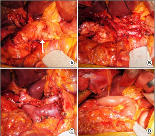
Fig. 1. Case 1 details. A 57-year-old male patient under- going pylorus-preserving pan- creaticoduodenectomy with ex- tended pancreatic transection technique for distal bile duct cancer. (A) An invagination fis- sure (arrow) is identified. (B) The pancreatic body was transected at the celiac axis level. (C) A small pancreatic duct located at the center of the pancreatic stump was identified. (D) Duct-to-mu- cosa pancreaticojejunostomy was performed.
proximal portion of the pancreatic body in a considerable proportion of patients undergoing PD. We intentionally transected the pancreatic body distal to this fusion fissure to completely remove the ventral pancreas. This was done with the hypothesis that there is a potential blood flow disturbance crossing the fusion fissure, and that extended pancreatic transection (EPT) can prevent PJ-associated complications.
In this study, we assessed the clinical usability of EPT on patients undergoing PD through a case-controlled study. The primary end-point was whether EPT induces reduction of PJ-associated complication. The secondary end-point was whether EPT induces no harmful effect on the remnant pancreatic function.
MATERIALS AND METHODS
Study design and patient selection
Based on the prior personal experience of one author (SH) on >200 cases of PD, we learned that EPT at the level of the celiac axis seems to be beneficial to prevent the incidence of major PF after PD. Thus, the EPT techni-
que has been selectively performed selectively for patients at high risk of PF, such as normally soft pancreatic paren- chyma with a small main pancreatic duct. After the EPT technique was deemed worthy of clinical application, it was routinely adopted to the patients undergoing PD be- ginning in August 2014.
To evaluate the clinical usability of EPT, we designed a case-controlled study. For the study group, the study pe- riod was set as the 2-year period from August 2014 to July 2016. During that time, EPT was applied to 19 con- secutive patients undergoing PD, pylorus-preserving PD (PPPD) or hepatopancreaticoduodenectomy (HPD) by a single surgeon (SH). These 19 patients were the study candidates and comprised the EPT group.
Considering the patient number of the study group, the patient number of the control group was estimated with a type-I error () of 0.10 and a type-II error () of 0.20 in addition to 20% difference in complication rates, by which the minimal patient number in the control group was 45. To objectively compare the incidence of PD-asso- ciated complications, we selected the control group pa- tients through a propensity score matching method.
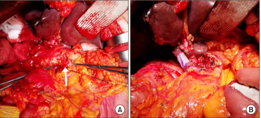
Fig. 2. Case 2 details. A 62-year-old male patient undergoing hepatopancreaticoduodenectomy including right hepatectomy, cau- date lobectomy, pylorus-preserving pancreaticoduodenectomy with extended pancreatic transection technique, and portal vein re- section and interposition graft for intrahepatic cholangiocarcinoma. (A) An invagination fissure (arrow) is identified. (B) The pancreatic body was transected at the celiac axis level.
Patient demographics, primary diseases, consistency of pancreatic parenchyma (soft nature only), diameter of the main pancreatic duct (≤3 mm) on imaging studies, types of pancreaticojejunal anastomosis (duct-to-mucosa re- construction with internal drainage tube only), and no ap- plication of EPT were the criteria. Through a review of the medical records and follow-up imaging, we identified 45 patients during the period from January 2008 and July 2014. The medical records of these patients were retro- spectively reviewed with special attention to perioperative complications after approval of the Institutional Review Board of our institution.
Determination of the pancreatic transection site In the control group, the conventional level of pancre- atic transection for PD was the medial border of the supe- rior mesenteric vein (SMV). Thus, only the confluence portion of the SMV-splenic vein was exposed. We did not dissect the caudal border of the remnant pancreas under the belief that preservation of all peripancreatic con- nective tissues is beneficial to make PJ secure.
In contrast, in the EPT group the ventro-caudal edge of the pancreatic body was gently dissected to identify an oblique-lying fusion fissure. The length, depth and loca- tion of this fissure varied, but all were located between the levels of the SMV and the celiac axis. Thus, the pan- creatic body was transected 1 cm distal to the fusion fis- sure, which was usually placed at the level of the celiac axis. EPT resulted in the additional removal of the pancre-
atic body by 3 cm in length comparing with the conven- tional pancreatic transection (Figs. 1 and 2).
Surgical procedure for pancreaticojejunostomy The same surgical technique was used for PJ regardless of EPT application. The surgical technique for end-to-side duct-to-mucosa PJ was as follows. Running sutures were made at the dorsal edge of the pancreatic stump with 5-0 Prolene. Multiple interrupted sutures with 6-0 Prolene were done for duct-to-mucosa anastomosis. A size-match- ed silastic tube 10-15 cm in length was inserted into the remnant pancreatic duct and then transfixed with 4-0 chromic catgut or 6-0 absorbable monofilament. Finally, running sutures at the ventral edge of the pancreatic stump with 5-0 Prolene were made.
Fibrin glue sealant was applied over the PJ site. The omental graft patch was attached around the PJ site if possible. Three suction-type cigarette drains were also in- serted around the PJ site. The procedures to make this drain were as follows. Multiple small side-holes were made at the 10 cm-length of each 25 cm-long and 10 mm-wide silastic Penrose drain and 50 cm-long and 4.8 mm-sized tube line of Jackson-Pratt drain tube. A Jackson-Pratt drain tube was inserted into the Penrose drain with overlapping of the side-hole portions, by which these two tubes were integrated as a conventional ciga- rette drain. A 4 mm-long skin incision was made and a set of Mosquito clamps was inserted to pull out the drain from the peritoneal side. The outside portion of this drain
Table 1. Comparison of demographic and surgical characteristics Variable
EPT group (n=19)
Control group (n=45)
p
Sex: Male:Female (n) Age (mean±SD, years) Body mass index (mean±SD) Diagnosis (n)
Distal bile duct cancer Perihilar bile duct cancer Ampulla of vater cancer Pancreas head cancer Gallbladder cancer
Intrahepatic cholangiocarcinoma Surgical procedures (n)
Pylorus-preserving PD Conventional PD HPD
11:8 63.7±6.3 27.1±3.8
8 3 4 2 1 1 13 2 4
29:16 65.2±7.1 28.4±3.2
26 3 8 6 2 0 34 6 5
0.62 0.27 1.66 0.25*
0.55**
EPT, extended pancreatic transection; PD, pancreaticoduodenectomy;
HPD, hepatopancreaticoduodenectomy
*Distal bile duct cancer versus others
**Pylorus-preserving PD versus others was tightly tied to prevent air leak at the skin level, while
the inner drainage tube was connected to a Jackson-Pratt type suction bag.13
Perioperative follow-up and definition of pancreatic leak
Multidetector dynamic abdominal computed tomog- raphy (CT) was performed at 5-7 days, 10-14 days and every week during admission. We set the hospitalization period to be 3 weeks after PD even in patients without any complication, thus all patients underwent CT scan at least three times after PD during admission. The status of PJ site was evaluated with comparison of the interval changes of CT findings, with special attention to the per- fusion status of the pancreatic stump.
For the diagnosis of pancreatic leak, the concentrations of amylase and lipase in the drains were measured daily or every other day for more than 10 days until drain removal. For the diagnosis of PF, an amylase concen- trations in the drain of more than three times of the serum value on day 3 or more after surgery was used to define patients with positive drain amylase. Postoperative pancre- atic leakage was graded according to the classification of the International Study Group on Pancreatic Fistula (ISGPF).14-16
For assessment of the endocrine function of the rem- nant pancreas, the 6-month incidence of de novo diabetes mellitus was assessed.
Imaging analysis for anatomy of the pancreas and pancreaticojejunostomy
The pancreatic resection rate was assessed through peri- operative measurement of the pancreas volume by using CT volumetry software (Petavision and Dr. Liver soft- ware). The reconstruction status of PJ was assessed with three-dimensional reconstruction software (Horos version 2.0; open-source free medical image viewer).
Statistical analyses
Numeric data are presented as the means with standard deviation or as medians with a range. Continuous varia- bles were compared using Student’s t-test. Variables re- porting incidences were compared using the 2 test or Fisher’s exact test. A p-value <0.05 was considered to be statistically significant. The eligible number of patients
in the control groups was calculated using MedCalc (ver- sion 15.11.4; MedCalc Software, Ostend, Belgium).
Statistical analyses were performed using SPSS version 22 (SPSS Inc., Chicago, IL, USA).
RESULTS
Patient demographics and surgical profiles The demographic characteristics and surgical profiles of the EPT and control groups are summarized in Table 1.
There were no significant differences in age, sex, body mass index, diagnosis and surgical procedures. In the EPT group, PPPD, conventional PD and HPD were done in 13, 2 and 4 patients, respectively.
Pancreatic fusion fissure anatomy
A small fusion fissure was identified at the ven- tro-caudal edge of the pancreatic body in all 19 patients undergoing EPT (Figs. 1 and 2). A noticeable fissure per- mitting easy separation of the pancreatic parenchyma for 5 mm or more in length was identified in 15 of 19 pa- tients (78.9%) in the study group.
Analysis of perioperative pancreatic leak complications
Incidences of postoperative complications focused on
Fig. 4. Computed tomography follow-up of case 2. (A) Preoperative image shows abundant pancreatic parenchyma with a small pancreatic duct. (B) Post-operation 1-week image shows secure attachment of the jejunal limb wall at the pancreatic stump.
(C) Post-operation 1-year image shows intact perfusion status of the remnant pancreas with mild pancreatic parenchymal atrophy.
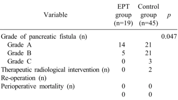
Table 2. Incidence of postoperative pancreatic leak Variable
EPT group (n=19)
Control group (n=45)
p
Grade of pancreatic fistula (n) Grade A
Grade B Grade C
Therapeutic radiological intervention (n) Re-operation (n)
Perioperative mortality (n)
14 5 0 0 0 0
21 21 3 2 0 0
0.047
EPT, extended pancreatic transection
Fig. 3. Computed tomography fol- low-up of case 1. (A) Preoperative image shows abundant pancreatic parenchyma with a small pancre- atic duct. (B) Post-operation 1-week image shows secure at- tachment of the jejunal limb wall at the pancreatic stump.
the incidence of PF in the study and control groups are summarized in Table 2. In the EPT group, postoperative PF was grade A, B and C in 14, 5 and zero patients, respectively. The incidences of PF grade B and C were significantly lower in the EPT group than in the control group (p=0.047). There was no case of reoperation and perioperative mortality within 3 months in the EPT and control groups.
Imaging analysis of pancreatic reconstruction We serially followed-up the status of the PJ site with dynamic CT scans to delineate the perfusion status of the pancreatic stump (Figs. 3 and 4). Pancreatic resection rates were compared between the study and control groups. Analysis of the 1-week CT scan images revealed secure approximation between the pancreatic stump and jejunal limb wall with unimpaired pancreatic perfusion in 17 of 19 patients (89.5%) in the EPT group and 33 of 45 patients (73.3%) in the control group.
Incidence of de novo diabetes mellitus and pancreatic exocrine insufficiency
Perioperative diabetes mellitus data are summarized at Table 3. Postoperative de novo diabetes mellitus involved 2 patients (10.5%) in the EPT group and 4 patients (8.9%), with no difference (p=0.60).
DISCUSSION
PD has been regarded as the standard procedures for
Table 3. Incidence of postoperative de novo diabetes mellitus (DM)
Variable Study group (n=19) Control group (n=45) p
Preoperative diagnosis of DM (n)
Postoperative diagnosis of DM at postoperative 6 moths (n) De novo DM (n)
5 (26.3%) 7 (36.8%) 2 (10.5%)
11 (24.4%) 15 (33.3%) 4 (8.9%)
0.87 0.79 0.60
various malignant lesions in the periampullary region.
Although the techniques of PD have improved consid- erably, a considerable proportion of patients still experi- ence major surgical complications. PJ leakage is the main cause of morbidity and mortality after PD. Pancreatic leak- age can result in various kinds of surgical complications, including pseudoaneurysm formation and anastomotic disruption.14,16 It also leads to prolonged hospitalization, increased health care costs and even mortality.17 There are many factors in literature that can influence pancreatic leakage after PD include age, gender, preoperative jaun- dice, operative time, intraoperative blood loss, type of pan- creatic reconstruction, anastomotic technique, consistency of the pancreatic stump, pancreatic duct diameter, use of somatostatin and surgeon’s experience.14,17,18
Surgical techniques for PD have evolved and matured over a long time. The majority of clinical studies regard- ing the surgical aspects of PD have primarily focused on the techniques of pancreatic reconstruction per se. Little attention has been paid to the site of pancreatic trans- action, which is the site of pancreatic reconstruction. The arterial blood supply of the pancreatic neck and body from the dorsal pancreatic artery and transverse pancreatic arteries is well described in detail in anatomy textbooks.
Yet, we have occasionally experienced scanty arterial bleeding at the pancreatic stump during transection, espe- cially at the ventro-caudal cross-sectional area. The imag- ing analysis on the PJ site in the present study revealed that this area at the pancreatic stump appears to be vulner- able to ischemic change.
Many hepato-pancreato-biliary surgeons believe that the pancreas is one integrated organ with an abundant arterial blood supply, through which any resection of pancreas in- cluding segmental resection of the pancreatic body is fea- sible without risk of ischemia. In contrast, anatomical var- iations in the pancreatic anatomy, such as accessory pan- creatic duct and annular pancreas, develop at the time of fusion of the dorsal and ventral pancreas during embry-
onic development. If the fusion process is not perfect, we presume that it can leave some rudimentary structures at the fusion site. We found a fissure with invagination of the pancreatic capsule at the pancreatic body. We pre- sumed it to be rudimentary evidence of embryonic pancre- atic fusion, and so arbitrarily named it fusion fissure. We hypothesized that arterial development beneath this fissure is less abundant than other pancreatic areas. This hypoth- esis led us to postulate that it may be beneficial to com- pletely remove the pancreatic parenchyma associated with this fissure. Such pancreatic fusion-associated variation can be overcome by the use of EPT.
The location of the main pancreatic duct is not at the center all along the pancreatic body. Its location is eccen- tric at the neck portion and tends to be more concentric at the body portion. This anatomical feature indicates that more concentrically located pancreatic duct stump can be obtained through pancreatic transection at the level of cel- iac axis like EPT comparing with the conventional pan- creatic transection. This concentric location must be bene- ficial to perform secure reconstruction of PJ. Thus, the theoretical advantages of EPT are the unimpeded pancre- atic stump blood supply and the creation of a more con- centric remnant pancreatic duct.
These theoretical advantages of EPT are validated in this study as being the primary end-point, which was whether EPT can effectively reduce the incidence of PJ-associated complication. The incidence of grade A PF was similar in the study and control groups, but the risk of grade B and C PF was significantly lower in the EPT group because grade A PF is often encountered during the early postoperative period but spontaneously resolved a few days later. Thus, we believe that EPT is beneficial to reduce PJ-associated complication effectively. At first, we had meticulously explored the presence of a fusion fis- sure and then determined the pancreatic transection line case by case. After performing EPT in first 10 cases, we recognized that the estimated pancreatic transection line
was uniformly located at the celiac axis level. So we de- termined the pancreatic transection line in advance and then searched for the fusion fissure in a retrograde fashion.
Our imaging analysis regarding the blood supply at the pancreatic stump revealed nearly no ischemic portion after EPT, but a small area of marginal ischemia was detected in 25% of patients in the control group. This finding is strong evidence that EPT is beneficial to prevent impaired blood supply at the pancreatic stump.
EPT has a potential disadvantage from the additional removal of the pancreatic parenchyma because it is in- tended only to secure pancreatic reconstruction, but is not related to the oncological aspect. Thus, we assessed whether EPT is detrimental to the exocrine and endocrine function of the remnant pancreas as the secondary end-point of the study. EPT resulted in additional pancre- atic resection of 10-15% compared with the control group.
No patient suffered from intractable steatorrhea in both the study and control groups. Under normal conditions in adults, approximately 1,000 ml of pancreatic juice is ex- creted daily. We previously reported that the mean daily amount of excreted pancreatic juice after PPPD through an external pancreatic drainage tube is 229±99 ml.19,20 Although the amount of pancreatic juice may be corre- lated with the volume and quality of the remnant pan- creas, we believe that additional pancreatic resection of 10-15% does not result in a significant reduction of pan- creatic secretion. Regarding the pancreatic endocrine function, 10.5% of patients developed de novo diabetes mellitus at postoperative 6 months. Considering that pan- creatic islets are more abundant at the pancreatic tail, ad- ditional pancreatic resection of 10-15% may not deterio- rate the pancreatic endocrine function significantly. The incidence of de novo diabetes mellitus was quite com- parable between the EPT and control groups.
Effective management of major PF is a major concern.
Firstly, effective drainage must be important, but post- operative insertion of percutaneous catheter seems to be often risky. We think that the conventional Jackson-Pratt type suction drain is not effective to drain thick or sticky materials that are a product of pancreatic leakage and sub- sequent necrosis. We devised a customized closed-suction drain with combination of Jackson-Pratt suction drain and cigarette drain tip. This drain works effectively for more
than 2-3 weeks in the situation of major PF.
The present study has several limitations. First, this is a retrospective case-controlled study although EPT was performed as a part of prospective trial. Second, the case number of study group was not large enough to demon- strate the fusion fissure as a new landmark of the pancre- atic anatomy. Further validation of our results regarding the benefit of EPT and anatomical significance of the fu- sion fissure is necessary through studies with large patient populations from multiple centers.
In conclusion, the results of our study demonstrate that EPT enables the complete removal of the ventral pan- creas, which contributes to the reduction of major PF without impairment of the remnant pancreatic function.
We believe that EPT is worthy of clinical application rou- tinely, or at least in patients with a known risk of PJ leak.
REFERENCES
1. Trede M, Schwall G. The complications of pancreatectomy. Ann Surg 1988;207:39-47.
2. Miedema BW, Sarr MG, van Heerden JA, Nagorney DM, McIlrath DC, Ilstrup D. Complications following pancreaticoduodenectomy.
Current management. Arch Surg 1992;127:945-949; discussion 949-950.
3. Yeo CJ. Management of complications following pancreaticoduodenectomy. Surg Clin North Am 1995;75:913-924.
4. Aranha GV, Aaron JM, Shoup M, Pickleman J. Current manage- ment of pancreatic fistula after pancreaticoduodenectomy.
Surgery 2006;140:561-568; discussion 568-569.
5. Strobel O, Brangs S, Hinz U, Pausch T, Hüttner FJ, Diener MK, et al. Incidence, risk factors and clinical implications of chyle leak after pancreatic surgery. Br J Surg 2017;104:108-117.
6. Fu SJ, Shen SL, Li SQ, Hu WJ, Hua YP, Kuang M, et al. Risk factors and outcomes of postoperative pancreatic fistula after pancreatico-duodenectomy: an audit of 532 consecutive cases.
BMC Surg 2015;15:34.
7. Relles DM, Burkhart RA, Pucci MJ, Sendecki J, Tholey R, Drueding R, et al. Does resident experience affect outcomes in complex abdominal surgery? Pancreaticoduodenectomy as an example. J Gastrointest Surg 2014;18:279-285; discussion 285.
8. Burkhart RA, Relles D, Pineda DM, Gabale S, Sauter PK, Rosato EL, et al. Defining treatment and outcomes of hep- aticojejunostomy failure following pancreaticoduodenectomy. J Gastrointest Surg 2013;17:451-460.
9. Xiong JJ, Altaf K, Mukherjee R, Huang W, Hu WM, Li A, et al.
Systematic review and meta-analysis of outcomes after intraoperative pancreatic duct stent placement during pancreaticoduodenectomy. Br J Surg 2012;99:1050-1061.
10. Haga Y, Wada Y, Takeuchi H, Ikejiri K, Ikenaga M. Prediction of anastomotic leak and its prognosis in digestive surgery. World J Surg 2011;35:716-722.
11. Guo P, Preuett B, Krishna P, Xiao X, Shiota C, Wiersch J, et al. Barrier function of the coelomic epithelium in the developing pancreas. Mech Dev 2014;134:67-79.
12. Godlewski G, Gaubert J, Cristol-Gaubert R, Radi M, Baecker V,
Travo P, et al. Moving and fusion of the pancreatic buds in the rat embryos during the embryonic period (carnegie stages 13-17) by a three-dimensional computer-assisted reconstruction. Surg Radiol Anat 2011;33:659-664.
13. Hwang S, Ha TY, Kim JS, Cheong O, Kim KH, Lee SG. Clinical application of sution-type cigarette drain for hepatopancreatoabiliary surgery. J Korean Surg Soc 2004;67:428-431.
14. Bassi C, Falconi M, Molinari E, Mantovani W, Butturini G, Gumbs AA, et al. Duct-to-mucosa versus end-to-side pancreaticojejunostomy reconstruction after pancreaticoduodenectomy: results of a pro- spective randomized trial. Surgery 2003;134:766-771.
15. Bassi C, Dervenis C, Butturini G, Fingerhut A, Yeo C, Izbicki J, et al. Postoperative pancreatic fistula: an international study group (ISGPF) definition. Surgery 2005;138:8-13.
16. Facy O, Chalumeau C, Poussier M, Binquet C, Rat P, Ortega-Deballon P. Diagnosis of postoperative pancreatic fistula.
Br J Surg 2012;99:1072-1075.
17. El Nakeeb A, Salah T, Sultan A, El Hemaly M, Askr W, Ezzat H, et al. Pancreatic anastomotic leakage after pancreaticoduodenectomy.
Risk factors, clinical predictors, and management (single center experi- ence). World J Surg 2013;37:1405-1418.
18. Strasberg SM, Drebin JA, Soper NJ. Evolution and current status of the Whipple procedure: an update for gastroenterologists.
Gastroenterology 1997;113:983-994.
19. Jung DH, Hwang S, Lee SG. An analysis on the amount of ex- creted pancreatic juice after pancreatoduodenectomy. Korean J Gastroenterol 2004;43:309-315.
20. Yoo D, Hwang S, Kim KH, Ahn CS, Moon DB, Ha TY, et al.
Pancreatic atrophy relative to external versus internal drainage of the pancreatic duct after pylorus-preserving pancreaticoduodenectomy. J Gastrointest Surg 2014;18:1604-1609.