J Korean Surg Soc 2009;76:388-391
□ 증 례 □
DOI: 10.4174/jkss.2009.76.6.388
388
책임저자: 장명철, 충남 천안시 동남구 안서동 산 16-5
330-714, 단국대학교병원 외과 Tel: 041-550-3930, Fax: 041-556-3878 E-mail: changmc@dankook.ac.kr
접수일:2008년 10월 7일, 게재승인일:2008년 11월 24일 본 논문의 요지는 2006년 대한외과학회 추계학술대회에서 포스터 전시되었음.
이 연구는 2008년도 단국대학교 대학연구비 지원으로 연구되었음.
제1형 신경섬유종증에서 발생한 양측성 이시성 유방암
단국대학교 의과대학 외과학교실
김선호ㆍ손원준ㆍ신동준ㆍ장명철
Bilateral Metachronous Breast Cancer in Neurofibromatosis Type 1
Sun Ho Kim, M.D., Won Jun Son, M.D., Dong Jun Sin, M.D., Myung-Chul Chang, M.D.
Department of Surgery, Dankook University College of Medicine, Cheonan, Korea
Neurofibromatosis type 1 (NF1) is an autosomal dominant disease, characterized by café-au-lait spot, axillary and inguinal freckle, peripheral neurofibroma and pigmented iris hamartoma. The various cancer incidences are increased in the NF1. But NF1 with breast cancer is rare. In this report we present a case of a 46˗year˗old NF1 female with a bilateral metachronous breast cancer. The patient has no BRCA2 mutation, but there are two unclassified variants in the exon 11 of BRCA1. The possibility of LOH of BRCA1 gene in the cancer tissue cannot be excluded. (J Korean Surg Soc 2009;76:388-391)
Key Words: Neurofibromatosis, Breast cancer
중심 단어: 선경섬유종증, 유방암서 론
제1형 신경섬유종증(neurofibromatosis type 1, NF1)은 상 염색체 우성으로 유전되며 담갈색반점(café au lait spot), 액 와 및 서혜부의 주근깨(freckle), 말초 신경섬유종, 색소성 홍채 과오종(pigmented iris hamartoma)을 특징으로 한다.
NF1에서는 태생기 신경 능선(embryogenic neural crest) 기원 종양을 포함한 다양한 악성종양이 발생할 수 있으며 만성 및 급성 골수백혈병의 위험도가 증가한다. 유방암은 전세 계적으로 여성에서 가장 흔한 암이지만 NF1에서 유방암이 발생한 보고는 드물다. 국내에서는 Kim 등(1)이 유방암과
소장 평활근육종이 발생한 예를 보고하였고, Lee 등(2)은 NF1에서 발생한 유방암 3예를 보고하였다. 저자들은 NF1 환자에서 이시성으로 발생한 양측성 유방암을 경험하였기 에 문헌고찰과 함께 보고한다.
증 례
46세의 기혼 여성이 2년 전부터 만져지기 시작한 무통성 의 유방 종양을 주소로 내원하였다. 환자는 10년 전 신경섬 유종증으로 진단 받았으며 환자 외 3명에서 신경섬유종증 의 가족력이 있었다(Fig. 1). 환자의 초경 연령은 18세이며 첫 출산은 27세로 현재 1명의 자녀가 있다. 평소 생리주기 는 규칙적이었다. 이전 유방질환의 과거력 없으며 가족 중 유방암 환자는 없었다. 환자의 전신 피부는 3∼10 mm 크기 의 결절이 산재하였으며, 담갈색반점과 액와부 기미가 다 수 관찰되었다(Fig. 2). 좌측 유방에 지름 4 cm 정도 종괴가 돌출되어 있었으며, 단단하고 주위와 유착되어 있었다. 압 통은 없었으며 액와림프절은 만져지지 않았다.
유방촬영상 전체적으로 비균질성 치밀 유방조직이었으
Sun Ho Kim, et al:Bilateral Metachronous Breast Cancer in Neurofibromatosis Type 1 389
Fig. 1. Pedigree of neurofibromatosis type 1. The older sister, fa- ther and grandmother are neurofibromatosis type 1. The ar- row indicates the patient.
Fig. 2. The chest and abdomen of patient. Small nodules less than 1 cm are scattered across the entire skin surface. Café- au-lait spots are shown in the right lower and left lower abdomen.
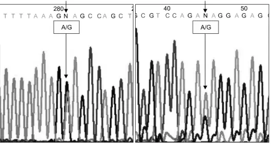
Fig. 3. The direct sequencing result of exon 11 of BRCA1. The left graph was an unclassi- fied variant of 3232A>G, resulted in Glutamic acid to Glycine change at codon 1,038 (E1038G). The right graph was an unclassified var- iant of 3667A>G, resulted in Lysine to Arginine change at codon 1,183 (K1183R).
며, 좌측 유두 내측으로 미세석회화를 동반한 종괴가 관찰 되어 BI-RADS category 5의 소견이었다. 초음파상 경계가 불규칙한 저에코성 종괴가 관찰되었다. 종괴의 초음파 유 도 핵 조직검사(core biopsy)상 침윤성 유방암으로 변형 근 치 절제술을 시행하였다. 수술소견상 크기는 4×5 cm였으며 대흉근과 피부에는 침범되지 않았다. 병리조직검사상 핵 및 조직등급이 좋은 전형적인 침윤성 유방암이었으며 액와 림프절 전이는 없었다. 면역조직검사에서 에스트로겐 수용 체와 프로게스테론 수용체는 양성, HER2 (1+)였다. 함께 절제된 피부의 다발성 결절은 신경섬유종이었다. 수술 후 CEF (Cyclophosphamide 75 mg/m2 PO days 1∼14, Epirubicin 60 mg/m2 IV days 1 & 8, 5-Fluouracil 500 mg/m2 IV days 1
& 8 for 6 cycles) 항암화학요법을 시행하였다.
타목시펜을 복용하며 추적 검사 시행 중 수술 2년 3개월 후 우측 유방의 유방촬영상 우측 유방 상외측으로 미세석 회화가 관찰되었다. 초음파 검사상 동일한 부위에서 1.5×0.5 cm 크기의 저에코성 종괴와 미세석회화가 관찰되 었으나, 만져지지는 않았다. 초음파 유도 핵 조직검사상 석 회화와 면포성 괴사(comedo necrosis)를 동반한 중등도의 관 상피내암으로 진단되어 변형 근치 절제술을 시행하였다.
병리조직검사상 종양의 대부분 관상피내암이었으나 2 mm 정도의 침윤암이 함께 관찰되었다. 액와림프절 전이는 없 었다. 면역조직검사 결과 에스트로겐 수용체 음성, 프로게
390 J Korean Surg Soc. Vol. 76, No. 6
스테론 양성, HER2 음성이었다. 수술 후 CMF (Cyclophos- phamide 100 mg/m2 PO days 1∼14, Methotrexate 40 mg/m2 IV days 1 & 8, 5-Fluouracil 600 mg/m2 IV days 1 & 8 for 6 cycles) 항암화학요법을 시행하였다. 현재 1년간 추적기간 중 재발의 증거 없이 관찰 중이다.
양측성 이시성 유방암으로 BRCA1 및 BRCA2 유전자 검 사를 시행하였다. BRCA1의 엑손 11에서 2개의 unclassified variant (3232A>G, 3667A>G)가 관찰되었으며(Fig. 3), 7개 의 이형접합의 다형성(heterozygous polymorphism)이 관찰되 었으나 임상적 의의는 없었다. BRCA2에서는 6개의 이형접 합의 다형성이 관찰되었으며 돌연변이는 관찰되지 않았다.
고 찰
제1형 신경섬유종증은 von Recklinghausen 병이라고도 알 려져 있으며 NF1 유전자의 돌연변이로 발생함이 알려져 있 다. NF1 유전자는 염색체 17q11.2에 위치하며 60개의 엑손 으로 이루어진 종양억제유전자이다. NF1 유전자가 생성하 는 neurofibromin은 2,818개의 아미노산으로 구성되어 있으 며 GTPase 활성화 단백질 부위가 존재하여 ras-GTP를 ras- GDP로 분해하며 결과적으로 ras 유전자의 활성을 감소시키 는 역할을 한다. 따라서 NF1 유전자의 기능상실은 p21ras를 활성화시켜 세포분열을 촉진한다.
NF1에서는 악성종양의 발생이 증가한다고 알려져 있는 데 Sorensen 등(3)에 따르면 일반인에 비해 암 발생률이 4배 높다고 하였다. NF1에서 유방암의 발생에 대하여는 이견이 많은데 Brasfield와 Das Gupta(4)는 54명의 NF1 여성에서 5 예의 유방암을 보고하였으나 대상군의 선정에 문제가 있다 고 알려져 있으며, Sorensen 등(3)에 따르면 NF1에서 유방종 양 발생은 신경계 종양 다음으로 흔하다고 하였으나 빈도 는 낮았다. NF1에서 유방암이 발생할 경우 젊은 나이에 발 생한다고 알려져 있는데 Rasmussen 등(5)은 20세 이전에 사 망한 NF1에서 유방암이 흔함을 보고하였고, Nakamura 등 (6)은 NF1에서 발생한 유방암중 18.5%가 35세 이전이라고 보고하였다.
최근 유방암과 신경섬유종증의 연관성에 대하여 연구되 고 있는데, Yaegashi 등(7)은 60예의 유방암 조직에서 NF2 유전자의 체성 돌연변이를 조사하여, 1예에서 정상 유방암 조직에서는 관찰되지 않는 12번 엑손 398번 코돈의 돌연변 이를 보고하였다. Ogata 등(8)은 유방암 세포주 MDA-MP- 231에서 neurofibromin의 발현이 거의 없는 반면 MAPK의
인산화 및 ras 활성화가 관찰되어 NF1 유전자의 기능소실 과 유방암 발생의 연관성에 대하여 시사하였다. Guran과 Safali(9)는 NF1 가계중 모녀에서 유방암이 발생하였으며 BRCA1, BRCA2 유전자 돌연변이는 없었으나, 유방암 조직 에서 NF1 유전자의 이형접합상실(loss of heterozygosity, LOH)이 있음을 보고하였다. Ceccaroni 등(10)은 NF1과 BRCA1의 유전자 돌연변이가 함께 나타나는 가계를 보고 하였는데, BRCA1 유전자는 NF1 유전자와 20 cm 떨어진 17q12∼q21 부위에 위치하기 때문에 돌연변이가 함께 발생 할 가능성이 높다. 본 증례에서는 BRCA2 유전자 돌연변이 는 관찰되지 않았으며, BRCA1에서는 2개의 unclassified variant가 관찰되었으나 임상적 의의는 없는 것으로 생각한 다. 또한 7개의 이형접합의 다형성이 관찰되었으나 많은 대 부분의 다형성은 동형접합(homozygote)이었기 때문에 LOH 가 함께 발생하였을 가능성을 완전히 배제할 수 없었다. 따 라서 향후 종양 조직에서 BRCA1 유전자에 대한 이형접합 상실에 대한 연구가 필요하리라 생각한다.
REFERENCES
1) Kim BG, Kim JJ, Cho JS, Park UC. Malignant metachronous cancers (breast cancer, small bowel leiomyosarcoma) asso- ciated with von Recklinghausen disease (NF 1). J Korean Surg Soc 1999;56:300-5.
2) Lee HJ, Chun YS, Lee NJ, Lee HS, Kim TH, Yoon HK, et al. Breast cancer in three women associated with von Recklinghausen's disease. J Korean Surg Soc 2008;74:217-21.
3) Sorensen SA, Mulvihill JJ, Nielsen A. Long-term follow-up of von Recklinghausen neurofibromatosis. Survival and malig- nant neoplasms. N Engl J Med 1986;314:1010-5.
4) Brasfield RD, Das Gupta TK. Von Recklinghausen's disease:
a clinicopathological study. Ann Surg 1972;175:86-104.
5) Rasmussen SA, Yang Q, Friedman JM. Mortality in neuro- fibromatosis 1: an analysis using U.S. death certificates. Am J Hum Genet 2001;68:1110-8.
6) Nakamura M, Tangoku A, Kusanagi H, Oka M, Suzuki T.
Breast cancer associated with Recklinghausen's disease: report of a case. Nippon Geka Hokan 1998;67:3-9.
7) Yaegashi S, Sachse R, Ohuchi N, Mori S, Sekiya T. Low in- cidence of a nucleotide sequence alteration of the neuro- fibromatosis 2 gene in human breast cancers. Jpn J Cancer Res 1995;86:929-33.
8) Ogata H, Sato H, Takatsuka J, De Luca LM. Human breast cancer MDA-MB-231 cells fail to express the neurofibromin protein, lack its type I mRNA isoform and show accumulation of P-MAPK and activated Ras. Cancer Lett 2001;172:159-64.
Sun Ho Kim, et al:Bilateral Metachronous Breast Cancer in Neurofibromatosis Type 1 391
9) Guran S, Safali M. A case of neurofibromatosis and breast cancer: loss of heterozygosity of NF1 in breast cancer. Cancer Genet Cytogenet 2005;156:86-8.
10) Ceccaroni M, Genuardi M, Legge F, Lucci-Cordisco E, Car-
rara S, D'Amico F, et al. BRCA1-related malignancies in a family presenting with von Recklinghausen's disease. Gynecol Oncol 2002;86:375-8.