Hepatic venous outflow obstruction after whole liver transplantation of large-for-size graft:
versatile intra-operative management
Chetana Lim1, Michael Osseis1, Antonella Tudisco1, Eylon Lahat1, Dobromir Sotirov1, Chady Salloum1, and Daniel Azoulay1,2,3
1Department of Hepatobiliary and Pancreatic Surgery and Liver Transplantation, Henri Mondor Hospital,
2Université Paris-Est UPEC, Créteil, 3INSERM, U955, Créteil, France
Backgrounds/Aims: Preservation of the native inferior vena cava using a large graft during adult whole liver trans- plantation is associated with a potential risk of hepatic venous outflow compression/obstruction, which may adversely affect both graft and short-term patient outcomes. Intraoperative placement of materials to restore adequate hepatic venous outflow can overcome this complication. Methods: Data of patients who underwent liver transplantation between 2011 and 2016 were retrospectively reviewed. All cases of hepatic venous outflow obstruction due to large graft size managed via intraoperative intervention were analyzed. The literature was searched for studies reporting adult cases of hepatic venous outflow obstruction following whole liver transplantation managed extrahepatically. Results: Three patients diagnosed with intraoperative hepatic venous outflow obstruction due to large graft size were managed via retro-hepatic placement of breast implants (2 cases) or abdominal pads (1 case). It was successfully carried out in all cases. Four studies including 15 patients were identified in the literature search. Different types of materials such as inflatable materials (Foley catheter, Blakemore balloon), surgical gloves or breast implants, were used. Conclusions:
Placement of inflatable materials leads to gradual deflation in the postoperative period, which might obviate the need for reoperation. Breast implants could be left in place indefinitely due to their bio-inert nature. (Ann Hepatobiliary Pancreat Surg 2018;22:321-325)
Key Words: Hepatic venous outflow; Breast implants; Abdominal pads; Surgical gloves; Inflatable materials
Received: February 6, 2018; Revised: July 3, 2018; Accepted: July 10, 2018 Corresponding author: Daniel Azoulay
Department of Hepatobiliary and Pancreatic Surgery and Liver Transplantation, Henri Mondor Hospital, 51 avenue de Lattre de Tassigny, 94010 Créteil, France
Tel: +33-149812548, Fax: +33-149812432, E-mail: daniel.azoulay@aphp.fr
Copyright Ⓒ 2018 by The Korean Association of Hepato-Biliary-Pancreatic Surgery
This is an Open Access article distributed under the terms of the Creative Commons Attribution Non-Commercial License (http://creativecommons.org/
licenses/by-nc/4.0) which permits unrestricted non-commercial use, distribution, and reproduction in any medium, provided the original work is properly cited.
Annals of Hepato-Biliary-Pancreatic Surgery ∙ pISSN: 2508-5778ㆍeISSN: 2508-5859
INTRODUCTION
When preserving the native inferior vena cava (IVC), adult whole liver transplantation (LT), using a large graft, combines the risks of hepatic venous outflow com- pression/obstruction (acute Budd-Chiari) and the large- for-size graft may cause compartmental syndrome1 in turn threatening both graft and short-term patient outcomes.
Other causes of hepatic venous outflow obstruction in- clude technical errors (e.g,, graft rotation or kinking due to excessively long supra-hepatic anastomosis) and car- diac causes (e.g., right heart insufficiency or tricuspid valve regurgitation).We report here three cases of hepatic venous outflow obstruction due to large graft size man-
aged via retro-hepatic placement of breast implants or ab- dominal pads to restore adequate hepatic venous outflow.
Alternative options are also discussed.
PATIENTS AND RESULTS
From 2011 to 2016, a total of 477 LTs were performed at our center. The liver graft weight was ≥1800 g in 38 (8%) cases (ranging from 1800 to 2000 g in 20 (4.2%), and ≥2000 g in 18 (3.8%) cases). In these recipients, the native IVC was resected in 7 (1.5%) and preserved in 31 (6.5%) cases. In the latter, hepatic venous outflow ob- struction due to IVC compression by a large-for-size graft occurred in 3 cases (3/31=9.7%). It was managed by the
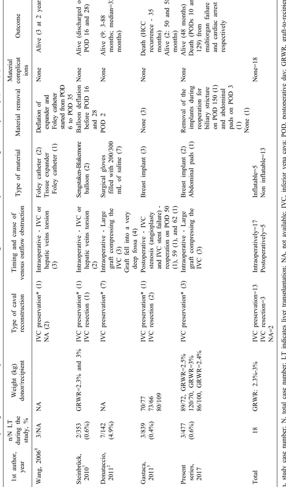
Table 1. Studies reporting adult cases of hepatic venous outflow obstruction following whole liver transplantation managed extrahepatically (>2005) (based on literature review) 1st author, year n/N LT during the study, %
Weight (kg) donor/recipientType of caval reconstructionTiming and cause of venous outflow obstruction Type of materialMaterial removalMaterial complicat ionsOutcome Wang, 20068 3/NA NAIVC preservation* (1) NA (2)Intraoperative - IVC or hepatic veins torsion (3) Foley catheter (2) Tissue expander Foley catheter (1) Deflation of expander and Foley catheter started from POD 6 to POD 35 None Alive (3 at 2 years) Steinbrück, 201072/353 (0.6%)GRWR=2.3% and 3%IVC preservation* (1) IVC resection (1)Intraoperative - IVC or hepatic veins torsion (2)
Sengstaken-Blakemore balloon (2)Balloon deflation before POD 16 and 28
NoneAlive (discharged on POD 16 and 28) Donataccio, 201127/142 (4.9%)NAIVC preservation* (7)Intraoperative - Large graft compressing the IVC (3) Graft fell into a very deep fossa (4)
Surgical gloves filled with 200/300 mL of saline (7)
POD 2NoneAlive (9: 3-88 months; median=32 months) Gastaca, 201133/839 (0.4%)70/77 73/66 80/109
IVC preservation* (1) IVC resection (2)Postoperative - IVC stenosis (angioplasty and IVC stent failure) - reoperation on POD 50 (1), 59 (1), and 62 (1) Breast implant (3)None (3)NoneDeath (HCC recurrence - 35 months) Alive (2: 50 and 58 months) Present series, 2017
3/477 (0.6%)89/72, GRWR=2.5% 120/70, GRWR=3% 86/100, GRWR=2.4%
IVC preservation* (3)Intraoperative - Large graft compressing the IVC (3)
Breast implant (2) Abdominal pads (1)Removal of the implants during reoperation for biliary stricture on POD 150 (1) and abdominal pads on POD 3 (1) None (1) NoneAlive (48 months) Death (PODs 10 and 129) from multiorgan failure and cardiac arrest respectively Total18GRWR: 2.3%-3%IVC preservation=13 IVC resection=3 NA=2
Intraoperatively=17 Postoperatively=5Inflatable=5 Non inflatable=13None=18 n, study case number; N, total case number; LT indicates liver transplantation; NA, not available; IVC, inferior vena cava; POD, postoperative day; GRWR, graft-to-recipient weight ratio; HCC, hepatocellular carcinoma *IVC preservation followed by cavo-caval or hepato-caval anastomosis
Fig. 1. (A) Intraoperative view after placement of a single bre- ast implant (yellow arrow) be- tween the diaphragm and the graft. (B and C) Computed to- mography (CT) scans were tak- en after liver transplantation.
placement of breast implants (2 cases) or abdominal pads (1 case) as described below (Table 1).
Case 1
A 55-year-old woman (body weight 72 kg; height 1.50 m; body mass index (BMI) 32 kg/m2) with hepatocellular carcinoma and cirrhosis due to human immunodeficiency virus and hepatitis C virus co-infection underwent com- bined liver-kidney transplantation. At the time of LT, her model for end-stage liver disease (MELD) score was 38.
The graft (graft weight 1772 g and graft-to-recipient weight ratio (GRWR) 2.5%) was harvested from a male donor (body weight 89 kg; height 1.89 m; BMI 28 kg/m2).
Graft implantation was initiated via latero-lateral cavo-caval anastomosis (LLCCA) following total hep- atectomy to preserve the native IVC. Severe graft con- gestion was observed at reperfusion. Doppler ultra- sonography (DUS) showed no hepatic venous blood flow without thrombosis, which was attributed to caval com- pression secondary to the weight of the large graft. DUS showed restoration of the hepatic venous flow when the liver was lifted downward and forward to the left side.
A single breast implant was placed in the right subphrenic space maintaining the liver in a position for optimal out- flow as confirmed by repeated DUS (Fig. 1A-C). No liver graft biopsy was performed intraoperatively. Surgery was completed by a skin-only closure. Postoperative DUS
showed patency of all vascular reconstructions. The pa- tient developed multiple complications including anasto- motic biliary stricture warranting endoscopic stenting, multiple kidney graft infections, and pneumonia. Three months later, computed tomography scan and angiography revealed a right hepatic vein stenosis with a pressure gra- dient of 13 mmHg across the stricture. Balloon angio- plasty was performed together with stenting of the right hepatic vein, which was followed by a decrease in the pressure gradient. The patient died on postoperative day 129 from multiple kidney graft infections and cardiac arrest. Before death, DUS revealed patent right hepatic vein and caval reconstruction.
Case 2
A 49-year-old man (body weight 70 kg; height 1.71 m;
BMI 24 kg/m2) with a MELD score of 26 at LT under- went combined liver-kidney transplantation for alcoholic cirrhosis and chronic kidney disease. The donor was a male (body weight 120 kg; height 1.78 m; BMI 38 kg/m2). The weight of the transplanted whole liver graft was 2086 g, and the GRWR was 3%. Total hepatectomy preserving the native IVC was performed and the graft was implanted with LLCCA. Severe graft congestion was observed at reperfusion. Intraoperative DUS showed no blood flow in the hepatic veins without thrombosis, and revealed complete restoration of hepatic vein flow upon
Fig. 2. (A) Computed tomog- raphy (CT) scan after liver transplantation showing the pla- cement of 2 breast implants (yellow arrow). (B) CT scan af- ter liver transplantation show- ing the placement of abdominal pads (red arrow).
lifting the liver forward and downward into the right sub- phrenic space to maintain the graft in midline position.
Two breast implants (Fig. 2A) were placed between the graft and the diaphragm to optimize the graft position for adequate hepatic venous outflow. Surgery was completed by a skin-only closure. Liver graft biopsy showed low graft macrovesicular steatosis. The patient developed post- operative acute kidney injury, pneumonia and systemic fungal infection. Postoperative DUS confirmed patency of all vascular and biliary reconstructions. Five months later, the patient underwent Roux-en-Y choledoco-jejunostomy for anastomotic biliary stricture. At the time of surgery, removal of the breast implants was uncomplicated and the patient tested negative for bacterial cultures. At 48 months, the patient is alive with normal kidney and liver function tests.
Case 3
A 53-year-old man (body weight 100 kg; height 1.78 m; BMI 32 kg/m2) with a MELD score of 40 underwent LT for autoimmune cirrhosis with a graft derived from a female donor (body weight 86 kg; height 1.73 m; BMI=29 kg/m2). The liver graft weighed 2400 g, and the GRWR was 2.4%. A total hepatectomy preserving the native IVC was performed and the graft was implanted with LLCCA.
Severe graft congestion was observed at reperfusion. Four abdominal pads were placed behind the right liver to re- store adequate hepatic venous outflow as demonstrated by DUS (Fig. 2B). Liver graft biopsy showed moderate mac- rovesicular steatosis. Surgery was completed by a skin-on- ly closure. Three days later, repeated laparotomy was per- formed to remove the abdominal pads. The patient under- went re-transplantation for early graft dysfunction on post- operative day 10. No hepatic venous outflow was detected
on DUS before re-transplantation. The patient died from multiorgan failure few hours after the re-transplantation.
DISCUSSION
Outflow obstruction syndrome after orthotopic whole LT is a rare but severe complication that may lead to the loss of graft and even recipient. Its incidence is less than 2% when the IVC is replaced6 and 3–4% when it is preserved.4 Indeed, we suspect, as in the present series (9.7%), that this rate is higher in cases involving large graft transplantation with LLCCA. Intraoperative DUS is critical in diagnosing outflow obstruction and its relief following optimal mobilization of the graft.
In the present cases (i.e., severe donor-recipient ana- tomical mismatch and need to maintain IVC graft in a midline position for adequate hepatic venous outflow), the intraoperative mode of presentation with graft congestion was managed via placement of breast implants or abdomi- nal pads. Two main strategies are involved in intra- operative management of outflow obstruction. The first strategy entails reoperation: temporary liver packing, in- flatable materials (of limited size),7,8 and surgical gloves (larger than usual inflatable material).2 The second strat- egy obviates the need for reoperation and entails the use of breast implants (of different sizes for possible ag- gregation, absolutely bio-inert and without the need for re- moval),3 or the use of a round ligament sutured at the an- terior abdominal wall with sufficient tension to fix the graft in midline position.8 Graft reduction may be another option. However, this procedure prolongs cold ischemia time and increases the risk of bleeding upon reperfusion and postoperative biliary leakage. Further, the excessively small size of the left liver even in case of a large-for-size
whole graft is another limitation. Finally, redo side-to-side cavo-cavostomy or additional end-to-end supra- and in- frahepatic cavo-cavostomy may be technically difficult and hazardous approaches.5
In the presence of potential large-for-size whole liver graft anticipated by a close communication between the liver procurement and the transplanting teams, our surgi- cal strategy is currently as follows. First, a liberal ve- no-venous bypass is performed to decrease the splanchnic congestion that may render graft implantation cumber- some and increase IVC compression. Second, resection of the recipient's IVC and end-to-end supra- and infrahepatic anastomosis is needed. Finally, a skin-only closure techni- que or vacuum-assisted wound closure represents a tem- porary solution for abdominal fascia closure if needed.
In conclusion, severe donor-recipient anatomical mis- match can happen. The transplant team should adapt the technique of transplantation with or without IVC preserva- tion and the decision to use a veno-venous bypass to the graft size. The timing of removal of the materials and their potential risk of infection remains open. Placement of inflatable materials leads to their gradual deflation dur- ing the postoperative period, which might obviate the need for reoperation. However, in addition to the avail- ability of various sizes, breast implants may be left in place indefinitely due to their bio-inert nature.
REFERENCES
1. Allard MA, Lopes F, Frosio F, Golse N, Sa Cunha A, Cherqui D, et al. Extreme large-for-size syndrome after adult liver trans- plantation: a model for predicting a potentially lethal compli- cation. Liver Transpl 2017;23:1294-1304.
2. Donataccio D, Grosso S, Donataccio M. A simple and new de- vice to avoid hepatic venous outflow obstruction in adult liver transplantation. Surg sci 2011;2:485-487.
3. Gastaca M, Valdivieso A, Ruiz P, Gonzalez J, Ventoso A, de Urbina JO. Venous outflow obstruction after orthotopic liver transplantation: use of a breast implant to maintain graft position.
Clin Transpl 2011;25:E320-326.
4. Navarro F, Le Moine MC, Fabre JM, Belghiti J, Cherqui D, Adam R, et al. Specific vascular complications of orthotopic liv- er transplantation with preservation of the retrohepatic vena cava: review of 1361 cases. Transplantation 1999;68:646-650.
5. Quintela J, Fernández C, Aguirrezabalaga J, Gerardo C, Marini M, Suarez F, et al. Early venous outflow obstruction after liver transplantation and treatment with cavo-cavostomy. Transplant Proc 2009;41:2450-2452.
6. Settmacher U, Nüssler NC, Glanemann M, Haase R, Heise M, Bechstein WO, et al. Venous complications after orthotopic liver transplantation. Clin Transplant 2000;14:235-241.
7. Steinbrück K, Fernandes RA Jr, Enne M, da Silva Gomes Martinho JM, da Silva Alves JA, Pacheco-Moreira LF. Ectopic placement of Sengstaken-Blakemore device to correct outflow obstruction in liver transplantation: case reports. Transplant Proc 2010;42:597-598.
8. Wang CC, Concejero AM, Yong CC, Chen YS, Wang SH, Lin CC, et al. Improving hepatic and portal venous flows using tis- sue expander and Foley catheter in liver transplantation. Clin Transplant 2006;20:81-84.