© 2016 The Korean Academy of Medical Sciences.
This is an Open Access article distributed under the terms of the Creative Commons Attribution Non-Commercial License (http://creativecommons.org/licenses/by-nc/4.0) which permits unrestricted non-commercial use, distribution, and reproduction in any medium, provided the original work is properly cited.
pISSN 1011-8934 eISSN 1598-6357
Hypopituitarism Presenting as Adrenal Insufficiency and
Hypothyroidism in a Patient with Wilson’s Disease: A Case Report
Wilson’s disease typically presents symptoms associated with liver damage or
neuropsychiatric disturbances, while endocrinologic abnormalities are rare. We report an unprecedented case of hypopituitarism in a patient with Wilson’s disease. A 40-year-old woman presented with depression, general weakness and anorexia. Laboratory tests and imaging studies were compatible with liver cirrhosis due to Wilson’s disease. Basal hormone levels and pituitary function tests indicated secondary hypothyroidism and adrenal insufficiency due to hypopituitarism. Brain MRI showed T2 hyperintense signals in both basal ganglia and midbrain but the pituitary imaging was normal. She is currently receiving chelation therapy along with thyroid hormone and steroid replacement. There may be a relationship between Wilson’s disease and hypopituitarism. Copper deposition or secondary neuronal damage in the pituitary may be a possible explanation for this theory.
Keywords: Hepatolenticular Degeneration; Hypopituitarism; Hypothyroidism; Adrenal Insufficiency
Hae Won Lee,1 Jin Du Kang,1 Chang Woo Yeo,1 Sung Woon Yoon,1 Kwang Jae Lee,1 and Mun Ki Choi2
1Department of Internal Medicine, Daedong Hospital, Busan, Korea; 2Department of Internal Medicine, New Tong Yeong Hospital, Tongyeong, Korea
Received: 17 February 2015 Accepted: 22 June 2015 Address for Correspondence:
Mun Ki Choi, MD
Department of Internal Medicine, New Tong Yeong Hospital, 192 Mujeon 7-gil, Tongyeong 53034, Korea
E-mail: cutegor@naver.com
http://dx.doi.org/10.3346/jkms.2016.31.8.1345 • J Korean Med Sci 2016; 31: 1345-1348
INTRODUCTION
Wilson’s disease is a rare autosomal recessive inherited disor- der of copper metabolism causing excessive copper accumula- tion in the liver, brain, cornea and several other organs in the body, requiring specific treatment to remove or detoxify tissue copper and to prevent reaccumulation (1). Most patients with Wilson’s disease present with acute or chronic liver conditions, while some present with major extrahepatic manifestations such as neurological disturbances, hematologic abnormalities, and psychiatric disorders. Others may show relatively rare condi- tions such as renal diseases, arthritis, cardiomyopathy, pancre- atitis, and endocrine manifestations including hypoparathy- roidism, abnormal menstruation, infertility, and hypoglycemia (1,2). Hypopituitarism has been reported sporadically in Wil- son’s disease, but cases we found clinically presented only as hypogonadotrophic hypogonadism or menstrual abnormali- ties. Here we report a case of a woman diagnosed with Wilson’s disease initially presenting with psychiatric symptoms, also ac- companied by hypopituitarism in the form of hypothyroidism and adrenal insufficiency.
CASE DESCRIPTION
A 40-year-old woman visited the department of psychiatry at our hospital on March 17, 2014 with depressive mood, general weakness, and loss of appetite. Her mother had been diagnosed with liver cirrhosis. She had received treatment for depression
at a local clinic for 3 years. Her symptoms worsened over the past year as she refused to talk or eat.
At admission, vital signs were stable and laboratory tests re- vealed the following results: hemoglobin 9.9 g/dL, hematocrit 30.5%, white blood cells 2,180/mm3, platelets 115,000/µL, total protein 5.4 g/dL, albumin 3.1 g/dL, BUN/creatinine 10.5/0.6 mg/dL, total bilirubin 1.1 mg/dL, direct bilirubin 0.47 mg/dL, AST/ALT 91/29 IU/L, prothrombin time 13.3 seconds (INR 1.22).
An abdominal ultrasound was performed for further evaluation of low hemoglobin, thrombocytopenia and abnormal liver func- tion test results, which showed liver cirrhosis and splenomega- ly. As the patient had no history of alcohol consumption or re- cent use of hepatotoxic drugs, additional blood and urine tests were done to distinguish the cause of liver cirrhosis. The hepati- tis B surface antigen, antibody, hepatitis C antibody, HIV and VDRL were all negative. Serum antinuclear antibody, Anti-sm- ooth muscle antibody, LKM-1 antibody and anti-mitochondrial antibody were all negative and IgG was 824 mg/dL being in the normal range. Serum copper was decreased to 28.88 µg/dL (nor- mal range: 90-130 µg/dL) and serum ceruloplasmin to less than 8 mg/dL (normal range: 20-40 mg/dL). The 24-hour urinary copper excretion was increased to 78.21 µg/day (normal range:
< 40 µg/day). Slit-lamp examination revealed a Kayser-Flesich- er ring, a band of golden pigment around the cornea which led to the final diagnosis of Wilson’s disease (Fig. 1).
Basal hormone levels were checked in order to exclude other causes of depression, appetite loss, and general weakness. LH, FSH, estradiol, testosterone, IGF-1, and prolactin were all with- CASE REPORT
Endocrinology, Nutrition & Metabolism
Lee HW, et al. • Adrenal Insufficiency and Hypothyroidism in Wilson’s Disease
1346 http://jkms.org http://dx.doi.org/10.3346/jkms.2016.31.8.1345 Table 1. Result of TRH stimulation test (TRH 400 μg IV)
THY function test
Time after TRH injection Normal reference Baseline 15 min 30 min 60 min 120 min
TSH, μIU/mL 0.227 1.498 2.621 2.816 1.993 0.4-4.5
T3, ng/dL 40.1 - - - - 60-180
fT4, ng/dL 0.72 - - 0.74 - 0.89-1.76
TRH, thyrotropin releasing hormone.
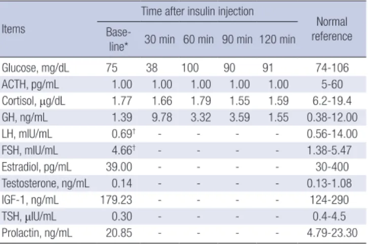
Table 2. Result of insulin tolerance test and baseline hormonal profile (regular insulin 0.1 U/kg IV)
Items
Time after insulin injection
Normal reference Base-
line* 30 min 60 min 90 min 120 min
Glucose, mg/dL 75 38 100 90 91 74-106
ACTH, pg/mL 1.00 1.00 1.00 1.00 1.00 5-60
Cortisol, μg/dL 1.77 1.66 1.79 1.55 1.59 6.2-19.4
GH, ng/mL 1.39 9.78 3.32 3.59 1.55 0.38-12.00
LH, mIU/mL 0.69† - - - - 0.56-14.00
FSH, mIU/mL 4.66† - - - - 1.38-5.47
Estradiol, pg/mL 39.00 - - - - 30-400
Testosterone, ng/mL 0.14 - - - - 0.13-1.08
IGF-1, ng/mL 179.23 - - - - 124-290
TSH, μIU/mL 0.30 - - - - 0.4-4.5
Prolactin, ng/mL 20.85 - - - - 4.79-23.30
ACTH, adrenocorticotropic hormone; GH, growth hormone; LH, luteinizing hormone;
FSH, follicle stimulating hormone; IGF-1, insulin-like growth factor 1; TSH, thyroid stimulating hormone.
*Baseline samples were taken after 8 hours of fasting; †Luteal phase.
Fig. 1. Copper deposits in the Descemet’s membrane of the cornea (Kayser-Fleischer ring).
Fig. 2. Bilateral symmetric increased signals of the basal ganglia on T2 weighted MR image (A) while no focal lesion was found in the pituitary gland (B).
A B
in normal range while thyroid function test results showed TSH 0.28 μIU/mL, fT4 0.72 ng/dL, and T3 50.85 ng/dL, all being be- low the lower limit. ACTH and cortisol were checked at 8 AM and 4 PM, being decreased to 1.00/1.72 μg/dL and 1.00/2.84 μg/dL, respectively. The thyrotropin releasing hormone (TRH) stimulation test and insulin tolerance test were performed to evaluate the pituitary-adrenal axis function. TSH levels showed only a marginal increase from 0.227 μIU/mL to 2.816 μIU/mL which was an insignificant response to TRH (Table 1), indicat- ing central hypothyroidism and not non-thyroidal illness syn- drome. ACTH remained as 1.00 pg/mL after provoking hypo- glycemia with insulin injection while cortisol merely rose from
1.77 µg/mL to 1.66, and GH from 1.39 mg/dL to 9.78 mg/dL (Table 2). This was determined as insufficient response to insu-
Lee HW, et al. • Adrenal Insufficiency and Hypothyroidism in Wilson’s Disease
http://jkms.org 1347
http://dx.doi.org/10.3346/jkms.2016.31.8.1345
lin-induced hypoglycemia. Results were consistent with sec- ondary hypothyroidism and adrenal insufficiency due to hypo- pituitarism.
After being admitted, the patient showed trouble swallowing food and other neurological symptoms that could not be ex- plained by depression such as rigidity, tremor and bradykine- sia. Brain MRI was done as an additional test for these symp- toms as well as to determine the presence of any pituitary tu- mors. T2-weighted images showed high signal intensity in both basal ganglia and in the midbrain, whereas no abnormality was found in the pituitary (Fig. 2).
Oral D-penicillamine (1.0 g/day) was given for copper chela- tion with pyridoxine (50 mg/day), while thyroid hormone and steroid replacement with levothyroxine (50 μg/day) and pred- nisolone (7.5 mg/day) were additionally used. Significant im- provements were seen in liver function. Increase in 24-hour urinary copper excretion was monitored which reached 922.8 μg/day after initiating treatment and was maintained in the range of 200-500 μg/day after 6 months. Anti-Parkinson agents were used for tremor and gait disturbance. The patient’s gener- al weakness, appetite loss and depressive mood improved as well. She is currently receiving treatment with the same regi- men and has had no side effects from medication or other new- ly developed symptoms for the last 7 months.
DISCUSSION
Hypopituitarism refers to decreased secretion of pituitary hor- mones as a result of abnormalities in the pituitary itself or the hypothalamus (3). It usually occurs due to a pituitary tumor or as a consequence of its treatment, and symptoms vary depend- ing on which hormone is deficient (3). Other causes include pi- tuitary apoplexy, Sheehan’s syndrome, stroke, traumatic brain injury, cerebral hemorrhage and infiltrative diseases such as sarcoidosis or hemochromatosis which are relatively rare and are often wrongfully labeled as idiopathic since imaging is usu- ally normal (3,4). In our case, hormone studies were consistent with secondary adrenal insufficiency and hypothyroidism due to hypopituitarism which we concluded to be associated with Wilson’s disease, since no other definite cause or coexisting disease was found. The patient had no headaches, visual field defects or disturbances and brain MRI was negative of any pi- tuitary lesion.
Among infiltrative diseases, hemochromatosis is known to cause pituitary dysfunction by depositing iron in the anterior pituitary, especially as hypogonadotrophic hypogonadism prob- ably due to the preference for iron of gonadotrophic cells (3). As for our case, we suggest copper deposition or secondary neuro- nal damage in the pituitary to explain the association between hypopituitarism and Wilson’s disease, but the mechanism could not be proven in this report. Recent studies showed that Wil-
son’s disease may be complicated by high iron concentrations in the brain as low ceruloplasmin levels and reduced ferroxi- dase activity lead to iron overload along with copper in exces- sive amounts, causing oxidative damage in a synergistic cas- cade (5). Paris et al. (6) proposed dopamine-dependent neuro- toxicity as another possible mechanism, decreasing survival of dopaminergic cells in the brain and contributing to the regional differences in copper accumulation or its consequent neuronal damage. In another report, a biomolecular basis of copper tox- icity was presented, demonstrating that both copper and iron induce genome damage and inhibit its repair process (7). Whe- ther these suggestions could be applied to the pituitary in Wil- son’s disease requires further investigation.
T2 hyperintense signals in the basal ganglia is the most com- mon finding on the brain MRI in Wilson’s disease, but interpre- tation of this lesion is ambivalent since copper deposition may be undetectable without the summation of underlying iron ac- cumulation. Secondary neuronal damage might occur without direct local copper accumulation (8). Also, it is not uncommon for pituitary imaging to be normal in hypopituitarism (4). De- spite normal pituitary imaging in our case, we could not rule out the possibility of copper infiltration as the cause for hypopi- tuitarism in Wilson’s disease.
Endocrinologic abnormalities are rare features in Wilson’s disease. Symptoms such as delayed puberty or amenorrhea were sporadically reported, as well as glucose intolerance and parathyroid function disorders (9,10). Krysiak et al. (1) reported galactorrhea and menstrual abnormalities in a patient with Wilson’s disease, presuming that the symptoms may have been caused by local deposits of copper in the pituitary and mam- mary glands. Frydman et al. (10) studied 16 patients with Wil- son’s disease to detect potential endocrine dysfunctions. In their report, most patients had low or borderline LH levels and a dynamic GnRH test revealed blunted LH and FSH responses.
Also regarding a possible association between pituitary dys- function and Wilson’s disease, a case of delayed puberty in an 18-year-old male with Wilson’s disease was reported, suggest- ing copper accumulation as the potential cause of decreased synthesis or secretion of GnRH in the hypothalamus or pitu- itary. Like in our study, the brain MRI was negative of any pitu- itary lesion (11). These cases presented pituitary dysfunction as hypogonadotrophic hypogonadism or menstrual abnormali- ties. To our knowledge, our study is an unprecedented case of Wilson’s disease coexisting with hypopituitarism presenting as TSH or ACTH deficiency in Korea, as other pituitary hormones were maintained within the normal range.
Meanwhile, common psychiatric symptoms in Wilson’s dis- ease are depression, incongruous behavior, and cognitive im- pairment (12). It may be challenging to differentiate these symp- toms from hypothyroidism or adrenal insufficiency which can be easily misdiagnosed or neglected if not examined for other
Lee HW, et al. • Adrenal Insufficiency and Hypothyroidism in Wilson’s Disease
1348 http://jkms.org http://dx.doi.org/10.3346/jkms.2016.31.8.1345 potential conditions (13). This study highlights the importance
of screening for hormone deficiency in Wilson’s disease espe- cially in patients presenting with psychiatric symptoms. We also suggest considering Wilson’s disease when evaluating pos- sible causes for idiopathic hypopituitarism. Regardless of wheth- er the two conditions are causal or coincidental, both require treatment as soon as practicable.
Further confirmation is needed on the endpoint of hormone therapy and on whether or not copper-chelating treatment alone brings pituitary function recovery, as pituitary dysfunction was successfully reversed by liver transplantation or therapeutic phlebotomy in some cases of hemochromatosis. Also, it would be worthwhile to reevaluate the pituitary function in 6 to 12 months after discontinuing hormone replacement. In conclu- sion, this case indicates a possible link between Wilson’s dis- ease and hypopituitarism presenting as altered thyroid homeo- stasis and pituitary-adrenal axis dysfunction.
DISCLOSURE
The authors have no potential conflicts of interest to disclose.
AUTHOR CONTRIBUTION
Conception and coordination of the study: Lee HW, Choi MK.
Case analysis and interpretation: Lee HW, Kang JD, Yeo CW, Yoon SO, Lee KJ, Choi MK. Manuscript preparation: Lee HW.
Revision and manuscript approval: all authors.
ORCID
Hae Won Lee http://orcid.org/0000-0002-7744-2187 Jin Du Kang http://orcid.org/0000-0003-2191-7718 Chang Woo Yeo http://orcid.org/0000-0001-5753-4191 Sung Oon Yoon http://orcid.org/0000-0001-7073-020X Kwang Jae Lee http://orcid.org/0000-0001-5468-6499 Mun Ki Choi http://orcid.org/0000-0002-5725-7128
REFERENCES
1. Krysiak R, Handzlik-Orlik G, Okopien B. Endocrine symptoms as the ini- tial manifestation of Wilson’s disease. Yale J Biol Med 2012; 85: 249-54.
2. Lee SY, Kim IH, Yoo SH, Kim DG. A case of colonic adenocarcinoma in a patient with Wilson’s disease. Gut Liver 2013; 7: 500-3.
3. Lewis AS, Courtney CH, Atkinson AB. All patients with ‘idiopathic’ hypo- pituitarism should be screened for hemochromatosis. Pituitary 2009; 12:
273-5.
4. Wilson V, Mallipedhi A, Stephens JW, Redfern RM, Price DE. The causes of hypopituitarism in the absence of abnormal pituitary imaging. QJM 2014; 107: 21-4.
5. Kim JM, Ko SB, Kwon SJ, Kim HJ, Han MK, Kim DW, Cho SS, Jeon BS. Fer- rous and ferric iron accumulates in the brain of aged Long-Evans Cinna- mon rats, an animal model of Wilson’s disease. Neurosci Lett 2005; 382:
143-7.
6. Paris I, Dagnino-Subiabre A, Marcelain K, Bennett LB, Caviedes P, Cavie- des R, Azar CO, Segura-Aguilar J. Copper neurotoxicity is dependent on dopamine-mediated copper uptake and one-electron reduction of ami- nochrome in a rat substantia nigra neuronal cell line. J Neurochem 2001;
77: 519-29.
7. Mitra J, Guerrero EN, Hegde PM, Wang H, Boldogh I, Rao KS, Mitra S, Hegde ML. New perspectives on oxidized genome damage and repair inhibition by pro-oxidant metals in neurological diseases. Biomolecules 2014; 4: 678- 703.
8. Mironov A. MRI in Wilson’s disease of the brain. Neuroradiology 1991; 33 Suppl: 598-600.
9. Tallis GA, Kitchener MI, Thomas AC. Hyperparathyroidism in a patient with Wilson’s disease. Clin Chem 1990; 36: 568-70.
10. Frydman M, Kauschansky A, Bonne-Tamir B, Nassar F, Homburg R. As- sessment of the hypothalamic-pituitary-testicular function in male pa- tients with Wilson’s disease. J Androl 1991; 12: 180-4.
11. Kim DH, Kweon BC, Lee HK, Nam KH, Kim SJ, Lee DW, Park CG, Lee CG, Lee SM, Lee CK. Wilson’s disease with abnormal secretion of pitu- itary hormones. Korean J Med 1997; 52: 429-35.
12. Zimbrean PC, Schilsky ML. Psychiatric aspects of Wilson disease: a re- view. Gen Hosp Psychiatry 2014; 36: 53-62.
13. Charmandari E, Nicolaides NC, Chrousos GP. Adrenal insufficiency. Lan- cet 2014; 383: 2152-67.