ABSTRACT
Background: The impact of myocardial damage on the prognosis of patients with septic shock is not clearly elucidated because complex hemodynamic changes in sepsis obscure the direct relationship. We evaluated left ventricular (LV) conditions that reflect myocardial damage independently from hemodynamic changes in septic shock and their influence on the prognosis of patients.
Methods: We retrospectively enrolled 208 adult patients who were admitted to the intensive care unit and underwent echocardiography within 7 days from the diagnosis of septic shock. Patients who were previously diagnosed with structural heart disease or coronary artery disease were excluded. Left ventricular ejection fraction (LVEF) was divided into four categories: normal, ≥ 50%; mild, ≥ 40%; moderate, ≥ 30%; and severe dysfunction, < 30%.
Wall motion impairment was categorized into the following patterns: normal, diffuse, ballooning, and focal.
Results: There were 141 patients with normal LVEF. Among patients with impaired LV wall motion, the diffuse pattern was the most common (34 patients), followed by the ballooning pattern (26 patients). Finally, 102 patients died, and in-hospital mortality was significantly higher in patients with severe LV systolic dysfunction (hazard ratio [HR], 1.97; 95%
confidence interval [CI], 1.04–3.75; P = 0.039) and in patients with diffuse pattern of LV wall motion impairment (HR, 2.28; 95% CI, 1.19–4.36; P = 0.013) than in those with a normal LV systolic function.
Conclusion: Severe LV systolic dysfunction and diffuse pattern of LV wall motion impairment significantly affected in-hospital mortality in patients with septic shock. Conventional echocardiographic evaluation provides adequate information on the development of myocardial damage and accurately predicts the prognosis of patients with septic shock.
Keywords: Septic Shock; Septic Cardiomyopathy; Left Ventricular Systolic Function
INTRODUCTION
Sepsis is a subset of organ dysfunction caused by a dysregulated host response to infection1 and easily causes cardiomyocyte dysfunction.2 Although the clinical signs and symptoms of
Original Article
Received: Jun 7, 2019 Accepted: Dec 11, 2019 Address for Correspondence:
Je Hyeong Kim, MD, PhD
Department of Critical Care Medicine, Korea University Ansan Hospital, Korea University College of Medicine, 123 Jeokgeum-ro, Danwon-gu, Ansan 15520, Republic of Korea.
E-mail: [email protected]
© 2020 The Korean Academy of Medical Sciences.
This is an Open Access article distributed under the terms of the Creative Commons Attribution Non-Commercial License (https://
creativecommons.org/licenses/by-nc/4.0/) which permits unrestricted non-commercial use, distribution, and reproduction in any medium, provided the original work is properly cited.
ORCID iDs Sua Kim
https://orcid.org/0000-0003-3513-7261 Jung-Dong Lee
https://orcid.org/0000-0002-3825-065X Beong Ki Kim
https://orcid.org/0000-0003-2009-7658 Yong-Hyun Kim
https://orcid.org/0000-0003-1376-5128 Je Hyeong Kim
https://orcid.org/0000-0002-8995-7460 Funding
This study was supported by Korea University Ansan Hospital (Grant No. O1903631).
Sua Kim ,1,2 Jung-Dong Lee ,3 Beong Ki Kim ,4 Yong-Hyun Kim ,2 and Je Hyeong Kim 1,4
1 Department of Critical Care Medicine, Korea University Ansan Hospital, Korea University College of Medicine, Ansan, Korea
2Division of Cardiology, Department of Internal Medicine, Korea University Ansan Hospital, Ansan, Korea
3JB Lab and Clinic, Seoul, Korea
4Division of Pulmonology, Department of Internal Medicine, Korea University Ansan Hospital, Ansan, Korea
Association between Left Ventricular Systolic Dysfunction and Mortality in Patients with Septic Shock
Cardiovascular Disorders
Disclosure
The authors have no potential conflicts of interest to disclose.
Author Contributions
Conceptualization: Kim JH. Data curation: Lee JD. Formal analysis: Lee JD. Investigation: Kim BK, Kim YH. Methodology: Kim S. Validation:
Kim S. Writing - original draft: Kim S. Writing - review & editing: Kim JH.
heart failure are challenging to identify in patients with septic shock, when cardiac function is evaluated, results commonly show cardiac dysfunction. The prevalence rate has ranged from 64% to 84%.3,4
Several studies have evaluated cardiac function in patients with sepsis5 with different parameters; cardiac biomarkers and left ventricular (LV) systolic function, diastolic function, and right ventricular (RV) function via echocardiography.6-8 However, the decrease in LV afterload and preload directly influence the systolic and diastolic function of LV, and the general condition of the patient with septic shock affects cardiac biomarkers. Therefore the objective parameters used in evaluating cardiac function did not properly reflect the actual cardiac function and did not show significant association with the prognosis of patients with septic shock.9,10 Special echocardiographic parameters of global longitudinal strain and tricuspid annular systolic excursion may reflect the prognosis of patients with septic shock, but more evidence is required.11
We hypothesized that the impairment of LV systolic function may have a non-linear relationship with intrinsic myocardial damage in sepsis but still influence the prognosis of patients with septic shock. Therefore, we aimed to identify the echocardiographic features that can reflect cardiac dysfunction independently from the heterogeneous hemodynamic conditions of patients with septic shock and to investigate its association with in-hospital mortality.
METHODS
Study population
Patients admitted to the intensive care unit (ICU) in our hospital due to septic shock from January 01, 2015 to June 30, 2018, were retrospectively screened. Patients who were 18 years or older and who presented with septic shock and underwent echocardiography within a week from shock development were included in the study. We excluded patients with tachycardia (heart rate, ≥ 150 beats per minute), patients under post-operative condition, and those who were previously diagnosed with structural heart disease such as valvular heart disease, congenital heart disease, and cardiomyopathies or heart failure with more than moderate systolic dysfunction. Patients with coronary artery disease were also excluded.
Septic shock was diagnosed according to Sepsis-31; however, we also included patients without serum lactate level if they met the sequential organ failure assessment score, and required vaso-active agents, despite adequate volume resuscitation considering the inclusion period. The presence of coronary artery disease was defined by previous or current evaluation of coronary angiography or coronary computed tomography angiography findings showing more than 70% stenosis at any epicardial coronary artery with or without revascularization.
Data acquisition
Patient data including past medical history, cause of infection, vital signs, and laboratory findings along with cardiac enzyme levels were collected by reviewing their medical records. Then, Acute Physiology and Chronic Health Evaluation II (APACHE II) scores were calculated using the data. Infection sources were categorized into respiratory tract, urinary tract, biliary tract, gastrointestinal tract, and others. When there were several results for the cardiac enzymes, the maximum values which were measured within 3 days from the echocardiographic evaluation were collected for the study. Echocardiographic data were retrospectively reviewed. The primary outcome of the study was in-hospital mortality.
Echocardiography
Conventional echocardiography was performed within 7 days from ICU admission or from the development of septic shock. Echocardiographic measurements were performed using a commercially available ultrasound system (VIVID-Q; GE Vingmed Ultrasound AS, Horten, Norway). Left ventricular ejection fraction (LVEF) was calculated using the biplane Simpson's method from apical four- and two-chamber views. We categorized LV systolic function into four groups: normal for ejection fraction (EF) ≥ 50%; mild systolic dysfunction for EF ≥ 40%;
moderate dysfunction for EF ≥ 30%; and severe dysfunction for EF < 30%. Mitral inflow was obtained via pulse-wave Doppler echocardiography using the sample volume between mitral leaflet tips during diastole (E). Mitral annular tissue velocity (e′) was obtained using tissue Doppler imaging with the sample volume on the septal area of the mitral annulus during diastole. Then, LV filling pressure was substituted by E and e′ ratio.12 The patterns of LV wall motion impairment were categorized into four types: normal, diffuse wall motion impairment, ballooning of LV apex or mid walls (Takotsubo cardiomyopathy type)13 and focal wall motion impairment. Focal wall motion impairment was defined as regional wall motion abnormalities that do not follow the wall motion patterns of stress-induced cardiomyopathy, regardeless of whether it follows the territories of a coronary artery or not. To quantify the extent of wall motion impairment, wall motion score index was calculated.14
Statistical analysis
Continuous variables are expressed as mean ± standard deviation or median (from 1st to 3rd quantiles). Categorical variables are expressed as frequencies and percentages. Continuous variables between survivors and non-survivors were compared using the independent t-test and Wilcoxon rank-sum test. Categorical variables were compared using the χ2 test. Disease severity was assessed using the APACHE II score. Event-free survival was estimated using the Kaplan-Meier method and was compared using the log-rank test. Cox proportional hazard regression analysis was performed to evaluate the effect of LV systolic function and patterns of LV wall motion impairment on in-hospital mortality. The parameters that influenced patient survival in the Cox regression model were adjusted in the final model. Statistical analysis was performed using R 3.5.3 (The R Foundation for Statistical Computing, Vienna, Austria) for Windows. In all statistical analyses, a P value < 0.05 was considered statistically significant.
Ethics statement
The Institutional Review Board (IRB) of Korea University Ansan Hospital approved the study protocol and waived the requirement for informed consent (IRB No. 2019AS0031).
RESULTS
Baseline characteristics of all patients and comparison of values by survival
A total of 208 patients were finally enrolled in the study. The mean age of the participants was 68.7 ± 14.3 years. The mean APACHE II score was 21.3 ± 6.8. Pneumonia was the leading cause of septic shock (126, 60.6%), followed by urinary tract infection (28, 13.5%). The time interval from the development of shock to transthoracic echocardiography was 1 (1–3) day.In-hospital mortality was 102 (49.5%) in total. No significant difference was observed in age, gender, and underlying disease except for malignancies, which was not significant when Cox proportional hazard regression analysis was performed (Supplementary Table 1). The
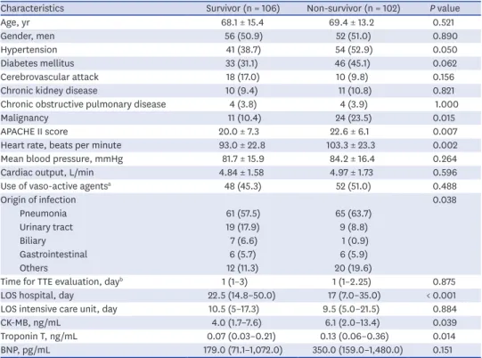
APACHE II score of the non-survivors was higher than that of the survivors, and the CK-MB and troponin-T levels were higher in the non-survivors than in survivors (Table 1).
Cardiac function of patients with septic shock
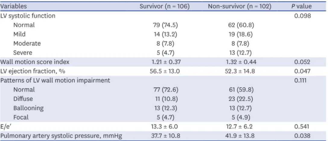
Among all patients, 141 (67.8%) had normal LV systolic function. The LV filling pressure as assessed using E/e′ and the pulmonary artery systolic pressure were slightly increased (13.1 ± 6.1 and 39.7 ± 12.5 mmHg, respectively). Among the patients with impaired LV wall motion, 34, 26, and 10 patients presented with diffuse pattern, ballooning pattern, and focal pattern of wall motion impairment, respectively. When parameters were compared according to patient survival, LVEF and pulmonary artery systolic pressure were significantly different between survivors and non-survivors (P = 0.047 and P = 0.017 respectively) in simple analysis (Table 2).
LV systolic dysfunction and in-hospital mortality in patients with septic shock
In univariate Cox proportional hazard regression analysis, severe LV systolic dysfunction with LVEF < 30% was significantly associated with higher in-hospital mortality (Supplementary Table 1). Kaplan-Meier curve analysis also showed significant log-rank test results (P = 0.037) (Fig. 1A), which was still significant when age, gender, APACHE II score, heart rate, use of vaso-active agent, and pulmonary artery systolic pressure were adjusted. Risk of in-hospital mortality of patients with severe LV systolic dysfunction with LVEF < 30% was significantly higher than that of patients with normal LV systolic function (hazard ratio [HR], 1.97; 95%confidence interval [CI], 1.04–3.75; P = 0.039), although the risk of in-hospital mortality did not change in patients with mild or moderate systolic dysfunction (Table 3).
Table 1. Baseline characteristics of patients with septic shock
Characteristics Survivor (n = 106) Non-survivor (n = 102) P value
Age, yr 68.1 ± 15.4 69.4 ± 13.2 0.521
Gender, men 56 (50.9) 52 (51.0) 0.890
Hypertension 41 (38.7) 54 (52.9) 0.050
Diabetes mellitus 33 (31.1) 46 (45.1) 0.062
Cerebrovascular attack 18 (17.0) 10 (9.8) 0.156
Chronic kidney disease 10 (9.4) 11 (10.8) 0.821
Chronic obstructive pulmonary disease 4 (3.8) 4 (3.9) 1.000
Malignancy 11 (10.4) 24 (23.5) 0.015
APACHE II score 20.0 ± 7.3 22.6 ± 6.1 0.007
Heart rate, beats per minute 93.0 ± 22.8 103.3 ± 23.3 0.002
Mean blood pressure, mmHg 81.7 ± 15.9 84.2 ± 16.4 0.264
Cardiac output, L/min 4.84 ± 1.58 4.97 ± 1.73 0.596
Use of vaso-active agentsa 48 (45.3) 52 (51.0) 0.488
Origin of infection 0.038
Pneumonia 61 (57.5) 65 (63.7)
Urinary tract 19 (17.9) 9 (8.8)
Biliary 7 (6.6) 1 (0.9)
Gastrointestinal 6 (5.7) 6 (5.9)
Others 12 (11.3) 20 (19.6)
Time for TTE evaluation, dayb 1 (1–3) 1 (1–2.25) 0.875
LOS hospital, day 22.5 (14.8–50.0) 17 (7.0–35.0) < 0.001
LOS intensive care unit, day 10.5 (5–17.3) 9.5 (5.0–21.5) 0.884
CK-MB, ng/mL 4.0 (1.7–7.6) 6.1 (2.0–13.4) 0.039
Troponin T, ng/mL 0.07 (0.03–0.21) 0.13 (0.06–0.36) 0.014
BNP, pg/mL 179.0 (71.1–1,072.0) 350.0 (159.0–1,480.0) 0.151
Data are presented as mean ± standard deviation or number (%).
APACHE II = acute physiology and chronic health evaluation II, TTE = transthoracic echocardiography, LOS = length of stay, BNP = B-natriuretic peptide.
aThe use of vaso-active agent when TTE was performed; bTime duration from the diagnosis of septic shock to echocardiographic evaluation.
Table 2. Comparison of cardiac function evaluated by transthoracic echocardiography by survival
Variables Survivor (n = 106) Non-survivor (n = 102) P value
LV systolic function 0.098
Normal 79 (74.5) 62 (60.8)
Mild 14 (13.2) 19 (18.6)
Moderate 8 (7.8) 8 (7.8)
Severe 5 (4.7) 13 (12.7)
Wall motion score index 1.21 ± 0.37 1.32 ± 0.44 0.052
LV ejection fraction, % 56.5 ± 13.0 52.3 ± 14.8 0.047
Patterns of LV wall motion impairment 0.111
Normal 77 (72.6) 61 (59.8)
Diffuse 11 (10.8) 23 (22.5)
Ballooning 13 (12.3) 13 (12.7)
Focal 5 (4.7) 5 (4.9)
E/e′ 13.3 ± 6.0 12.7 ± 6.2 0.541
Pulmonary artery systolic pressure, mmHg 37.7 ± 10.8 41.9 ± 13.8 0.038 Data are presented as mean ± standard deviation or number (%).
LV = left ventricular.
A
0.8 1.0
0.2
00 10 20 30 40 50
0.6 0.4
Cumulative survival rate, %
Days from the ICU admission
Log-rank test, P = 0.037 Log-rank test, P = 0.022
71 40 28
116 57
Normal
13 9 7
23 11
Mild
10 3 3
13 5
Moderate
9 3 1
13 5
Severe
Normal Ballooning Diffuse Focal
B
0.8 1.0
0.2
00 10 20 30 40 50
0.6 0.4
Cumulative survival rate, %
Days from the ICU admission
113 70 57 41 29
22 15 8 7 4
22 15 11 5 4
8 3 2 2 2
Moderate Normal Mild
Severe Diffuse
Normal Ballooning Focal
Fig. 1. Kaplan-Meier curve for mortality in patients with septic shock. The overall survival in patients with septic shock was compared by the LV systolic function (A) and by the pattern of LV wall motion impairment (B).
ICU = intensive care unit, LV = left ventricular.
Table 3. The relationship between LV systolic function and in-hospital mortality; the result of multivariable Cox proportional hazard regression analysis adjusted for covariates
Variables HR (95% CI) P value
LV systolic functiona
Normal 1.00 (Ref) -
Mild 1.55 (0.82–2.93) 0.179
Moderate 1.65 (0.73–3.70) 0.229
Severe 1.97 (1.04–3.75) 0.039
Wall motion score indexa 1.64 (0.99–2.73) 0.063
Patterns of LV wall motion impairmentb
Normal 1.00 (Ref) -
Diffuse 2.28 (1.19–4.36) 0.013
Ballooning 1.14 (0.55–2.39) 0.722
Focal 1.33 (0.39–4.53) 0.643
LV = left ventricular, HR = hazard ratio, CI = confidence interval.
aAdjusted for age, gender, APACHE II score, heart rate, use of vaso-active agent, and pulmonary artery systolic pressure; bAdjusted for age, gender, APACHE II score, heart rate, use of vaso-active agent, pulmonary artery systolic pressure, and severe LV systolic dysfunction.
Patterns of wall motion impairment and in-hospital mortality in patients with septic shock
When patients were divided according to patterns of LV wall motion impairment, disease severity characterized by APACHE II score was not different. However, cardiac function, evaluated with B-type natriuretic peptide level and E/e′, was significantly lower and LV systolic function was greater in patients with normal LV wall motion (Table 4). In addition, the overall survival of patients was different according to the patterns of LV wall motion impairment, with lower overall survival in patients with the diffuse pattern of LV wall motion impairment in the Kaplan-Meier curve (log-rank test, P = 0.022) (Fig. 1B). The result did not change even when other covariates were adjusted with multivariate Cox regression analysis.
The risk of in-hospital mortality was higher in patients with the diffuse pattern of LV wall motion impairment than in those with normal LV wall motion in multivariate analysis (HR, 2.28; 95% CI, 1.19–4.36; P = 0.013). Ballooning pattern wall motion impairment or focal wall motion impairment did not affect in-hospital mortality (Table 3).
DISCUSSION
We evaluated the cardiac function in patients with septic shock and identified the association between the development of myocardial dysfunction and in-hospital mortality via echocardiographic evaluation. In patients with septic shock, LV systolic dysfunction was common, and LV end-diastolic filling pressure and pulmonary arterial systolic pressure were slightly increased. In such condition, significant decrease in LV systolic function with an LVEF < 30% and diffuse pattern of LV wall motion impairment significantly influenced in- hospital mortality.
The development of myocardial dysfunction, referred to as septic cardiomyopathy, is one of the common findings in patients with septic shock. However, the characteristics of septic cardiomyopathy are challenging to define because different patterns of cardiac dysfunction Table 4. Comparison of demographic data and myocardial function of patients with septic shock by the pattern of LV wall motion impairment
Variables Normal (n = 138) Diffuse (n = 34) Ballooning (n = 26) Focal (n = 10) P value P valuea
Age, yr 67.3 ± 14.7 69.1 ± 13.5 74.0 ± 11.6 74.2 ± 15.9 0.094 0.147
Gender, men 72 (52.2) 24 (70.6) 8 (30.8) 4 (40) 0.019 0.004
APACHE II score 20.5 ± 7.2 22.3 ± 6.3 23.3 ± 4.8 22.8 ± 6.3 0.160 0.520
Heart rate, per min 96.7 ± 24.3 108.3 ± 19.1 94.2 ± 24.7 90.7 ± 13.1 0.045 0.019
Mean blood pressure, mmHg 80.3 ± 14.8 88.5 ± 19.7 89.0 ± 14.6 83.4 ± 17.7 0.011 0.905
Cardiac output, L/min 5.20 ± 1.66 4.76 ± 1.39 3.87 ± 1.55 3.91 ± 0.91 0.001 0.033
Use of vaso-active agents 74 (53.6) 14 (41.2) 9 (34.6) 3 (30) 0.136 0.789
Troponin T, ng/mL 0.28 ± 0.96 0.55 ± 0.94 0.59 ± 0.90 0.37 ± 0.31 0.445 0.894
CK-MB, ng/mL 11.9 ± 30.8 8.8 ± 10.5 14.0 ± 13.0 7.3 ± 10.4 0.874 0.176
BNP, pg/mL 505.5 ± 907.6 1,984.5 ± 1,693.6 235.1 ± 4,195.2 1,769.3 ± 1,560.7 0.006 0.771
LV systolic function < 0.001 0.356
Normal 136 (98.6) 0 (0.0) 0 (0.0) 5 (50.0)
Mild 4 (2.9) 16 (47.1) 9 (34.6) 4 (40.0)
Moderate 0 (0.0) 8 (23.5) 7 (26.9) 1 (10.0)
Severe 0 (0.0) 10 (29.4) 8 (30.8) 0 (0.0)
Ejection fraction, % 63.0 ± 7.1 37.9 ± 8.4 38.3 ± 10.5 51.6 ± 10.4 < 0.001 0.874
Wall motion score index 1.00 ± 0.00 2.00 ± 0.00 1.68 ± 0.27 1.28 ± 0.22 < 0.001 < 0.001
E/e′ 11.6 ± 4.7 15.4 ± 7.8 16.3 ± 6.7 19.5 ± 8.9 < 0.001 0.692
PASP, mmHg 39.5 ± 12.7 38.9 ± 10.9 41.7 ± 14.4 40.4 ± 9.6 0.844 0.410
LV = left ventricular, APACHE II = acute physiology and chronic health evaluation II, BNP = b-type natriuretic peptide, PASP = pulmonary artery systolic pressure.
aComparison between patient groups with diffuse wall motion impairment and with ballooning wall motion impairment.
are observed with varying degrees in patients with septic shock. Furthermore, septic shock affects the objective parameters that are used in evaluating cardiac function even when there is no myocardial damage; afterload and preload decrease in septic shock due to the decrease in peripheral vascular resistance.15 This decrease in afterload exaggerates LV systolic function while the decrease in preload reduces LV filling pressure, as reflected in E/e′, and influences stroke volume. With such complexity, conventional parameters, such as LVEF and E/e′, were not associated with the prognosis of patients with septic shock in previous studies. Then, further studies evaluated other parameters, such as myocardial performance index,16 mitral annular posterior systolic excursion, tricuspid annular posterior systolic excursion (TAPSE),17 and global longitudinal strain (GLS) as for prognostic factors in septic shock.11,18 Although TAPSE and GLS showed promising results, they are still challenging parameters because they are not usually assessed and commonly reported in routine echocardiographic evaluations, moreover, they require additional high-quality images to get the values and needs more evidences.
With the interest in the development of myocardial damage in septic shock, LVEF was firstly focused as a prognostic factor4 but failed to reveal the significant influence on the prognosis because of the hemodynamic complexity of septic shock. Septic shock patients with normal LVEF may be masking the decreased LV systolic function because of decreased peripheral vascular resistance or may have normal LV systolic function with minimal changes in peripheral vascular resistance. Patients with hyperdynamic left ventricles may have relatively overestimated LV systolic function because of severely decreased peripheral vascular resistance. All these situations were reflected in previous studies with inconsistent relationships between LVEF and patient prognosis.3,6,9 However, we assumed that although the relationship between LVEF and the prognosis of the patient might not be linear, there might be a specific range of LVEF which reflects intrinsic myocardial damage, irrespective of the change in peripheral vascular resistance. A significant decrease in LVEF, with or without decreased peripheral vascular resistance, may indicate an absolute decrease in LV systolic function and remarkably influence the prognosis of patients with septic shock. In our study, that specific range was an LVEF < 30%.
When evaluating cardiac function in patients with sepsis, parameters that can reflect intrinsic cardiac function independently from hemodynamic conditions may have advantages. GLS may be better than LVEF as mentioned above.11,18 However, high-quality additive images, and more evidence for practical use are required. Therefore, the result of our study that showed the clinical implication of LVEF in patients with septic cardiomyopathy is significant.
There is no consensus on the definition and clinical spectrum of septic cardiomyopathy, which is attributed to its complex and multifactorial pathophysiology. The type of wall motion impairment may also have been challenging to define in septic cardiomyopathy considering that most studies that evaluated the systolic function of LV in patients with septic shock did not present the patterns of wall motion impairment. We also expected the diffuse type in patients with septic shock,19 however, only 34 (16.3%) patients presented diffuse wall motion impairment followed by 26 (12.5%) patients with the ballooning pattern and 10 (4.8%) with focal pattern. The different patterns may differentiate the cause of the myocardial dysfunction in septic shock, but there is a lack of evidence to exclude the possibilities of combination of pathophysiologies for the cardiac dysfunction in the septic shock. Therefore, we may conclude that the dominant type of cardiomyopathy in patients with diffuse LV wall motion impairment is septic cardiomyopathy and that in patients with ballooning pattern of LV wall motion impairment is stress induced cardiomyopathy rather than differentiating
patients into septic cardiomyopathy and stress induced cardiomyopathy. However, in patients with ballooning type wall motion impairment, women patients were dominant and 11 of 15 patients who underwent follow-up echocardiography showed improvement of LV systolic function. This implies that patients with the ballooning type of wall motion impairment may follow the characteristics of stress induced cardiomyopathy.
The present study had several limitations, primarily due to its retrospective nature. First, echocardiography was not performed in all patients with septic shock in our hospital.
Therefore, the severity of the disease in the study population, who had echocardiography data, may be higher than the severity of average patients with septic shock. And, we were not able to predict the prevalence of septic cardiomyopathy nor to characterize the pattern of cardiac dysfunctions in septic cardiomyopathy. Second, although we excluded patients with coronary artery disease, our study population may still have included patients with coronary artery disease, which might have influenced LV systolic function and patient mortality. Third, the time interval from shock development to echocardiographic evaluation differed between cases. For this reason, the change in LV systolic function at the hyperacute stage of septic shock and its influence on patient outcome may have been missed. Fourth, we did not routinely evaluate central venous pressure. Therefore, we could not obtain data about peripheral vascular resistance and could not analyze the effect of peripheral vascular resistance on the change of LV function in the study patients.
LV systolic dysfunction was commonly observed in patients with septic shock. An LVEF <
30% and diffusely impaired LV wall motion were significantly associated with higher in- hospital mortality. The relationship between LV systolic dysfunction and the prognosis of septic shock became more distinct in this study. However, further evaluation in prospective settings is required for assessing of the prevalence and patterns of cardiac dysfunction considering peripheral vascular resistance.
SUPPLEMENTARY MATERIAL
Supplementary Table 1
The relationship between demographic and echocardiographic data and in-hospital mortality; the result of univariate Cox proportional hazard regression analysis Click here to view
REFERENCES
1. Singer M, Deutschman CS, Seymour CW, Shankar-Hari M, Annane D, Bauer M, et al. The third international consensus definitions for sepsis and septic shock (sepsis-3). JAMA 2016;315(8):801-10.
PUBMED | CROSSREF
2. Ehrman RR, Sullivan AN, Favot MJ, Sherwin RL, Reynolds CA, Abidov A, et al. Pathophysiology, echocardiographic evaluation, biomarker findings, and prognostic implications of septic cardiomyopathy: a review of the literature. Crit Care 2018;22(1):112.
PUBMED | CROSSREF
3. Pulido JN, Afessa B, Masaki M, Yuasa T, Gillespie S, Herasevich V, et al. Clinical spectrum, frequency, and significance of myocardial dysfunction in severe sepsis and septic shock. Mayo Clin Proc 2012;87(7):620-8.
PUBMED | CROSSREF
4. Parker MM, Shelhamer JH, Bacharach SL, Green MV, Natanson C, Frederick TM, et al. Profound but reversible myocardial depression in patients with septic shock. Ann Intern Med 1984;100(4):483-90.
PUBMED | CROSSREF
5. Beesley SJ, Weber G, Sarge T, Nikravan S, Grissom CK, Lanspa MJ, et al. Septic cardiomyopathy. Crit Care Med 2018;46(4):625-34.
PUBMED | CROSSREF
6. Lanspa MJ, Shahul S, Hersh A, Wilson EL, Olsen TD, Hirshberg EL, et al. Associations among left ventricular systolic function, tachycardia, and cardiac preload in septic patients. Ann Intensive Care 2017;7(1):17.
PUBMED | CROSSREF
7. Landesberg G, Gilon D, Meroz Y, Georgieva M, Levin PD, Goodman S, et al. Diastolic dysfunction and mortality in severe sepsis and septic shock. Eur Heart J 2012;33(7):895-903.
PUBMED | CROSSREF
8. Vallabhajosyula S, Sakhuja A, Geske JB, Kumar M, Poterucha JT, Kashyap R, et al. Role of admission troponin-T and serial troponin-T testing in predicting outcomes in severe sepsis and septic shock. J Am Heart Assoc 2017;6(9):e005930.
PUBMED | CROSSREF
9. Sevilla Berrios RA, O'Horo JC, Velagapudi V, Pulido JN. Correlation of left ventricular systolic dysfunction determined by low ejection fraction and 30-day mortality in patients with severe sepsis and septic shock:
a systematic review and meta-analysis. J Crit Care 2014;29(4):495-9.
PUBMED | CROSSREF
10. McLean AS, Huang SJ, Hyams S, Poh G, Nalos M, Pandit R, et al. Prognostic values of B-type natriuretic peptide in severe sepsis and septic shock. Crit Care Med 2007;35(4):1019-26.
PUBMED | CROSSREF
11. Chang WT, Lee WH, Lee WT, Chen PS, Su YR, Liu PY, et al. Left ventricular global longitudinal strain is independently associated with mortality in septic shock patients. Intensive Care Med 2015;41(10):1791-9.
PUBMED | CROSSREF
12. Lang RM, Badano LP, Mor-Avi V, Afilalo J, Armstrong A, Ernande L, et al. Recommendations for cardiac chamber quantification by echocardiography in adults: an update from the American Society of Echocardiography and the European Association of Cardiovascular Imaging. Eur Heart J Cardiovasc Imaging 2015;16(3):233-70.
PUBMED | CROSSREF
13. Ghadri JR, Wittstein IS, Prasad A, Sharkey S, Dote K, Akashi YJ, et al. International expert consensus document on Takotsubo syndrome (Part I): clinical characteristics, diagnostic criteria, and pathophysiology. Eur Heart J 2018;39(22):2032-46.
PUBMED | CROSSREF
14. Schiller NB, Shah PM, Crawford M, DeMaria A, Devereux R, Feigenbaum H, et al. Recommendations for quantitation of the left ventricle by two-dimensional echocardiography. American Society of Echocardiography Committee on Standards, Subcommittee on Quantitation of Two-Dimensional Echocardiograms. J Am Soc Echocardiogr 1989;2(5):358-67.
PUBMED | CROSSREF
15. Vieillard-Baron A. Septic cardiomyopathy. Ann Intensive Care 2011;1(1):6.
PUBMED | CROSSREF
16. Nizamuddin J, Mahmood F, Tung A, Mueller A, Brown SM, Shaefi S, et al. Interval changes in myocardial performance index predict outcome in severe sepsis. J Cardiothorac Vasc Anesth 2017;31(3):957-64.
PUBMED | CROSSREF
17. Price LC, Wort SJ, Finney SJ, Marino PS, Brett SJ. Pulmonary vascular and right ventricular dysfunction in adult critical care: current and emerging options for management: a systematic literature review. Crit Care 2010;14(5):R169.
PUBMED | CROSSREF
18. Ng PY, Sin WC, Ng AK, Chan WM. Speckle tracking echocardiography in patients with septic shock: a case control study (SPECKSS). Crit Care 2016;20(1):145.
PUBMED | CROSSREF
19. Sato R, Nasu M. A review of sepsis-induced cardiomyopathy. J Intensive Care 2015;3(1):48.
PUBMED | CROSSREF