Computed Tomography and Magnetic Resonance Images of Adrenocortical Oncocytoma Cases
We present two cases of adrenocortical oncocytomas that were well-delineated on multi- detector computed tomography and magnetic resonance imaging. The images showed a well-enhanced large mass with multiple stippled calcifications in a 10-yr-old girl who was consulted due to precocious puberty. A well-enhanced solid mass with necrotic
components was incidentally noticed in a 54-yr-old man. These lesions were resected and diagnosed as adrenocortical oncocytomas through immunohistochemical studies and electron microscopy. Adrenocortical oncocytomas are rare disease entities, therefore, we report these interesting, rare adrenocortical oncocytomas here with radiologic findings, and discuss differential diagnosis.
Keywords: Adrenal Glands; Neoplasms-Oncocytoma; Computed Tomography; Magnetic Resonance Imaging
Jung-Hee Yoon,1 Seong-Sook Cha,2 and Seong Kuk Yoon3
1Department of Radiology, Inje University Haeundae Paik Hospital, Busan; 2Department of Radiology, Inje University Busan Paik Hospital, Busan; 3Department of Radiology, Dong-A University Hospital, Busan, Korea
Received: 27 July 2013 Accepted: 15 November 2013 Address for Correspondence:
Jung-Hee Yoon, MD
Department of Radiology, Haeundae Paik Hospital, Inje University College of Medicine, 875 Haeundae-ro, Haeundae- gu, Busan 612-896, Korea
Tel: +82.51-797-0355, Fax: +82.51-797-0397 E-mail: radyjh@hanmail.net
http://dx.doi.org/10.3346/jkms.2014.29.3.445 • J Korean Med Sci 2014; 29: 445-451
INTRODUCTION
Adrenocortical oncocytomas are benign tumors consisting of oncocytes in which the cytoplasm becomes eosinophilic due to accumulation of abnormal mitochondria. Oncocytomas devel- op in various organs and are frequently found in the salivary gland, kidneys, thyroid gland, parathyroid gland, and hypophy- sis (1). Adrenocortical oncocytomas have been rarely reported;
the first reported cases were documented by Kakimoto et al. (2) in 1986. Since then, only 110 cases of adrenocortical oncocyto- mas have been reported in the English-language literature, which has primarily focused on pathologic studies, especially of functioning adrenocortical oncocytomas. Adrenocortical on- cocytomas are extremely rare; only 9 cases have been reported (3-8), of which there were 3 cases that displayed androgenic hormone being secreted in female patients. One case produced interleukin-6, 4 cases showed Cushing syndrome, and one man was found with gynecomastia and elevated serum prolactin and estradiol. These oncocytomas must be treated with great care to exclude metastasis from the extra-adrenal primary site or primary adrenal carcinoma, which is a more common phe- nomenon. In these present cases, one case of functioning on- cocytoma presented with precocious puberty, and the other case was an incidentally detected nonfunctioning oncocytoma that was found via multi-detector computed tomography (CT) and magnetic resonance (MR) imaging.
CASE DESCRIPTION Case 1
In May 2012, a 10-yr-old girl presented to our outpatient clinic for the evaluation of precocious puberty, which was diagnosed elsewhere. The patient reported no specific past history and no specific clinical symptoms, such as flushing, fever, or abdomi- nal pain. A physical examination revealed elevated breast buds and the start of menstruation. The results of laboratory evalua- tions showed increased levels of androstenedione (23.1, nor- mal range: 1.7-2.7 ng/dL) and estradiol (46.61, normal range:
0.2-3.0 ng/mL). Testosterone was mildly increased (1.64, nor- mal range: 0.03-0.68 ng/mL), cortisol was slightly decreased (5.49, normal range: 6.2-19.4 µg/dL), and normal ranges of fol- licle stimulating hormone, luteinizing hormone, and adreno- corticotropic hormone were observed. There were no chromo- somal anomalies. Abdominal CT scans detected a well-circum- scribed, oval-shaped, 6-cm mass in the left adrenal gland, lo- cated superior to the left kidney, which demonstrated stippled calcifications on a precontrast scan. After the injection of a non- ionic contrast medium (Omnipaque 350®, GE Healthcare, USA), volume of 100 mL of contrast medium was injected at a rate of 3 mL/s via an antecubital vein, heterogeneous well enhance- ment was visualized during the 1-min delayed scan, and en- hancement washout was demonstrated on the 15-min delayed scan (Fig. 1). The CT Hounsfield unit (HU) was 40 HU on pre- contrast, 140 HU at 1 min, and 70 HU at 15 min. The absolute CASE REPORT
Oncology & Hematology
Yoon J-H, et al. • Images of Adrenocortical Oncocytoma
washout value ([Enhanced CT HU-Delayed CT HU]/[Enhanced CT HU-Unenhanc ed CT HU] × 100) was approximately 70% (a value of more than 60% indicates adrenal adenoma), and the relative washout value ([Enhanced CT HU-Delayed CT HU]/
Enhanced CT HU × 100) was 50% (a value of more than 40%
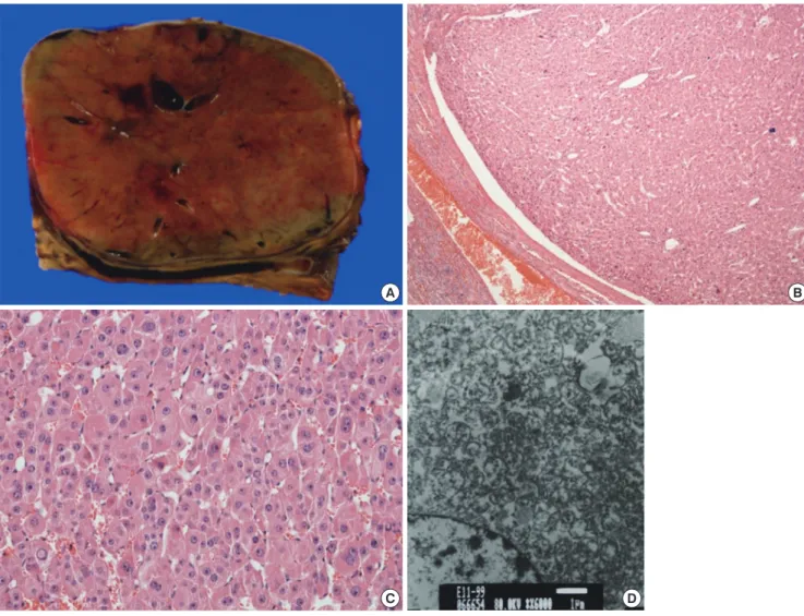
indicates adrenal adenoma) (9). Initially we thought that func- tional adrenal adenoma should be included in the differential diagnosis. A left adrenalectomy was performed. During the op- eration, there was no evidence of hypertension associated with palpation of the mass or direct invasion to the surrounding or- gans. The tumor was approximately 6 × 4 cm, well-encapsulat- ed and dark brown in color (Fig. 2A). The tumor consisted of multiple small hemorrhagic vascular lakes with old blood. The microscopic examination revealed that the neoplasm consisted of polygonal cells with abundant eosinophilic cells and granu- lar cytoplasm. Nuclear cellular atypia with enlarged nuclei were identified (× 200, × 400 High Power Field [HPF], hematoxylin and eosin [H&E], Fig. 2B and C). An electron microscopic study
was performed, and electron-dense inclusion and closely packed mitochondria with Golgi complex were found (× 400 HPF, Fig.
2D). Thus, we concluded that the final diagnosis in this case was adrenocortical oncocytoma. The patient had an uneventful postoperative course and was doing well one year after surgery without new lesions.
Case 2
In June 2010, an incidentally detected left adrenal mass was found in a 54-yr-old man via CT performed at another hospital.
Abdominal CT scans detected a lobulating contoured, well-de- marcated, well-enhancing solid mass in the left adrenal gland, which contained the central necrotic portion.
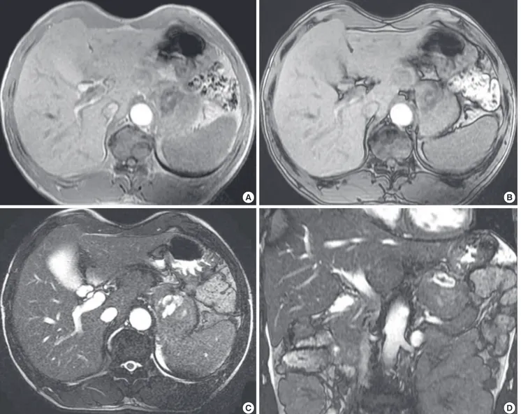
MR images were obtained with a 1.5 T unit using a contrast agent (Gadovist®, Bayer Healthcare, Germany). On a T2-weight- ed gradient echo image (TR/TE: 3.7/1.6), the mass was primari- ly of slightly high signal intensity with a hyperintense central portion. This mass primarily demonstrated hypointensity on A
C
B
D Fig. 1. Adrenocortical oncocytoma in a 10-yr-old girl (Case 1), abdominal multi-detector CT findings. (A) A well-circumscribed, oval-shaped mass in the left adrenal gland with stippled calcifications on a precontrast scan. (B) Thoroughly heterogeneous enhancement during the 1-min delayed axial scan. (C) On a coronal reconstructed image of B. (D) Enhancement washout in the 15-min delayed scan.
Yoon J-H, et al. • Images of Adrenocortical Oncocytoma
T1-weighted gradient echo images (TR/TE: 175.0/5.0), without a definite signal drop (suggesting a fat component) on the op- posed-phase correlated with the in phase. The previously high- ly hyperintense focus on T2-weighted images showed hypoin- tensity on the T1-weighted images, suggesting a necrotic por- tion, and a central hemorrhagic component was revealed with- in the necrosis by demonstrating a hyperintense foci on a T1- weighted image and hypointense foci on a T2-weighted image (Fig. 3). Contrast-enhanced MR images heterogeneously dem- onstrated the enhancement of the solid tumor portion at the 1-min, peak enhancement at the 3-min, and slight washout at the 5-min delayed phases. Intratumoral hemorrhagic necrosis was not constantly enhanced (Fig. 4). The blood vanillylman- delic acid, total metanephrine, epinephrine, and norepineph- rine were within the normal ranges.
Based on these observations, adrenal carcinoma and metas- tasis were included with the differential diagnosis. The patient
underwent meticulous examination for the primary origin, in- cluding colonoscopy, gastroscopy, and chest CT; there was no evidence of a primary focus. The most probable preoperative diagnosis was adrenal carcinoma. The patient underwent lapa- roscopic adrenalectomy, and histology suggested adrenal on- cocytoma with uncertain malignant potential.
The tumor was a soft oval mass measuring 9 × 6 × 5 cm in di- mensions and 116 g in mass. The outer surface was well encap- sulated by a thin fibrous capsule, and it was focally attached to a normal adrenal gland. Upon sectioning, the mass showed a brownish-yellow, fish-flesh-like cut surface with a multifocal brownish hemorrhagic necrosis and focal cystic changes (Fig.
5A and B).
The tumor was composed of epithelial cells with abundant acidophilic cytoplasm, with nuclear pleomorphism and a dif- fuse growth pattern, as shown using a light microscope (Fig.
5C), and the oncocytic cytoplasm was filled with a large num- A
C
B
D
Fig. 2. Adrenocortical oncocytoma in a 10-yr-old girl (Case 1), gross and microscopic findings. (A) The tumor was approximately 6 × 4 cm, well-encapsulated and dark brown in color, and it consisted of multiple small hemorrhagic vascular lakes with old blood. (B, C) A microscopic examination revealed that the neoplasm was comprised of polygonal cells with abundant eosinophilic cells and granular cytoplasm. A nuclear cellular atypia with enlarged nuclei was identified (B: × 200 HPF, C: × 400 HPF, H&E). (D) By electron microscopy, electron dense inclusions, as well as closely packed mitochondria with Golgi complexes, were found ( × 400 HFP).
Yoon J-H, et al. • Images of Adrenocortical Oncocytoma
Fig. 3. Adrenocortical oncocytoma in a 54-yr-old man (Case 2), MR images. The mass primarily demonstrates hypointensity on in-phase T1WI MR imaging (A) without a definite signal drop on the opposed-phase T1WI MR imaging (B), with a central hemorrhagic necrotic component. On T2WI MR imaging (C, axial; D, coronal), this mass is primarily of slightly high signal intensity with a central hyperintense portion.
A B
C D
ber of mitochondria, as well as some lysosomes, Golgi bodies, small lipid particles, and glycogen particles (Fig. 5D), the histo- logical features of which were consistent with adrenocortical oncocytoma. The tumor showed a low mitotic count ( < 5/50 HPFs), the absence of atypical mitosis, and venous invasion, but it also showed focal necrosis and inconspicuous capsule in- vasion, the histological features of which were consistent with uncertain malignant potential (borderline) behavior. Electron microscopy showed that microscopic and immunohistochemi- cal findings were compatible with an adrenocortical oncocyto- ma. Because of this finding, we performed a careful follow-up study to exclude the possibility that the adrenal tumor was ma- lignant over a three-year follow-up period. This workup includ- ed multiple abdominal ultrasounds examinations, as well as chest and abdominal CT scans. All of the imaging findings were unremarkable. We concluded that the final diagnosis in this case
was primary adrenocortical oncocytoma.
DISCUSSION
Adrenocortical oncocytomas are classified as benign, border- line malignant potential, and malignant according to the Lin- Weiss-Bisceglia criteria (10). Proposed major criteria (high mi- totic rate greater than 5 per 50 HPF, atypical mitoses, venous in- vasion) and minor criteria (large size and huge weight, necrosis, capsular invasion, sinusoidal invasion) in distinguishing malig- nant tumors are discussed. Their proposed working rules for diagnostic categorization of adrenocortical tumors are defined, with the presence of 1 major criterion indicating malignancy, 1 to 4 minor criteria indicating uncertain malignant potential (bor- derline), and the absence of all major and minor criteria indica- tive of benignancy (10). According to the proposed these crite-
Yoon J-H, et al. • Images of Adrenocortical Oncocytoma
Fig. 4. Adrenocortical oncocytoma in a 54-yr-old man (Case 2), contrast-enhanced MR images. (A) Precontrast (B-D) Thoroughly heterogeneous enhancement of a solid tumor- al portion at the 1-min (B), peak enhancement at 3-min (C), and slight enhancement washout at the 5-min delayed phases (D). Intratumoral hemorrhagic necrosis is visualized as a constantly non-enhancing portion.
A B
C D
ria, our first case should be considered as benign, because there are no major or minor criteria, and our second case should be considered as uncertain malignant potential (borderline) due to focal capsular invasion and necrosis. Adrenocortical oncocy- tomas were reported as large neoplasms (> 5 cm) that did not contain macroscopic evidence of necrosis or hemorrhage (1, 7, 11). One exception was the case reported by Nguyen et al. (1), which arose from heterotopic adrenocortical tissue separate from the adrenal glands within the retroperitoneum; that tu- mor measured only 3 cm and contained central necrosis. Our first case showed typical findings as a solid tumor without evi- dence of necrosis or hemorrhage and confirmed with benign oncocytoma, but our second case demonstrated atypical imag- ing findings as a large tumor with central hemorrhagic necrosis and surgically confirmed with borderline malignancy. Adreno- cortical oncocytoma that arisen in the adrenal gland is very rare
and usually benign and nonfunctioning, incidentally discov- ered by abdominal imaging performed to investigate an unre- lated problem, and have arisen in patients ranging in age from 27 to 72 yr without a male or female predominance (12). Most to the best of our knowledge, only 3 cases of functioning adre- nocortical oncocytomas have been reported in childhood.
The most widely studied oncocytoma from an imaging view- point has been the renal oncocytoma. Renal oncocytomas ap- pear as solid masses on CT and characteristically have central radiating scars when large. The MR signal characteristics are nonspecific; currently, the tumor cannot be reliably differenti- ated from the more common renal cell carcinoma (13). The cla- ssic central radiating scar that has been described in renal on- cocytomas is not inevitably present in adrenal oncocytomas.
Recently segmental enhancement inversion during corticome- dullary phase and early excretory phase was found to be a char-
Yoon J-H, et al. • Images of Adrenocortical Oncocytoma
acteristic enhancement pattern of small (smaller than 4 cm) re- nal oncocytoma at biphasic multidetector CT (14), but there was no definite segmental enhancement inversion in our cases.
We thought that the tumor mass of our two cases were larger in size than that of previously reported cases.
The imaging characteristics of our second case were very dif- ferent from the previously reported cases but well correlated with the histopathology, heterogeneous signal intensity in MR imaging represent hemorrhagic necrosis detected on patholog- ic examination. Fibrous encapsulation is characteristic finding for adrenal oncocytomas and was found in both benign and malignant variants, and could be well detected by both CT and MR imaging. Given the large size (> 6 cm), heterogeneous ap- pearance and presence of necrosis or calcification in the tumor, adrenocortical carcinoma was obviously the most likely preop- erative diagnosis. Non-functioning ones can be heterogeneous enhancement with central necrosis and hemorrhage, calcifica-
tion may be present occasionally. Thus unfortunately, the im- aging features of adrenocortical oncocytomas did not allow its differentiation from adrenocortical carcinomas (15). Cytopa- thology of adrenocortical oncocytoma by diagnosis of fine nee- dle aspiration biopsy was reported (4). However, the morpho- logic features and ancillary studies of find-needle aspiration cy- tology material do not help in indicating the biological behavior of these tumors. Surgical resection remains the only tool for the definitive diagnosis.
Recently, MR imaging has become increasingly useful in char- acterizing adrenal masses, particularly using chemical shift im- aging (in-phase and opposed-phase) to use the fat content of adenomas and lack of fat within metastases; opposed-phase imaging allows noninvasive distinction between these two enti- ties with a high degree of accuracy (16). Adrenocortical carci- noma, which was a diagnostic consideration in our case, often can be differentiated from adenoma on CT or MR imaging bas- A
C
B
D Fig. 5. Adrenocortical oncocytoma in a 54-yr-old man (Case 2), gross and microscopic findings. (A, B) The tumor was an oval-shaped soft mass that was encapsulated by a thin fibrous capsule and a multifocal brownish hemorrhage with a focal cystic change. (C) The tumor was composed of epithelial cells with abundant acidophilic cytoplasm, nu- clear pleomorphism, and a diffuse growth pattern, as observed using a light microscope. (D) By electron microscopy, the oncocytic cytoplasm was filled with a large number of mitochondria, as well as some lysosomes, Golgi bodies, small lipid particles, and glycogen particles.
Yoon J-H, et al. • Images of Adrenocortical Oncocytoma
ed on its size (i.e., generally > 5 cm), central necrosis, contrast enhancement, and frequent invasion of local organs and exten- sion into the inferior vena cava (17). In the second case present- ed here, the tumor was heterogeneous hypointense with re- spect to the liver on T1-weighted and slightly hyperintense on T2-weighted imaging, and displayed mild homogeneous en- hancement, this mass did not loss signal intensity on opposed- phase gradient echo imaging, a finding exhibited by most adre- nal adenomas. These imaging characteristics correlated with the pathologic findings, which included a lack of significant amount of lipid. The lack of lipid is not typical of adrenocortical cells but may be secondary to the large amount of mitochon- dria, which instead pack the cytoplasm of oncocytic cells (18).
In summary, two rare primary adrenocortical oncocytomas with radiologic findings are presented with a focus on dynamic CT and contrast-enhanced MR imaging and histopathologic findings with immunohistochemical studies and electron mi- croscopy.
Adrenocortical oncocytoma is rare disease entity, but can be included in the differential diagnosis of especially large, well- defined solid adrenal tumors. However, non-specific imaging features do not warrant a definitive preoperative diagnosis and unfortunately do not change the patient’s management.
ORCID
Jung-Hee Yoon http://orcid.org/0000-0001-5152-6668 Seong-Sook Cha http://orcid.org/0000-0002-4753-008X Seong Kuk Yoon http://orcid.org/0000-0001-7182-1701 REFERENCES
1. Nguyen GK, Vriend R, Ronaghan D, Lakey WH. Heterotopic adrenocor- tical oncocytoma: a case report with light and electron microscopic stud- ies. Cancer 1992; 70: 2681-4.
2. Kakimoto S, Yushita Y, Sanefuji T, Kondo A, Fujishima N, Kishikawa M, Matsumoto K. Non-hormonal adrenocortical adenoma with oncocyto- ma-like appearances. Hinyokika Kiyo 1986; 32: 757-63.
3. Geramizadeh B, Norouzzadeh B, Bolandparvaz S, Sefidbakht S. Func- tioning adrenocortical oncocytoma: a case report and review of litera- ture. Indian J Pathol Microbiol 2008; 51: 237-9.
4. Akatsu T, Kameyama K, Araki K, Ashizawa T, Wakabayashi G, Kitajima M. Functioning adrenocortical oncocytoma: the first documented case producing interleukin-6 and review of the literature. J Endocrinol Invest 2008; 31: 68-73.
5. Gołkowski F, Buziak-Bereza M, Huszno B, Bałdys-Waligórska A, Stefań
ska A, Budzyński A, Okoń K, Chrzan R, Urbanik A. The unique case of adrenocortical malignant and functioning oncocytic tumour. Exp Clin Endocrinol Diabetes 2007; 115: 401-4.
6. Xiao GQ, Pertsemlidis DS, Unger PD. Functioning adrenocortical onco- cytoma: a case report and review of the literature. Ann Diagn Pathol 2005; 9: 295-7.
7. Erlandson RA, Reuter VE. Oncocytic adrenal cortical adenoma. Ultra- struct Pathol 1991; 15: 539-47.
8. Wong DD, Spagnolo DV, Bisceglia M, Havlat M, McCallum D, Platten MA. Oncocytic adrenocortical neopla: a clinicopathologic study of 13 new cases emphasizing the importance of their recognition. Hum Pathol 2011; 42: 489-99.
9. Korobkin M, Brodeur FJ, Francis IR, Quint LE, Dunnick NR, Londy F.
CT time-attenuation washout curves of adrenal adenomas and nonad- enomas. AJR Am J Roentgenol 1998; 170: 747-52.
10. Bisceglia M, Ludovico O, Di Mattia A, Ben-Dor D, Sandbank J, Pasqui- nelli G, Lau SK, Weiss LM. Adrenocortical oncocytic tumors: report of 10 cases and review of the literature. Int J Surg Pathol 2004; 12: 231-43.
11. El-Naggar AK, Evans DB, Mackay B. Oncocytic adrenal cortical carcino- ma. Ultrastruct Pathol 1991; 15: 549-56.
12. Kitching PA, Patel V, Harach HR. Adrenocortical oncocytoma. J Clin Pa- thol 1999; 52: 151-3.
13. Defossez SM, Yoder IC, Papanicolaou N, Rosen BR, McGovern F. Non- specific magnetic resonance appearance of renal oncocytomas: report of 3 cases and review of the literature. J Urol 1991; 145: 552-4.
14. Kim JI, Cho JY, Moon KC, Lee HJ, Kim SH. Segmental enhancement in- version at biphasic multidetector CT: characteristic finding of small re- nal oncocytoma. Radiology 2009; 252: 441-8.
15. Shah RK, Oto A, Ozkan OS, Ernst RD, Hernandez JA, Chaudhary HB, Koroglu M. Adrenal oncocytoma: US and CT findings. JBR-BTR 2004;
87: 180-2.
16. Mitchell DG, Crovello M, Matteucci T, Petersen RO, Miettinen MM. Be- nign adrenocortical masses: diagnosis with chemical shift MR imaging.
Radiology 1992; 185: 345-51.
17. McLoughlin RF, Bilbey JH. Tumors of the adrenal gland: findings on CT and MR imaging. AJR Am J Roentgenol 1994; 163: 1413-8.
18. Gandras EJ, Schwartz LH, Panicek DM, Levi G. Case report: adrenocor- tical oncocytoma: CT and MRI findings. J Comput Assist Tomogr 1996;
20: 407-9.