Radiofrequency Ablation of Papillary Thyroid
Microcarcinoma:
A 10-Year Follow-Up Study
갑상선 미세유두암의 고주파 절제술 후 10년 경과 관찰
Yoo Kyeong Seo, MD1 , Seong Whi Cho, MD1* , Jung Suk Sim, MD2 , Go Eun Yang, MD1 , Woojin Cho, MD3
1Department of Radiology, Kangwon National University Hospital, Chuncheon, Korea Departments of 2Radiology and 3Otolaryngology and Head and Neck Surgery, Withsim Clinic, Seongnam, Korea
Purpose To investigate the efficacy and safety of radiofrequency ablation (RFA) for papillary thyroid microcarcinoma (PTMC) after > 10 years of follow-up.
Materials and Methods This study included five patients who underwent RFA to treat PTMCs (five lesions, mean diameter 0.5 cm, range 0.4–0.7 cm) between November 2006 and December 2009. The inclusion criteria were histopathologically confirmed PTMCs, a single PTMC lesion without extrathyroidal extension, no metastasis, and ineligibility or refusal to undergo surgery.
RFA was performed by a single radiologist using a radiofrequency generator and an internally cooled electrode. We retrospectively analyzed the procedure-induced complications, serial changes in ablated tumors, recurrence, and local as well as lymph node metastasis based on data obtained from medical records and radiological images.
Results The mean follow-up period was 130.6 months (range 121–159 months). Three patients underwent a single RFA session, and two patients underwent two RFA sessions. We observed no procedure-induced complications. Three tumors completely disappeared after ablation, and ablation of the other two tumors resulted in the formation of a small scar that showed long-term stability (mean duration 16.8 months, range 12–27 months). At the last follow-up, no patient showed recurrence or lymph node metastasis, and serum thyroglobulin levels were within normal limits in all patients.
Conclusion RFA may be effective and safe to treat low-risk PTMC in patients who refuse or are ineligible for surgery.
Index terms Papillary Thyroid Microcarcinoma; Thyroid Neoplasm; Radiofrequency Ablation
Received June 25, 2020 Revised September 11, 2020 Accepted October 15, 2020
*Corresponding author Seong Whi Cho, MD Department of Radiology, Kangwon National University Hospital, 156 Baengnyeong-ro, Chuncheon 24289, Korea.
Tel 82-33-258-2329 Fax 82-33-258-2221
E-mail [email protected] This is an Open Access article distributed under the terms of the Creative Commons Attribu- tion Non-Commercial License (https://creativecommons.org/
licenses/by-nc/4.0) which permits unrestricted non-commercial use, distribution, and reproduc- tion in any medium, provided the original work is properly cited.
ORCID iDs Yoo Kyeong Seo https://
orcid.org/0000-0001-5118-491X Seong Whi Cho
https://
orcid.org/0000-0001-5844-0584 Jung Suk Sim
https://
orcid.org/0000-0001-6803-3544 Go Eun Yang
https://
orcid.org/0000-0002-8689-8127 Woojin Cho
https://
orcid.org/0000-0002-9059-648X
INTRODUCTION
The incidence of thyroid carcinomas is rapidly increasing (1-3), mainly due to enhanced early diagnosis of small cancers that are mostly known to be indolent (4-8). Among them, low-risk papillary thyroid microcarcinomas (PTMCs) with a maximum diameter of ≤ 1 cm, without gross extra-thyroidal extension, lymph node (LN) metastasis, distant metastasis, or aggressive histopathological features are the subject of active discussion for treatment op- tions (3, 8-11). The current trend of treatment options for low-risk PTMC is becoming more conservative considering the indolence of the tumor and the long life expectancy of patients (4, 12-15). For example, reducing the extent of surgical excision from total thyroidectomy to thyroidectomy is suggested (13-15). Furthermore, active surveillance without immediate sur- gery is considered one of the main options for managing low-risk PTMC (16). Based on this trend, attempts have been made to treat low-risk PTMC by thermal ablation, which can pre- serve most of the thyroid parenchyma (17-19).
According to a recent meta-analysis, the results of thermal ablation for PTMC were favor- able, reporting less than 0.5% LN metastasis, no distant metastasis, and a 1.5% proportion of delayed surgery (20-22). However, the mean follow-up period was only 39 ± 25 months. The study with the longest follow-up was retrospective and only evaluated for a mean of 49.2 ± 4.5 months of follow-up (20-24). In addition, the follow-up periods for thermal ablation stud- ies were still insufficient compared to the studies of active surveillance as an option for treat- ing low-risk PTMC. Therefore, studies reporting results for more than 10 years of follow-up for radiofrequency ablation (RFA) treatment of PTMC are needed.
MATERIALS AND METHODS
This retrospective study was approved by the Institutional Review Boards of Kangwon Na- tional University Hospital (IRB No. KNUH-2020-06-004), and written informed consent was obtained from all patients prior to RFA.
STUDY POPULATION
Between November 2006 and December 2009, five patients with PTMC were treated with RFA. The inclusion criteria for RFA were: 1) cytologically or pathologically proven papillary thyroid carcinoma confirmed by fine needle aspiration or core needle biopsy under ultraso- nographic guidance, 2) maximum tumor diameter of ≤ 1 cm, 3) no evidence of gross ex- trathyroidal extension, 4) no evidence of LN or distal metastasis, 5) unifocal carcinoma, and 6) ineligibility or refusal for general anesthesia and/or surgery. The characteristics of the patients in this study are shown in Table 1.
PRE-RFA EVALUATION
Ultrasonographic scans were performed for all patients using real-time ultrasonographic systems with linear probes of 5–13 MHz (Accuvix XG and Accuvix V10, Samsung Medison Co.
LTD., Seoul, Korea) and 5–12 MHz (Envisor, Philips Healthcare, Andover, MA, USA). The maximum tumor diameter, tumor location (including the distance between nodule margin
and nearest capsule), total number of thyroid carcinomas, and LN metastasis were evaluated using ultrasonographic evaluation.
All included tumors were diagnosed as papillary carcinoma by fine needle aspiration cytol- ogy or core-needle biopsy. All patients underwent blood testing for thyroid function and co- agulation profiles.
RFA PROCEDURE
A single radiologist performed the RFA procedure using generators (RF 300, Apro-Korea, Gunpo, Korea; SSP-2000, Taewoong Medical, Gimpo, Korea) and straight-type modified inter- nally cooled electrodes with active tip lengths of 5 mm and 7 mm (Well-Point RF Electrode, STARmed, Goyang, Korea; CoATherm electrode, Apro-Korea). The RFA experience of this ra- diologist was 4 years at that time. Grounding pads were attached to the hips or thighs depend- ing on the patient. We used the following standard techniques suggested by the Korean Soci- ety of Thyroid Radiology (25). Patients were placed in a supine position with the necks fully extended. Under local anesthesia with 2% lidocaine, we used a trans-isthmic approach and moving-shot technique. We made every effort to avoid blood vessel injury by ultrasonography with Doppler imaging. Tumors near the nerves and capsule were ablated using the hydrodis- section technique (26). The procedure was initiated with an output radiofrequency power of 20 W. If no evaporation was observed from the tip of the electrode for 10 s, the output power was increased by 5 W. The highest output power was 50 W. As the procedures were performed to treat the malignant tumors, we ablated the entire tumor volume and surrounding thyroid parenchyma as much as possible without injury to nearby critical tissues or organs. During the procedure, we frequently checked the patients’ voices and blinking. All procedures were performed on an outpatient basis, and patients were offered a resting time of 1 to 6 hours to recover after the procedure. None of the patients required overnight observation following the procedure. The RFA parameters are described in detail in Table 2.
POST-RFA FOLLOW-UP
RFA was tolerable in all five patients. All patients underwent post-RFA ultrasonography to check for immediate complications before discharge. The first follow-up evaluation was per- formed 1 month after the procedure. The patients were then followed up for intervals of 3–6 Table 1. Basic Characteristics of Enrolled Patients
Patient 1 2 3 4 5
Age at time of initial ablation 47 29 36 39 51
Sex Female Female Female Female Female
Reason for RFA instead of surgery Refusal Ineligible (cardiac) Refusal Refusal Refusal
Tumor diameter (mm) 7 4 5 5 4
Tumor location Mid-left Mid-right Mid-right Mid-right Mid-left
Tumor distance from nearest capsule (mm) 2 4 1 1 0
Method of diagnosis FNA FNA FNA CNB CNB
Follow-up period (months) 159 125 125 124 121
CNB = core needle biopsy, FNA = fine needle aspiration, RFA = radiofrequency ablation
months for 1 year and an average of once per year thereafter. Follow-up evaluations included ultrasonography, depending on the clinical status, to evaluate the maximum diameter of nodules, recurrence, LN metastasis, early (within 30 days) and late (later than 30 days) com- plications, and thyroid function (20, 27). Ten years after the initial RFA procedure, all patients underwent ultrasonography, thyroid function tests, serum thyroglobulin level, and neck CT to evaluate recurrence and/or metastasis.
RESULTS
The mean maximum diameter was 0.5 cm (range, 0.4–0.7 cm) of the five PTMCs from the enrolled five patients. Three of the PTMCs were located in the right thyroid lobe and the oth- er two were located in the left thyroid lobe. The mean distance from the nearest thyroid cap- sule was 1.6 mm (range, 0–4 mm). Despite a tumor (in patient 5) showing a protrusion from the lateral capsule of the left lobe, the tumor was totally ablated using the hydrodissection technique (26). There were no procedure-related complications in any patient. All patients were followed up for > 10 years. The mean follow-up period was 130.6 months (range, 121–159 months).
Treatment was completed after a single RFA session in three patients, while two patients (patients 1 and 4) underwent two sessions. The second RFA sessions of patients 1 and 4 were performed 23 and 51 months, respectively, after the first session was performed. Three tu- mors completely disappeared, whereas two tumors showed small irregular hypoechoic le- sions with tiny spots at the site of RFA (Fig. 1A, B). Despite confirmation of tumor absence by fine needle aspiration, the two tumors were additionally ablated with the patients’ agreement to avoid recurrence. Final evaluation showed minimal residual scar-like changes (Fig. 1C).
The mean time to reach final tumor status (i.e., complete disappearance or minimal and long-term stable scar formation) was 16.8 months (range, 12–27 months).
There were no early or delayed complications during the follow-up period. At the last > 10- year follow-up evaluation, there was no tumor recurrence or LN metastasis in any patient.
No patient underwent delayed surgery. Furthermore, the serum thyroglobulin levels re- mained within the normal range in all patients (Table 3).
DISCUSSION
This study is the first to present follow-up results of more than 10 years for five PTMCs Table 2. Data from the Radiofrequency Ablation Procedures
Patient 1 2 3 4 5
Session 1 2 1 1 1 2 1
Active electrode tip length (mm) 7 5 5 7 5 7 5
Maximum energy strength (watts) 20 30 20 50 35 50 30
Total energy delivery (kJ) 0.73 0.24 1.27 0.69 0.52 1.07 1.83
Ablation time (seconds) 107 27 202 33 53 47 207
Hospitalization time (hours) 2 1 5 4 4 4 6
treated by RFA. No patients experienced local tumor recurrence, LN metastasis, or distant metastasis. There were no early or delayed complications and no delayed surgery during the 10-year follow-up period. Therefore, RFA is safe and effective for 10-year control of low-risk PTMC.
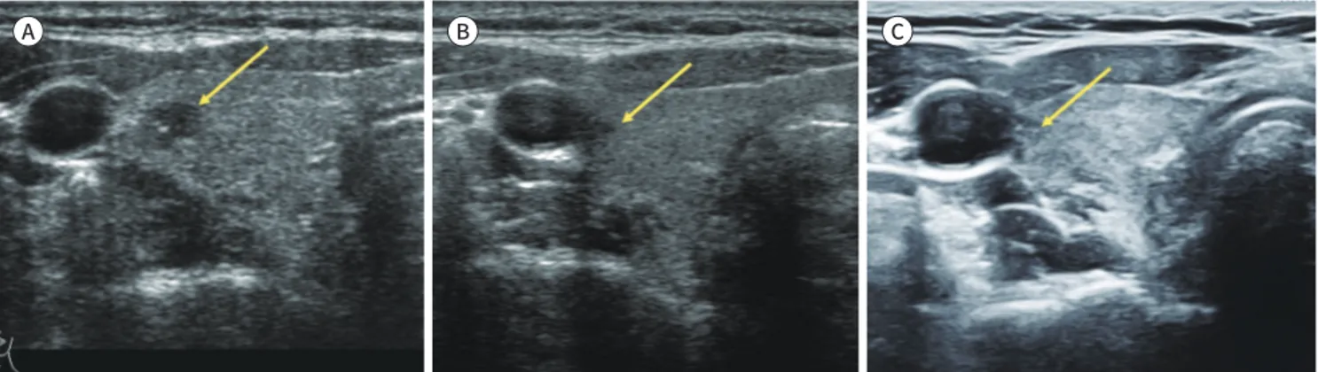
The prognosis for papillary thyroid carcinoma is excellent, and the recent treatment trend is gradually changing from total thyroidectomy to lobectomy (12). However, due to the intrin- sic disadvantages of the operation itself (9), active surveillance has been proposed as the first- line treatment for low-risk PTMC (28, 29). However, patient anxiety due to the presence of a mass limits active surveillance. In a systematic review and meta-analysis by Cho et al. (20), 8.7% to 32% of patients elected for surgery during active surveillance, whereas only 1.1% of the patients underwent surgery after RFA. Active surveillance of low-risk PTMC after thermal Fig. 1. Ultrasonographic images of patient 4.
A-C. Pre-RFA (A), 46 months after the first RFA session (B), and 34 months after the second RFA session (C). Arrow in (A) shows the papillary thyroid microcarcinoma before ablation. Arrows in (B) and (C) show tiny remnant hypoechoic spots near the site of RFA. Histopathological evaluation of a fine-needle aspiration biopsy specimen after obtaining image (B) shows no malignant cells.
RFA = radiofrequency ablation
A B C
Table 3. Follow-Up Results
1 2 3 4 5
Total follow-up period
(months) 159 125 125 123 121
Number of RFA sessions 2 1 1 2 1
Timing of second RFA
(months) 23 Not performed Not performed 51 Not performed
Final result Minimal and
stable scar
Complete disappearance
Complete disappearance
Minimal and stable scar
Complete disappearance Time to reach final status
(months) 14 12 12 27 19
Local recurrence at last
follow-up None None None None None
LN metastasis at last
follow-up None None None None None
Tg level at last follow-up
(ng/mL) 5.6 NA 21.0 NA 16.4
LN = lymph node, NA = not available, RFA = radiofrequency ablation, Tg = thyroglobulin
ablation has been considered effective and safe in several published reports (13, 29, 30).
Zhang et al. (30) reported a prospective 1-year follow-up study after treatment with RFA for papillary thyroid cancer. They found that the malignant lesions disappeared or decreased in volume in 96% of patients, and there was no local recurrence or neck LN metastasis (1, 30).
Kim et al. (25) and Jeong et al. (29) reported excellent and safe outcomes after a follow-up of 4 years in six patients and 19.3 months in nine patients, respectively, after RFA for thyroid cancer.
Recently, thermal ablation has been found to be an effective and safe treatment for pa- tients with low-risk PTMC in studies with extended follow-up periods. Lim et al. (21) per- formed RFA for 152 biopsy-proven PTMC and followed up for an average of 39 ± 25 months.
All nodules were treated, and there were no recurrences or complications. Cho et al. (17) col- lected 84 PTMC cases in the same cohort that were followed for > 5 years and reported that RFA treatment remained effective and safe. Li et al. (31) reported a retrospective review of patients with PTMC treated with microwave ablation (n = 168) and surgery (n = 143) after a follow-up of 824 ± 452 days. A prospective study was conducted by Zhang et al. (32) in pa- tients treated with RFA (n = 94) and surgery (n = 80) after > 5 years of follow-up. In all these meaningful comparative studies, thermal ablation was not inferior to surgery in any respect, such as treatment effects and complications.
However, some researchers disagree with the use of RFA for patients with PTMC. Ma et al. (33) reported that 12 patients showed incomplete treatment after thermal ablation of pap- illary thyroid carcinoma. They claimed that thermal ablation was insufficient and the risk of recurrence and metastasis was high. After careful review of the details of the 12 patients, we found that none of the 12 incompletely treated patients met the inclusion criteria of our and other well-designed studies (20, 30, 31). All 12 patients had multifocal lesions and/or lesions >
1 cm in size (33). On the contrary, we included patients using the strict criteria of maximum diameter ≤ 1 cm, no evidence of gross extra-thyroidal extension, no evidence of LN or distal metastasis, or unifocal carcinoma. Therefore, strict inclusion criteria should be secured to obtain favorable long-term results of RFA after treatment of PTMC (34-36).
Residual hypoechoic scar-like lesions were observed in two cases. Both cases were exam- ined by cytology, and only benign cells were detected. No changes in the shapes of the hy- poechoic lesions were seen one year after the second RFA session, and there were no chang- es in the ultrasonographic findings for > 5 years in both cases. We now think that these findings were post-ablation scars. Kim et al. (25) reported similar findings after RFA for PTMC (one of six cases), with benign findings on histological examination and no ultrasono- graphic changes for > 2 years (14). Lim et al. (21) also reported a small scar-like lesion on fol- low-up ultrasonographic imaging after RFA for recurrent thyroid cancer (18).
The small sample size of 5 patients is the major limitation of this study. The results of long- term studies with more than 10 years of follow-up are expected to prove the effectiveness and safety of RFA treatment for low-risk PTMC.
In conclusion, we suggest that RFA may be an alternative to surgery for patients with low- risk PTMC.
Author Contributions
Conceptualization, all authors; data curation, all authors; formal analysis, S.Y.K., C.S.W., S.J.S.; in- vestigation, S.Y.K., C.S.W., S.J.S.; methodology, all authors; project administration, C.S.W., S.J.S.; re- sources, S.J.S.; software, S.Y.K.; supervision, C.S.W.; validation, all authors; visualization, S.Y.K., S.J.S.;
writing—original draft, S.Y.K.; and writing—review & editing, all authors.
Conflicts of Interest
The authors have no potential conflicts of interest to disclose.
Funding None
REFERENCES
1. Haugen BR, Alexander EK, Bible KC, Doherty GM, Mandel SJ, Nikiforov YE, et al. 2015 American Thyroid As- sociation Management Guidelines for adult patients with thyroid nodules and differentiated thyroid can- cer: the American Thyroid Association Guidelines Task Force on thyroid nodules and differentiated thyroid cancer. Thyroid 2016;26:1-133
2. Liu Y, Su L, Xiao H. Review of factors related to the thyroid cancer epidemic. Int J Endocrinol 2017;2017:
5308635
3. Sakai T, Sugitani I, Ebina A, Fukuoka O, Toda K, Mitani H, et al. Active surveillance for T1bN0M0 papillary thyroid carcinoma. Thyroid 2019;29:59-63
4. Shin JH, Baek JH, Chung J, Ha EJ, Kim JH, Lee YH, et al. Ultrasonography diagnosis and imaging-based management of thyroid nodules: revised Korean Society of Thyroid Radiology Consensus Statement and Recommendations. Korean J Radiol 2016;17:370-395
5. Sanabria A, Kowalski LP, Shah JP, Nixon IJ, Angelos P, Williams MD, et al. Growing incidence of thyroid car- cinoma in recent years: factors underlying overdiagnosis. Head Neck 2018;40:855-866
6. Russ G. Risk stratification of thyroid nodules on ultrasonography with the French TI-RADS: description and reflections. Ultrasonography 2016;35:25-38
7. Chen AY, Jemal A, Ward EM. Increasing incidence of differentiated thyroid cancer in the United States, 1988-2005. Cancer 2009;115:3801-3807
8. Ito Y, Miyauchi A, Inoue H, Fukushima M, Kihara M, Higashiyama T, et al. An observational trial for papillary thyroid microcarcinoma in Japanese patients. World J Surg 2010;34:28-35
9. Oda H, Miyauchi A, Ito Y, Yoshioka K, Nakayama A, Sasai H, et al. Incidences of unfavorable events in the management of low-risk papillary microcarcinoma of the thyroid by active surveillance versus immediate surgery. Thyroid 2016;26:150-155
10. Cho SJ, Suh CH, Baek JH, Chung SR, Choi YJ, Chung KW, et al. Active surveillance for small papillary thyroid cancer: a systematic review and meta-analysis. Thyroid 2019;29:1399-1408
11. Tufano RP, Clayman G, Heller KS, Inabnet WB, Kebebew E, Shaha A, et al. Management of recurrent/persis- tent nodal disease in patients with differentiated thyroid cancer: a critical review of the risks and benefits of surgical intervention versus active surveillance. Thyroid 2015;25:15-27
12. Ahmadi S, Gonzalez JM, Talbott M, Reed SD, Yang JC, Scheri RP, et al. Patient preferences around extent of surgery in low-risk thyroid cancer: a discrete-choice experiment. Thyroid 2020;30:1044-1052
13. Kim JH, Baek JH, Sung JY, Min HS, Kim KW, Hah JH, et al. Radiofrequency ablation of low-risk small papil- lary thyroidcarcinoma: preliminary results for patients ineligible for surgery. Int J Hyperthermia 2017;33:
212-219
14. Sun J, Liu X, Zhang Q, Hong Y, Song B, Teng X, et al. Papillary thyroid carcinoma treated with radiofrequen- cy ablation in a patient with hypertrophic cardiomyopathy: a case report. Korean J Radiol 2016;17:558-561 15. Tong M, Li S, Li Y, Li Y, Feng Y, Che Y. Efficacy and safety of radiofrequency, microwave and laser ablation for
treating papillary thyroid microcarcinoma: a systematic review and meta-analysis. Int J Hyperthermia 2019;36:1278-1286
16. Ito Y, Miyauchi A. Active surveillance of low-risk papillary thyroid microcarcinomas in Japan and other countries: a review. Expert Rev Endocrinol Metab 2020;15:5-12
17. Cho SJ, Baek SM, Lim HK, Lee KD, Son JM, Baek JH. Long-term follow-up results of ultrasound-guided ra- diofrequency ablation for low-risk papillary thyroid microcarcinoma: more than 5-year follow-up for 84 tu- mors. Thyroid 2020;30:1745-1751
18. Bongers PJ, Greenberg CA, Hsiao R, Vermeer M, Vriens MR, Lutke Holzik MF, et al. Differences in long-term quality of life between hemithyroidectomy and total thyroidectomy in patients treated for low-risk differ- entiated thyroid carcinoma. Surgery 2020;167:94-101
19. Choi JY, Lee KE, Chung KW, Kim SW, Choe JH, Koo do H, et al. Endoscopic thyroidectomy via bilateral axil- lo-breast approach (BABA): review of 512 cases in a single institute. Surg Endosc 2012;26:948-955 20. Cho SJ, Baek JH, Chung SR, Choi YJ, Lee JH. Thermal ablation for small papillary thyroid cancer: a system-
atic review. Thyroid 2019;29:1774-1783
21. Lim HK, Cho SJ, Baek JH, Lee KD, Son CW, Son JM, et al. US-guided radiofrequency ablation for low-risk papillary thyroid microcarcinoma: efficacy and safety in a large population. Korean J Radiol 2019;20:1653- 1661
22. Zhou W, Ni X, Xu S, Zhang L, Chen Y, Zhan W. Ultrasound-guided laser ablation versus surgery for solitary papillary thyroid microcarcinoma: a retrospective study. Int J Hyperthermia 2019;36:897-904
23. Choi Y, Jung SL, Bae JS, Lee SH, Jung CK, Jang J, et al. Comparison of efficacy and complications between radiofrequency ablation and repeat surgery in the treatment of locally recurrent thyroid cancers: a single- center propensity score matching study. Int J Hyperthermia 2019;36:359-367
24. Choi Y, Jung SL. Efficacy and safety of thermal ablation techniques for the treatment of primary papillary thyroid microcarcinoma: a systematic review and meta-analysis. Thyroid 2020;30:720-731
25. Kim JH, Baek JH, Lim HK, Ahn HS, Baek SM, Choi YJ, et al. 2017 thyroid radiofrequency ablation guideline:
Korean Society of Thyroid Radiology. Korean J Radiol 2018;19:632-655
26. Park HS, Baek JH, Park AW, Chung SR, Choi YJ, Lee JH. Thyroid radiofrequency ablation: updates on inno- vative devices and techniques. Korean J Radiol 2017;18:615-623
27. Papini E, Guglielmi R, Bizzarri G, Graziano F, Bianchini A, Brufani C, et al. Treatment of benign cold thyroid nodules: a randomized clinical trial of percutaneous laser ablation versus levothyroxine therapy or follow- up. Thyroid 2007;17:229-235
28. Kong SH, Ryu J, Kim MJ, Cho SW, Song YS, Yi KH, et al. Longitudinal assessment of quality of life according to treatment options in low-risk papillary thyroid microcarcinoma patients: active surveillance or immedi- ate surgery (interim analysis of MAeSTro). Thyroid 2019;29:1089-1096
29. Jeong SY, Baek JH, Choi YJ, Chung SR, Sung TY, Kim WG, et al. Radiofrequency ablation of primary thyroid carcinoma: efficacy according to the types of thyroid carcinoma. Int J Hyperthermia 2018;34:611-616 30. Zhang M, Luo Y, Zhang Y, Tang J. Efficacy and safety of ultrasound-guided radiofrequency ablation for
treating low-risk papillary thyroid microcarcinoma: a prospective study. Thyroid 2016;26:1581-1587 31. Li J, Liu Y, Liu J, Yang P, Hu X, Qian L. A comparative study of short-term efficacy and safety for thyroid mi-
cropapillary carcinoma patients after microwave ablation or surgery. Int J Hyperthermia 2019;36:640-646 32. Zhang M, Tufano RP, Russell JO, Zhang Y, Zhang Y, Qiao Z, et al. Ultrasound-guided radiofrequency abla-
tion versus surgery for low-risk papillary thyroid microcarcinoma: results of over 5 years’ follow-up. Thy- roid 2020;30:408-417
33. Ma B, Wei W, Xu W, Wang Y, Guan H, Fan J, et al. Surgical confirmation of incomplete treatment for primary papillary thyroid carcinoma by percutaneous thermal ablation: a retrospective case review and literature review. Thyroid 2018;28:1134-1142
34. Kim EK. Re: papillary thyroid carcinoma treated with radiofrequency ablation in a patient with hypertro- phic cardiomyopathy: a case report. Korean J Radiol 2016;17:965
35. Valcavi R, Piana S, Bortolan GS, Lai R, Barbieri V, Negro R. Ultrasound-guided percutaneous laser ablation of papillary thyroid microcarcinoma: a feasibility study on three cases with pathological and immunohis- tochemical evaluation. Thyroid 2013;23:1578-1582
36. Vaccarella S, Franceschi S, Bray F, Wild CP, Plummer M, Dal Maso L. Worldwide thyroid-cancer epidemic?
The increasing impact of overdiagnosis. N Engl J Med 2016;375:614-617
갑상선 미세유두암의 고주파 절제술 후 10년 경과 관찰
서유경1 · 조성휘1* · 심정석2 · 양고은1 · 조우진3
목적 이 연구는 갑상선 미세유두암을 고주파 절제술로 치료한 후 10년 이상 경과 관찰한 환 자들의 결과를 통해 효과와 안전성을 평가하고자 했다.
대상과 방법 2006년 11월부터 2009년 12월까지 갑상선 미세유두암을 고주파 절제술로 치료 받은 환자 5명을 대상으로 하였다. 이 환자들은 모두 조직검사를 통해 갑상선 미세유두암으 로 진단을 받았고, 병변은 갑상선 내에 국한되어 있었으며, 전이의 증거가 없었고, 수술이나 전신마취가 의학적으로 부적합하거나 수술을 거부한 환자들이었다. 고주파 절제술은 고주 파 발생기와 냉각기를 사용하여 한 명의 영상의학과 의사가 시행하였다. 우리는 시술과 연관 된 부작용, 소작된 종양의 변화, 재발 여부, 국소 또는 림프절 전이 등에 대해 초음파 영상 소 견과 의무 기록을 토대로 분석하였다.
결과 평균 경과 관찰기간은 130.6개월(범위, 121~159개월) 이었다. 세 명의 환자는 한 번의 고 주파 절제술을, 두 명의 환자는 두 번의 시술을 받았다. 다섯 명의 환자 모두 시술과 연관된 부작용은 보이지 않았다. 다섯 개 중 세 개의 종양은 시술 후 완전히 사라졌으며 두 개의 종 양은 최소한의 흔적으로 남아 평균 16.8개월의 경과 관찰기간 동안 큰 변화가 없었다. 가장 최근의 경과관찰에서 다섯 명의 환자 모두 국소 전이나 림프절 전이는 보이지 않았고 갑상선 글로불린(thyroglobulin)의 수치도 정상 범위였다.
결론 고주파 절제술은 수술에 부적합하거나 수술을 거부하는 저위험 갑상선 미세유두암 환 자들에 대해서 효과적이고 안전한 치료법이 될 수 있다.
1강원대학교병원 영상의학과, 위드심의원 2영상의학과, 3이비인후과