Fibrocartilaginous Intramedullary Bone Forming Tumor of the Distal Femur Mimicking Osteosarcoma
Fibrocartilaginous dysplasia (FCD) has occasionally led to a misdiagnosis and wrong decision which can significantly alter the outcome of the patients. A 9-yr-old boy presented with pain on his left distal thigh for 6 months without any trauma history. Initial radiographs showed moth eaten both osteolytic and osteosclerotic lesions and biopsy findings showed that the lesion revealed many irregular shaped and sclerotic mature and immature bony trabeculae. Initial diagnostic suggestions were varied from the conventional osteosarcoma to low grade central osteosarcoma or benign intramedullary bone forming lesion, but close observation was done. This study demonstrated a case of unusual fibrocartilaginous intramedullary bone forming tumor mimicking osteosarcoma, so that possible misdiagnosis might be made and unnecessary extensive surgical treatment could be performed. In conclusion, the role of orthopaedic oncologist as a decision maker is very important when the diagnosis is uncertain.
Key Words: Fibrocartilaginous Dysplasia; Osteosarcoma; Orthopaedic Oncologist;
Diagnosis Sang-Heon Song,1 Hanna Lee,2
Hae-Ryong Song,2 Myo-Jong Kim,3 and Jong-Hoon Park3
1Department of Orthopaedic Surgery, Soonchunhyang University Bucheon Hospital, Bucheon; 2Institute for rare diseases and Department of Orthopaedic Surgery, Korea University Medical Center, Guro Hospital, Seoul;
3Department of Orthopaedic Surgery, Korea University Medical Center, Anam Hospital, Seoul, Korea
Received: 11 September 2012 Accepted: 11 January 2013 Address for Correspondence:
Jong-Hoon Park, MD
Department of Orthopaedic Surgery, Korea University Medical Center, Anam Hospital, 145 Anam-ro, Seongbuk-gu, Seoul 136-705, Korea
Tel: +82.2-920-6609, Fax: +82.2-924-2471 E-mail: pjh1964@hanmail.net
This study was supported by a grant from the Korea Healthcare Technology R&D Project, Ministry for Health, Welfare & Family Affairs, Republic of Korea (A110416).
http://dx.doi.org/10.3346/jkms.2013.28.4.631 • J Korean Med Sci 2013; 28: 631-635
INTRODUCTION
Osteosarcoma is the most common malignant tumor of the bone (1). The population affected is predominantly children, teenagers, and young adults aged 10-30 yr (2). In most case, typical radiographic features clearly demonstrate the aggressive bone forming nature of the lesion, with long-bone metaphyseal location, mixed areas of lysis and sclerosis, cortical destruction, various periosteal reactions and abundant extraskeletal soft tis- sue mass (3, 4).
However, there can be a diagnostic dilemma. Kumar et al. (5) reported a rare form of low grade central osteosarcoma which shares some radiological and histopathological resemblance with fibrous dysplasia (FD). They warned that misdiagnosis of low grade central osteosarcoma as benign lesion may lead in- adequate treatment resulting in a more malignant recurrent bone tumor. In the contrary, some cases of aggressive benign bone forming tumors including fibrocartilaginous dysplasia (FCD) or fibrocartilaginous mesenchymoma which resemble with low grade central osteosarcoma or chondrosarcoma aris- ing in FD (5-8) has occasionally led to a misdiagnosis and wrong decision which can significantly alter the outcome of the patients.
In this study, we present the radiologic and pathologic fea- tures of a fibrocartilaginous intramedullary bone forming tu- mor of the distal femur mimicking osteosarcoma, so that possi- ble misdiagnosis might be made and unnecessary extensive surgical treatment could be performed.
CASE DESCRIPTION
A 9-yr-old boy presented with pain on his left distal thigh for 6 months without any trauma history (date of initial consultation:
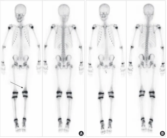
2009-12-31). On physical examination, no palpable mass, no tenderness was identified and the range of motion was full. A plain radiograph was taken (Fig. 1A, B) and it showed moth eaten both osteolytic and osteosclerotic lesions in the left distal femoral metaphysis. But there was no periosteal reaction or no cortical destruction. Magnetic resonance imaging (MRI) showed T1 low and T2 heterogenous signal intensity intramedullary le- sion in the metaphysis which had not crossed the growth plate and no definite cortical destruction was also identified (Fig. 1C, D, E, F). A radiologist reported that this case was consistent with an osteosarcoma but the possibility of benign lesion could not be exclud ed. A whole body bone scan (Fig. 2A) showed
Fig. 1. Initial anterior-posterior and lateral plain radiographs show moth eaten both osteolytic and osteosclerotic lesions in the metaphysis without definite sign of periosteal re- action or cortical destruction. (A) anterior-posterior and (B) lateral radiograph at initial visit. Magnetic resonance imaging showed T1 low and T2 heterogenous signal intensity intramedullary lesion in the metaphysis which had not crossed the growth plate and no definite cortical destruction was also identified. (C) T1-axial cut, (D) T2-axial cut, (E) T1- sagittal cut and (F) T2-coronal cut. AXI, axial; FS, fat suppression; SAG, sagittal; COR, coronal.
A B E
C
F T1AXI (FS) D
T1SAG
T2AXI
T2COR
A B
Fig. 2. Initial whole body bone scan showed mild increased uptake in the left distal femur. Whole body bone scan at the time of 36 months after the initial visit shows no defi- nite interval changes of extent and morphology. (A) whole body bone scan at initial presentation and (B) whole body bone scan at 36 months after the initial visit.
mild increased uptake in the left distal femur. Complete blood count, erythrocyte sedimentation rate, C-reactive protein level and blood che mistry were all within normal limits. An incision- al biopsy was performed in the belief of possible malignancy.
Pathologic specimen (Fig. 3) showed that the lesion revealed many irregular shaped and sclerotic mature and immature bony trabeculae with moderately cellular stroma composed of relatively uniform spindle cells and cartilaginous components without significant cellular atypia. A “diagnosis” was not easily
made because of conflicting debates for the diagnosis were is- sued from the conventional osteosarcoma to low grade central osteosarcoma or benign intramedullary bone forming lesion after case conference discussion and consultations with foreign pathologic specialists. We informed all of the findings to his parents and decided to have a close observation without any further invasive procedures or surgical treatments, because the boy had a just minimal symptom of intermittent pain and the parents agreed.
A B
Fig. 3. Inicisional biopsy was done and pathologic specimen showed irregular shaped and scletoric mature and immature bony trabeculae with relatively uniform spindle cells and cartilaginous components without significant cellular atypia. Hematoxylin and eosin stained (A) low- (original magnification, ×400) and (B) high- power (original magnifica- tion, ×1,000)
Fig. 4. Plain radiograph at the time of 36 months after the initial visit shows no definite interval changes of extent and morphology. (A) anterior-posterior and (B) lateral radio- graph at the time of 36 months after the initial visit. Magnetic resonance imaging at the time of 36 months after the initial visit shows no definite interval changes of extent and morphology. (C) T1-axial cut, (D) T2-axial cut, (E) T1-sagittal cut and (F) T2-coronal cut. AXI, axial; SAG, sagittal; COR, coronal.
A B E
C
F D T1AXI
T1SAG
T2AXI
T2COR
Thirty-six months after initial visit, radiologic findings from the plain radiographs (Fig. 4A, B) and MRI findings (Fig. 4C, D, E, F) showed no significant interval changes of extent and mor- phology. A whole body bone scan showed mild decreased up- take in comparison to the initial scan (Fig. 2B). Now he is 12-yr- old and clinically he is pain free and has no restriction of any activities until now. There is no sign of growth disturbance. He will be followed annually.
DISCUSSION
Generally, it is widely recognized that a small number of osteo- sarcoma cases are considerably difficult to interpret radiogra- phically. According to Rosenberg et al. (3), the subtle, rare, and misleading plain film features of some types of bone tumors, such as osteosarcomas simulating osteoblastomas, intracortical osteosarcomas, etc., may produce a confusing radiologic pic- ture. They reported that several factors contributed to the mis- leading radiolographic patterns of osteosarcomas. These in- cluded histological low-grade, lytic, or minimally sclerotic le- sions, early detection, and confinement to the intramedullary canal, benign-appearing periosteal reactions, and rare intraos- seous locations. On the contrary, some of benign bone tumor can be misdiagnosed as osteosarcoma or malignancy. A case in this study showed radiologic and pathologic findings which might be shown as intramedullary osteosarcoma. Initial plain radiograph showed moth eaten osteolytic and osteosclerotic le- sions in distal femur and MRI showed heterogenous signal in- tensity lesion which can be thought as malignant lesion, so ra- diologist warned the possibility of osteosarcoma. Also patho- logic findings showed many irregular shaped and sclerotic ma- ture and immature bony trabeculae with moderately cellular stroma composed of spindle cells and cartilaginous compo- nents, so that pathologist reported similar warning as radiolo- gist. However, the decision was made by the orthopaedic on- cologist by the reason of no definite periosteal reaction, no defi- nite cellular atypia, minimally increased uptake in bone scan and mainly due to that massive surgical procedure like wide ex- cision and limb salvage surgery using tumor prosthesis would cause irreversible damage to 9-yr-old patient. Finally the clinical situations over than 36 months of close observation showed that this case could be diagnosed as benign fibrocartilaginous bone forming tumor.
Fibrous dysplasia (FD), a dysplastic process of the bone form- ing mesenchyme, is histologically characterized by a benign appearing spindle cell fibrous stroma containing scattered, ir- regularly shaped trabeculae of immature woven bone, lacking osteoblasts, that appear directly from the stroma (9-11). Occa- sionally, nodules of cartilage can be present in cases of polyos- totic or monostotic forms of FD (6, 9, 12, 13). The term “fibro- cartilaginous dysplasia” has been applied by some authors for
cases that exhibit abundant cartilage (6, 7, 9, 14, 15). Fibrocarti- laginous dysplasia is a variant of fibrous dysplasia in which car- tilaginous differentiation is identified. FCD occurs in the lower extremities, especially in the proximal lesion of the femur. Ra- diologically, FCD is similar to conventional FD, the lesion is well demarcated. Cortical expansion can be seen, however, the cortex is always intact. The differential diagnosis of FCD include:
enchondroma, chondrosarcoma and well-differentiated intra- medullary osteosarcoma and FCD has occasionally led to a misdiagnosis of chondrosarcoma arising in FD or low grade central osteosarcoma (5-8). The distinction between fibrocarti- laginous bone forming tumor and other benign and malignant cartilaginous tumors is critical in the management of these pa- tients (9, 10). Our case showed somewhat similar but more ag- gressive initial radiologic findings than previously reported cas- es of FCD. Bone scintigraphy (BS) using technetium-99m meth- ylene disphosphate (MDP), had been regarded as sufficiently accurate and adequate for detecting malignant bone tumors.
However, one of the drawbacks of the BS is its insensitivity in detecting some cases, which are located intramedullary with- out cortical bone involvement (16). Our case also showed intra- medullary lesion without definite cortical involvement, so that BS showed minimally increased uptake. We can consider that this finding may not be used as a differential point between malignant bone tumor and benign tumor in this case.
In conclusion, this study demonstrated a case of unusual ag- gressive nature of fibrocartilaginous intramedullary bone form- ing tumor mimicking osteosarcoma, so that possible misdiag- nosis might be made and unnecessary extensive surgical treat- ment could be performed. It is suggested that the role of ortho- paedic oncologist as a decision maker is very important in clini- cal situation, especially when the diagnosis is uncertain.
ACKNOWLEDGMENTS
We wish to thank Dr. Scott Nelson for his excellent pathologic comment about this case.
REFERENCES
1. Meyers PA, Gorlick R. Osteosarcoma. Pediatr Clin North Am 1997; 44:
973-89.
2. Savitskaya YA, Rico-Martínez G, Linares-González LM, Delgado-Cedil- lo EA, Téllez-Gastelum R, Alfaro-Rodríguez AB, Redón-Tavera A, Ibar- ra-Ponce de León JC. Serum tumor markers in pediatric osteosarcoma:
a summary review. Clin Sarcoma Res 2012; 2: 9.
3. Rosenberg ZS, Lev S, Schmahmann S, Steiner GC, Beltran J, Present D.
Osteosarcoma: subtle, rare, and misleading plain film features. AJR Am J Roentgenol 1995; 165: 1209-14.
4. Campanacci M, Cervellati G. Osteosarcoma: a review of 345 cases. Ital J Orthop Traumatol 1975; 1: 5-22.
5. Kumar A, Varshney MK, Khan SA, Rastogi S, Safaya R. Low grade cen-
tral osteosarcoma: a diagnostic dilemma. Joint Bone Spine 2008; 75:
613-5.
6. Pelzmann KS, Nagel DZ, Salyer WR. Case report 114. Skeletal Radiol 1980; 5: 116-8.
7. Ishida T, Dorfman HD. Massive chondroid differentiation in fibrous dys- plasia of bone (fibrocartilaginous dysplasia). Am J Surg Pathol 1993; 17:
924-30.
8. De Smet AA, Travers H, Neff JR. Chondrosarcoma occurring in a pa- tient with polyostotic fibrous dysplasia. Skeletal Radiol 1981; 7: 197-201.
9. Reigel DH, Larson SJ, Sances A Jr, Hoffman NE, Switala KJ. A gastric acid inhibitor of cerebral origin. Surgery 1971; 70: 161-8.
10. Kyriakos M, McDonald DJ, Sundaram M. Fibrous dysplasia with carti- laginous differentiation (“fibrocartilaginous dysplasia”): a review, with an illustrative case followed for 18 years. Skeletal Radiol 2004; 33: 51-62.
11. Bianco P, Riminucci M, Majolagbe A, Kuznetsov SA, Collins MT, Mank- ani MH, Corsi A, Bone HG, Wientroub S, Spiegel AM, et al. Mutations
of the GNAS1 gene, stromal cell dysfunction, and osteomalacic changes in non-McCune-Albright fibrous dysplasia of bone. J Bone Miner Res 2000; 15: 120-8.
12. Sanerkin NG, Watt I. Enchondromata with annular calcification in as- sociation with fibrous dysplasia. Br J Radiol 1981; 54: 1027-33.
13. Harris WH, Dudley HR Jr, Barry RJ. The natural history of fibrous dys- plasia. An orthopaedic, pathological, and roentgenographic study. J Bone Joint Surg Am 1962; 44-A: 207-33.
14. Hermann G, Klein M, Abdelwahab IF, Kenan S. Fibrocartilaginous dys- plasia. Skeletal Radiol 1996; 25: 509-11.
15. Drolshagen LF, Reynolds WA, Marcus NW. Fibrocartilaginous dyspla- sia of bone. Radiology 1985; 156: 32.
16. Taoka T, Mayr NA, Lee HJ, Yuh WT, Simonson TM, Rezai K, Berbaum KS. Factors influencing visualization of vertebral metastases on MR im- aging versus bone scintigraphy. AJR Am J Roentgenol 2001; 176: 1525-30.

