Head and neck infection initially spreads to oral vesti- bule or myofascial space. However, if such initial infection progresses and spreads near the pharynx, airway obstruc- tion may occur and it can cause death in serious cases. If infection spreads to the carotid sheath, the damaged vessel may cause bleeding or nerve disorder.1,2Contrast-enhanced computed tomography in an appropriate phase helps the diagnosis of the infection and its spreading route, leading to successful treatment. If infection spreads to the retro- pharyngeal space, it progresses quickly through the supe- rior and posterior to the mediatinum causing severe com- plications and the progress may continue on to the prever- tebral space, diaphragm, and thorax. Even with antibiotics and surgical treatment, mediastinitis is a severe compli- cation with 35-50% of fatality rate. Also, if the infection spreads in the superficial level, necrotizing fasciitis may occur and it can cause gangrenes on the skin and its fol- lowing complications such as systemic shock. Therefore, the early diagnosis of infection and appropriate treatment
is very important in head and neck infections.
In this report, we introduce a patient with extensive chro- nic mandibular osteomyelitis with necrotizing fasciitis and airway obstruction by deep neck infection in the lateral pharyngeal space, subscapular space, and carotid sheath, who was successfully treated by early diagnosis, continu- ous incision and drainage, active antibiotic treatment and follow up dressing through the appropriate use of radiogra- phic images, along with a review of literature.
Case Report
A 77-year-old female visited our clinic with a chief com- plaint of painful swelling in the submental area which had begun 2 days ago. The patient had been treated for pulmo- nary edema caused by asthma and aspiration pneumonia from 7 years ago, and she was currently being treated for pyelitis, cerebral infarction, dementia and herpes zoster.
Clinical examination showed several root rests, gingival swelling on the entire dentition, redness and pus discharge in the upper and lower anterior teeth, induration, redness, and tenderness to palpation on the submental area. Also, bilateral bone exposure on the lingual mandible suggested osteomyelitis. Trismus, dysphasia and dyspnea were not
Application of radiographic images in diagnosis and treatment of deep neck infections with necrotizing fasciitis: a case report
Young-Joo Kim, Ju-dong Kim, Hye-In Ryu, Yeon-Hee Cho, Jun-Ha Kong, Joo-Young Ohe, Yong-Dae Kwon, Byung-Joon Choi, Gyu-Tae Kim*
Department of Oral and Maxillofacial Surgery, Dental School, Kyung Hee University, Seoul, Korea
*Department of Oral and Maxillofacial Radiology, Dental School, Kyung Hee University, Seoul, Korea ABSTRACT
The advent and wide use of antibiotics have decreased the incidence of deep neck infection. When a deep neck infec- tion does occur, however, it can be the cause of significant morbidity and death, resulting in airway obstruction, mediastinitis, pericarditis, epidural abscesses, and major vessel erosion. In our clinic, a patient with diffuse chronic osteomyelitis of mandible and fascial space abscess and necrotic fasciitis due to odontogenic infection at the time of first visit came. We successfully treated the patient by early diagnosis using contrast-enhanced CT and follow up dressing through the appropriate use of radiographic images. (Imaging Sci Dent 2011; 41 : 189-93)
KEY WORDS : Abscess; Fasciitis, Necrotizing; Osteomyelitis; Tomography, X-Ray, Computed
Received July 30, 2011; Revised August 25, 2011; Accepted August 30, 2011 Correspondence to : Prof. Gyu-Tae Kim
Department of Oral and Maxillofacial Radiology, Dental School, Kyung Hee Univer- sity, Hoegi-dong, Dongdaemun-gu, Seoul 130-701, Korea
Tel) 82-2-958-9406, Fax) 82-2-965-1256, E-mail) latinum.omfr@khu.ac.kr
Copyright ⓒ 2011 by Korean Academy of Oral and Maxillofacial Radiology
This is an Open Access article distributed under the terms of the Creative Commons Attribution Non-Commercial License (http://creativecommons.org/licenses/by-nc/3.0) which permits unrestricted non-commercial use, distribution, and reproduction in any medium, provided the original work is properly cited.
Imaging Science in Dentistry∙pISSN 2233-7822 eISSN 2233-7830
found.
On the initial visit, clinical and radiographic examina- tions including panoramic radiograph were carried out. On the panoramic radiograph, several dental caries were ob- served, as well as periapical abscess on the right maxillary lateral incisor and right maxillary first molar. Extraction sockets were also observed. Ill-defined bony destructive change on the left mandibular canine area with sclerotic change of surrounding trabecular bone was found (Fig. 1).
The patient was tentatively diagnosed with submental space abscess. Incision and drainage was carried out under local anesthesia and a drain was inserted lingually, how-
ever pus drainage was poor.
On the next day, dysphasia and dyspnea were intensifi- ed. Soft tissue neck anteroposterior view and oxygen satu- ration was examined. On the radiographic image, the air- way was severely narrowed (Fig. 2). The oxygen satura- tion was 89%. Immediate endotracheal intubation was car- ried out and the oxygen saturation was stabilized at 96%, with no other problem on vital sign. Blood test revealed high inflammation values with white blood cell at 18,110 /mm3and C-reactive protein at 33.23 mg/dL. Infusion and antibiotic treatment (amoxicillin-potassium clavulanate, isepamicin sulfate, and metronidazole) was carried out.
Contrast-enhanced CT images showed non-enhanced low attenuated region with the irregular periphery of non-homo- geneous enhancement on the right submandibular space (Fig. 3). Also, these findings were found similarly in the bilateral submental and the left submandibular spaces.
There was enhanced soft tissue swelling in the nasophar- ynx, oropharynx, and the inferior pharynx. The airway was deviated to the left and had decreased diameter. Extraoral incision and drainage on the submental area was immedi- ately carried out under general anesthesia. Necrotizing fasciitis was suspected, and the necrotized area was remov- ed. Drains were inserted in bilateral submandibular and submental area. The patient was transferred to the inten- sive care unit without removing the endotracheal tube.
After surgery, saline with povidone-iodine irrigation in the drain insertion area was carried out twice a day. The necrotizing area in the submental region was increased.
Blood test revealed that there was no difference in the in- flammation level. However, the follow-up contrast-enhanc- ed CT revealed that pus had spread widely to the posterior area of the right sternocleidomastoid muscle, retropharyn- geal space, parapharyngeal space, hyoid bone area, and
Fig. 1.Panoramic radiograph shows ill-defined bone destruction on left mandibular canine area. The lesion has ragged border and shows scle- rotic change on the surrounding tra- becular bone.
Fig. 2. Soft tissue neck AP view shows that the airway is severely narrowed by soft tissue (arrow).
prevertebral space (Fig. 4). Due to the increasing necrotiz- ing area and the deep spread of the infection, the previous drains were removed and further submental necrotized area was excised under general anesthesia. An incision was made 6 cm below the anterior border of the right sternoclei- domastoid muscle, and the pus formation space was reach- ed and irrigated. Drains were inserted from the superior border of the left clavicle to the left mandibular border, from the submental area to intraoral area and to the right mandible border, and from the anterior border of the right sternocleidomastoid muscle to the interclavicular area.
Intraoral drains were inserted in the right submandibular area and in the right pterygomandibular space.
After the second surgery, saline with povidone-iodine irrigation in the drain insertion areas was continued twice a day. Another follow-up contrast-enhanced CT showed reduced pus formation in the submental area, right para- pharyngeal space, and retropharyngeal space. Blood test
resulted in decreased inflammation, however the edema in the airway and the pus formation around the hyoid bone were still found. Third surgery was carried out under gen- eral anesthesia. The drain in the right sternocleidomastoid muscle area was removed. An incision was made on the hyoid bone area. When the hyoid bone was completely reached, a small amount of pus discharge was observed.
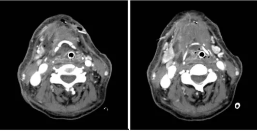
After vigorous irrigation, additional drains were inserted in the right sternocleidomastoid muscle incision area in the direction of the hyoid bone, the interclavicular area and above the right clavicle. After surgery, the drains were irri- gated with saline mixed with antibiotics (Ceftriaxone 1 g, Hanmi Pharm, Seoul, Korea) twice a day. The other fol- low-up contrast-enhanced CT (Fig. 5) showed that pus for- mation in the right submandibular and submental area had almost completely disappeared, as well as the parapha- ryngeal and the retropharyngeal space. The edema in the airway was also reduced, therefore the endotracheal tube
Fig. 3.Preoperative contrast-enhanced CT images show submandibular and sublingual abscess formation. Inflammation of subcutaneous fat is found on submental and neck area.
Fig. 4.Contrast-enhanced CT ima- ges at two days after first operation show the increased extent of right submandibular abscess to preverte- bral space and posterior to the right sternocleidomastoid muscle, and cellulitis and myositis of left sub- mental and submandibular.
was removed. The patient state was stabilized and she was transferred to the general ward.
The number of the drains was reduced as the patient’s state improved. Blood test revealed decreasing inflamma- tion state. The frequency of irrigation on the drain area reduced to once a day. Appointment for treatment plans regarding the mandibular osteomyelitis that was observed on the follow-up CT was scheduled.
Discussion
Among odontogenic infections that lead to deep space abscess, mediastinitis and necrotizing fasciitis of the neck are known to be secondary complications which can be life threatening. Patients with odontogenic infection who require incision and drainage in the oral maxillofacial re- gion are normally affected with more than one myofascial space. Generally, submandibular, submental, and parapha- ryngeal space are affected.3When head and neck infection spreads to the fascia and the subcutaneous tissue, necrotiz- ing fasciitis may occur and sometimes follow a deeper infection path. The more caudal the infection spreads, the more likely the severe complications such as mediastinitis may occur.
Necrotizing fasciitis of head and neck is caused by infec- tion of several fatal bacteria. Infection spreads along the fascia and affects the subcutaneous tissues, myofascia, skin and muscles.4 This type of infection is very rare in head and neck region and odontogenic infection causes it even rarer. If immediate treatment is not applied, the infection spreads quickly and becomes fatal. The fatality rate was
reported to be 20-75%.5Deep neck infection can also arise from infections of the teeth, glands, and neck lymph nodes, however odontogenic causes are few.6 Such infections spread to the parapharyngeal and retropharyngeal space to obstruct the airway, or take an upper path to form brain abscess, or when it spreads along the carotid sheath, fatal long-term complications such as bleeding due to vessel damage may occur. Bonapart et al7 stated that the main routes of neck infection spreading to the mediastinum were the prevertebral space, which connected to the ante- rior part of the mediastinum, and the retropharyngeal and viscerovascular space, which lead to the posterior part of the mediastinum and the diaphragm. In this report, the infection had spread to the parapharyngeal space and the prevertebral space, but not to the mediastinum.
For evaluation of deep neck space infection, ultrasound, MRI, and radiographic examinations such as soft tissue neck X-ray, CT may be useful. The soft tissue neck X-ray is the most convenient method, and it is especially useful for evaluating the retropharyngeal space and the preverte- bral space since it allows the measurement between the anterior part of the vertebrae and the posterior wall of the pharynx. In the present case, when the patient was display- ing dyspnea, the soft tissue neck anteroposterior view al- lowed the evaluation of the airway obstruction and its fol- lowing treatment. However, using the soft tissue neck X- ray alone for the diagnosis, gives 33% of false negative rate, meaning that other imaging evaluation method is re- quired for a more accurate clinical diagnosis.8
Ultrasonography has been used to detect abscess cavi- ties, although the anatomic detail provided by CT makes it superior in confirming the presence of an abscess.9MRI can provide more detailed anatomic structures, and can detect infected spaces which have been indefinable on CT images. However, the high cost, time for image processing, and the cooperation required from the patients are the dis- advantages.10
Contrast-enhanced CT images clearly delineate the posi- tion and size of the lesion as well as its relationship with the adjacent anatomic structures. Also, it is useful in eval- uating any changes in the upper airway in patients with trismus or with parapharyngeal or retropharyngeal space abscess. The images provide valuable information in decid- ing the method for maintaining the airway such as endo- tracheal intubation, cricothryoidotomy, or tracheostomy.11 On CT images, the attenuation level of space abscess is between that of water and soft tissues. When a contrast medium is used, a rim enhancement around the abscess area is observed. Swelling of the adjacent muscles signifies
Fig. 5. Contrast-enhanced CT image at 10 days after third opera- tion shows that the abscess in the submandibular and sublingual space is almost dissolved.
the spreading of inflammation in the surrounding area.12 In the present case, the contrast-enhanced CT was used to identify the spreading of pus into deeper spaces after ini- tial treatment, and for guiding extensive incision and dra- inage under general anesthesia to prevent further expan- sion. Also, follow-up CTs were used to evaluate the edema of the airway and to determine the timing for endotracheal extubation.
For effective treatment of deep space abscess, early accu- rate diagnosis and its appropriate treatment mandatory, and for necrotizing fasciitis, absolute understanding of the ana- tomical structures in the infection area and active surgical treatment along with intensive antibiotic treatment is nec- essary.
In our case, since the patient state was in severe, the ur- gent infection was treated with antibiotics for a long term, and sequestrectomy and decortication of the exposed bone in the oral cavity was sufficient for satisfying results ins- tead of extensive mandibulectomy.
In conclusion, early diagnosis and treatment is important for a good prognosis of deep neck infection. When the infection spreads to the deep neck area, especially contrast- enhanced CT would be useful to observe every fascial space and to locate abscesses effectively in the head and neck area.
References
1. Wolf H, Rusan M, Lambertsen K, Ovesen T. Necrotizing fas- ciitis of the head and neck. Head Neck 2010; 32 : 1592-6.
2. Reynolds SC, Chow AW. Life-threatening infections of the peripharyngeal and deep fascial spaces of the head and neck.
Infect Dis Clin North Am 2007; 21 : 557-76
3. Zeitoun IM, Dhanarajani PJ. Cervical cellulitis and mediastini- tis caused by odontogenic infections: report of two cases and review of literature. J Oral Maxillofac Surg1995; 53 : 203-8.
4. Whitesides L, Cotto-Cumba C, Myers RA. Cervical necrotiz- ing fasciitis of odontogenic origin: a case report and review of 12 cases. J Oral Maxillofac Surg 2000; 58 : 144-51.
5. Bahu SJ, Shibuya TY, Meleca RJ, Mathog RH, Yoo GH, Sta- chler RJ, et al. Craniocervical necrotizing fasciitis: an 11-year experience. Otolaryngol Head Neck Surg 2001; 125 : 245-52.
6. Sakaguchi M, Sato S, Ishiyama T, Katsuno S, Taguchi K. Cha- racterization and management of deep neck infections. Int J Oral Maxillofac Surg 1997; 26 : 131-4.
7. Bonapart IE, Stevens HP, Kerver AJ, Rietveld AP. Rare com- plications of an odontogenic abscess: mediastinitis, thoracic empyema and cardiac tamponade. J Oral Maxillofac Surg 1995; 53 : 610-3.
8. Rosenthal M, Oreadi D, Kraus J, Bedi H, Stark PC, Shastri K.
Comparison of preoperative computed tomography and surgi- cal findings in maxillofacial infections. J Oral Maxillofac Surg 2011; 69 : 1651-6.
9. Hilbert G, Vargas F, Valentino R, Gruson D, Chene G, Bébéar C, et al. Comparison of B-mode ultrasound and computed tomography in the diagnosis of maxillary sinusitis in mechani- cally ventilated patients. Crit Care Med 2001; 29 : 1337-42.
10. Munoz A, Castillo M, Melchor MA, Gutiérrez R. Acute neck~ infections: prospective comparison between CT and MRI in 47 patients. J Comput Assist Tomogr 2001; 25 : 733-41.
11. Flynn TR. The swollen face. Severe odontogenic infections.
Emerg Med Clin North Am 2000; 18 : 481-519.
12. Yonetsu K, Izumi M, Nakamura T. Deep facial infections of odontogenic origin: CT assessment of pathways of space invol- vement. AJNR Am J Neuroradiol 1998; 19 : 123-8.