Postoperative renal functional changes assessed by 99m Tc-DTPA scintigraphy and predictive factors after miniaturized percutaneous nephrolithotomy and retrograde intrarenal surgery: An
observational 1-year follow-up study
Jae Hyun Jung1 , Sangjun Yoo2 , Juhyun Park2 , Min Chul Cho2 , Hwancheol Son2 , Hyeon Jeong2 , Seung Hoon Ryang1 , Sung Yong Cho1,2
1Department of Urology, Seoul National University Hospital, Seoul, 2Department of Urology, SMG-SNU Boramae Medical Center, Seoul, Korea
Purpose: We evaluated the comparative effect of miniaturized percutaneous nephrolithotomy (mini-PCNL) and retrograde intra- renal surgery (RIRS) on perioperative kidney function by use of diethylenetriamine penta-acetic acid (99mTc-DTPA) scintigraphy and identified significant predictors associated with deterioration or amelioration of renal function after surgery.
Materials and Methods: All 70 patients who underwent mini-PCNL or RIRS between 2012 and 2016 were monitored by 99mTc- DTPA scintigraphy preoperatively. Patients with abnormal renal function were monitored from 3 to 12 months postoperatively.
Logistic regression analyses were conducted to estimate the predictors of aggravated renal dysfunction and improvement.
Results: The difference in preoperative renal function between the contralateral and the operative side was >10% in 57 patients (81.4%). Among those in the group with abnormal renal function, 40 (70.2%), 10 (17.5%), and 7 (12.3%) patients showed stability, deterioration, and improvement in renal function at postoperative year 1, respectively. Functional changes did not differ accord- ing to the type of surgery. A high level of serum creatinine preoperatively (p=0.060) and a history of previous stone procedures (p=0.051) showed borderline significance for prediction of deterioration in renal function.
Conclusions: RIRS and mini-PCNL had similar effects and favorable outcomes on renal function during a 1-year follow-up period.
High baseline serum creatinine levels and a history of procedures warrant careful attention.
Keywords: Kidney calculi; Kidney function tests; Minimally invasive surgical procedures; Urinary calculi
This is an Open Access article distributed under the terms of the Creative Commons Attribution Non-Commercial License (http://creativecommons.org/licenses/by-nc/4.0) which permits unrestricted non-commercial use, distribution, and reproduction in any medium, provided the original work is properly cited.
Received: 15 July, 2019 • Accepted: 2 October, 2019
Corresponding Author: Sung Yong Cho https://orcid.org/0000-0001-9271-6951
Department of Urology, Seoul National University Hospital, 101 Daehak-ro, Jongno-gu, Seoul 03080, Korea TEL: +82-2-2072-2428, FAX: +82-2-742-4665, E-mail: [email protected]
ⓒ The Korean Urological Association
www.icurology.org
Investig Clin Urol 2020;61:59-66.
https://doi.org/10.4111/icu.2020.61.1.59 pISSN 2466-0493 • eISSN 2466-054X
INTRODUCTION
Active treatment of renal stones is indicated for stones causing urinary obstructions and hematuria or uncontrolled
pain, for growing stones, for stones persisting longer than 2 years, and in patients at high risk for stone formation or urinary tract infection [1-3]. However, these indications have not yet considered the assessment of renal function. In pa-
tients with kidney stones, the risks for renal insufficiency are multifactorial and include multiple stone episodes, co- morbidity, urinary tract infection, and urinary obstruction [1,2,4,5]. Information pertaining to renal function facilitates appropriate treatment decisions and the timing of manage- ment in patients diagnosed with nephrolithiasis and at high risk for renal insufficiency.
Recently, minimally invasive procedures such as retro- grade intrarenal surgery (RIRS) and miniaturized percu- taneous nephrolithotomy (mini-PCNL) have been reported as feasible and safe options for various kidney stones [6,7].
These surgical techniques are conducted after complete evaluation of their effect on renal function. Bilen et al. [8]
investigated altered renal function at 3 months after con- ventional PCNL and found that the estimated glomerular filtration rate (eGFR) reflected stable or significantly im- proved status in patients with late-stage chronic kidney dis- ease (CKD). The positive effect of minimally invasive renal surgery on relative renal function has also been reported in studies [9] using diethylenetriamine penta-acetic acid (99mTc- DTPA) and technetium-99m dimercaptosuccinic acid (99mTc- DMSA) at postoperative 3 months. Although El-Nahas et al.
[10] and Canes et al. [11] investigated the long-term effects of conventional PCNL on renal functional outcomes based on eGFRs for more than 12 months, studies evaluating the long-term impact of mini-PCNL and RIRS on separate renal function have rarely been reported.
Therefore, the purpose of the present study was to evaluate the effect of mini-PCNL or RIRS on perioperative kidney function as determined by 99mTc-DTPA scintigraphy at 1 year postoperatively and to identify significant predic- tors associated with deterioration or amelioration of renal function after surgery.
MATERIALS AND METHODS
1. Study population and data sources
Between 2012 and 2016, patients older than 19 years who underwent minimally invasive surgery for kidney stones were included in an observational study. The authors ob- tained informed consent from the patients to collect their information. The authors followed the European Association of Urology Guidelines for determining active surgical treat- ment [3]. Cases diagnosed with preoperative hydronephrosis without complete urinary tract obstruction were included.
Patients with febrile urinary tract infection, aged less than 20 years, with bleeding tendency, pregnancy, or urogenital anomalies including horseshoe kidney, solitary kidney, bi- lateral stones, or three or more percutaneous tracts were
excluded from the database. Finally, patients monitored by
99mTc-DTPA for more than 12 months were selected. Patients with positive results on urine culture were treated with an- tibiotics and sterile urine was routinely ensured before both surgical procedures.
2. Surgical methods
1) Mini-PCNLPatients in the supine or prone position underwent mini- PCNL under general anesthesia. The percutaneous tract was established by using a combination of ultrasonography and fluoroscopy by a single experienced urologist (Cho SY). A 0.035-mm Terumo guidewire (Terumo Group, Tokyo, Japan) and a Superstiff guidewire (Boston Scientific, Miami, FL, USA) were inserted into the renal pelvis through the tract.
The tract was dilated using an UltraxxTM balloon dilator (Cook Medical, Bloomington, IN, USA) up to 18 Fr followed by insertion of a 15-Fr Miniature nephroscope (Richard Wolf, Knittlingen, Germany). Otherwise, the authors used a 16.5-Fr MIP M nephroscopic system (Karl Storz, Tuttlingen, Germany). The PCNL cases were performed with a single tract of 16.5 to 18 Fr in 90% of cases. Two tracts were made in the rest of the cases with the first tract being 16.5 to 18 Fr and the second tract being 12 Fr using an MIP S neph- roscopic system (Karl Storz). A holmium:yttrium-aluminum- garnet (YAG) laser with a 550-µm fiber (Trimedyne, Irvine, CA, USA or Lumenis Ltd., Yokneam, Israel) was used for stone fragmentation. Fragmented stones were removed with a 5-Fr grasping Alligator forceps (Richard Wolf), if neces- sary. The authors routinely inserted a 6-Fr ureteral JJ stent for about 1 week. Finally, a 16-Fr urethral Foley catheter was inserted. In most cases, no percutaneous nephrostomy tube was placed.
2) RIRS
Patients in the dorsal lithotomy position underwent RIRS under general anesthesia. After cystoscopic or uretero- scopic examination, an 11/13-Fr ureteral access sheath was inserted until it reached the ureteropelvic junction using a 0.035-mm Terumo guidewire, 5-Fr ureteral catheter, and Su- perstiff guidewire. A Flex-X2 or X2STM flexible ureteroscope (Karl Storz) or URF-P5TM or URF-V2TM flexible ureteroscope (Olympus, Tokyo, Japan) was inserted through the ureteral access sheath. A holmium:YAG laser with a 365- or 200-µm laser fiber was used to fragment stones. Fragmented stones were removed with a stone basket. The authors routinely used a 6-Fr ureteral JJ stent after lithotripsy. Finally, a 16- Fr urethral Foley catheter was inserted.
3. Clinical parameters
The authors evaluated the patients’ preoperative medi- cal history and physical examination, urine microscopy with culture, 24-hour urine collection, serum hemoglobin (mg/dL), creatinine and electrolyte levels, plain radiography (kidneys, ureters, bladder [KUB]), computed tomography (CT), and
99mTc-DTPA scintigraphy results. eGFR was evaluated by using the Modification of Diet in Renal Disease (MDRD) [12].
Stone characteristics were assessed and the volume of stones was determined as the sum of each stone volume calculated as follows: 0.523×length×width×height (mm3). Anatomical factors included the presence of hydronephrosis without complete urinary tract obstruction and duplication, the pres- ence of diverticular stones, and Seoul National University Renal Stone Complexity (S-ReSC) scores [13] based on the categorization of the kidney collecting system including renal pelvis, major calyces, and minor calyces. Perioperative parameters included operative time (minute), stone-free rate, complications, indwelling duration of a ureteral JJ stent (day), admission days (day), and use of an access sheath in RIRS cases. During the postoperative follow-up at 3 months, a KUB and noncontrast CT scan was routinely performed to investigate the recurrence or presence of a residual stone.
99mTc-DTPA scintigraphy was performed 3 and 12 months postoperatively in the group with abnormal renal function.
The authors defined stone-free status as follows: absence of residual stones or stones <2 mm on postoperative images at 3 months. The modified Clavien–Dindo system [14] was used for the classification of perioperative complications. Further follow-up and management were individualized.
4. Investigation of predictors of altered renal function
Separate renal function was assessed on the operative side (Fn_Op) and on the contralateral side (Fn_Con). The normal group was defined according to the differences in measured absolute values between the contralateral and the operative sides (GAP, Fn_Con/Fn_Op) ≤10%; the abnormal group was defined by a GAP >10%. In the abnormal group, if the reassessed separate renal function showed a GAP lower or higher than 10% compared with the preoperative values, it was defined as “improvement” or “deterioration,”
respectively [15]. The 10% cutoff level in this study was an arbitrary value. Additionally, if the GAP was higher than 10% without deterioration in renal function upon reassess- ments, it was defined as functionally “stable.” The authors determined significant predictors of change in renal func- tion on the basis of the patients’ characteristics, stone data, and perioperative characteristics.
5. Statistical analysis
Statistical analyses were performed by using IBM SPSS Statistics version 22.0 (IBM Corp., Armonk, NY, USA). Inde- pendent t-test or Mann–Whitney U test was used to analyze the comparative results between the two surgical groups.
Chi-square and Fisher’s exact tests were used to analyze cat- egorical variables. Univariate and multivariate logistic re- gression analyses with a stepwise approach were performed to determine the predictors of change in renal function and stone-free status. A p-value of 0.05 was considered statisti- cally significant.
6. Ethics statement
The approved number of the Institutional Review Board (IRB) for this observational study design was 16-2015-11 in the SMG-SNU Boramae Medical Center (Seoul, Republic of Korea) and 1901-104-1005 in the Seoul National University Hospital (Seoul, Republic of Korea). The study was per- formed in accordance with the Declaration of Helsinki. All patients signed informed consent to participate.
RESULTS
1. Demographics
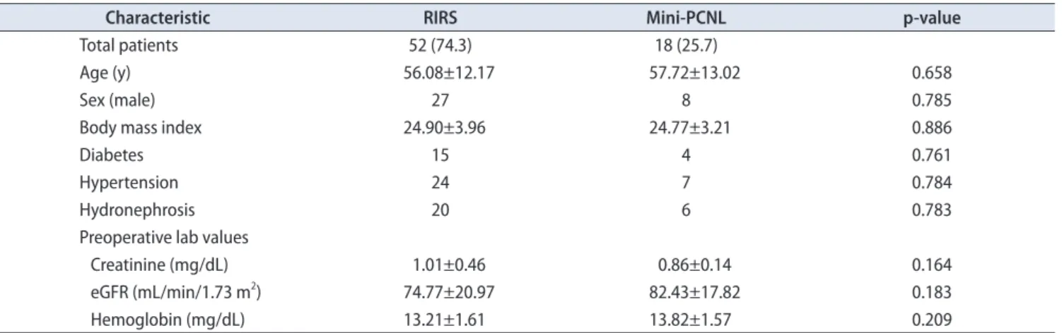
A total of 70 patients with a mean age of 56.3±12.4 years were enrolled (Table 1). Age, sex, body mass index (BMI), and eGFR were not significantly different between the two pa- tient populations. The preoperative eGFR was lower than 60 mL/min/1.73 m2 in 13 cases (18.6%). The overall stone-free rate was 81.4%. Five and 20 patients underwent ureteral stenting and extracorporeal shock-wave lithotripsy, respectively, and 4 and 2 patients had undergone RIRS and PCNL previously.
2. Complications
Transfusion was performed in a single case of mini- PCNL, and Foley catheter re-insertion as a result of blood clot obstruction and ureteral stent repositioning. No addi- tional management was needed for these conditions.
3. Perioperative renal functional outcomes
About one-fifth of the total cases (13/70, 18.6%) were in- cluded in the group with normal renal function (Table 2).In the group with abnormal function, the mean Fn_Op was 55.5%. The preoperative GAP was 24.4%, 6.2%, and 28.6% in the total, normal, and abnormal groups, respectively. Among the patients in the abnormal group, more than two-thirds (40/57, 70.2%) of the patients were stable and seven patients (12.3%) were improved at postoperative 12 months (Table 2).
4. Prediction of aggravated kidney function
Univariate analysis was conducted to estimate the sig- nificant predictors of postoperative renal aggravation. Age, BMI, comorbidities, previous stone procedure, stone volume, laterality, diameter, number, S-ReSC, Hounsfield unit, preop- erative serum creatinine, and hydronephrosis were assessed.At 3 months postoperatively, none of the predictors showed p-values <0.05. At postoperative 12 months, the preoperative creatinine level (p=0.079) and a history of previous stone procedures (p=0.079) showed borderline significance (Supple- mentary Table 1). In a multivariate analysis, preoperative creatinine level (p=0.060) and a history of previous stone procedures (p=0.051) showed borderline significance at post- operative 12 months (Supplementary Table 1).
5. Prediction of renal function improvement at postoperative follow-up
Univariate analysis was performed to determine the sig- nificant predictors of postoperative renal function improve- ment. Operative time and presence of remnant stones were additional factors assessed to predict aggravated kidney dys- function. None of the predictors showed p-values lower than 0.05 at 3 and 12 months postoperatively and were thus not included in the multivariate analysis (Supplementary Table 2).
6. Comparison of abnormal renal function on the basis of surgical method
Age, creatinine, eGFR, and hemoglobin level did not dif- fer significantly between the RIRS and mini-PCNL groups (Table 3). Stone characteristics between the two procedures differed significantly. Although the number of stones was not significantly different between the two procedures, the Table 1. Patient characteristics (n=70)
Characteristic RIRS Mini-PCNL p-value
Total patients 52 (74.3) 18 (25.7)
Age (y) 56.08±12.17 57.72±13.02 0.658
Sex (male) 27 8 0.785
Body mass index 24.90±3.96 24.77±3.21 0.886
Diabetes 15 4 0.761
Hypertension 24 7 0.784
Hydronephrosis 20 6 0.783
Preoperative lab values
Creatinine (mg/dL) 1.01±0.46 0.86±0.14 0.164
eGFR (mL/min/1.73 m2) 74.77±20.97 82.43±17.82 0.183
Hemoglobin (mg/dL) 13.21±1.61 13.82±1.57 0.209
Values are presented as number (%), mean±standard deviation, or number only.
RIRS, retrograde intrarenal surgery; mini-PCNL, miniaturized percutaneous nephrolithotomy; eGFR, estimated glomerular filtration rate.
Table 2. Perioperative renal functional outcomes according to altered separate renal function (A) Preoperative separate renal function
Variable Operative kidney (%) Contralateral kidney (%)
Total patients (n=70) 37.8±8.67 62.2±8.67
Normal group (n=13, 18.6%)a 46.9±1.21 53.1±1.21
Abnormal group (n=57, 81.4%)b 35.7±8.27 64.3±8.28
(B) GAPc in the separate renal functions: operative kidney versus contralateral kidney
Variable Preoperative (%) Postoperative 3 mo (%) Postoperative 12 mo (%)
Abnormal group (n=57) 28.6±16.55 33.8±22.47 32.6±24.50
Improved (n=7) 19.6±9.10 9.0±3.22 3.6±3.65
Stable (n=40) 31.0±17.94 33.5±18.98 30.0±17.95
Aggravated (n=10) 25.2±12.35 56.4±25.41 63.3±24.53
Values are presented as mean±standard deviation.
a:The difference between the preoperative separate renal function of the operative kidney and that of the contralateral kidney was <10%. b:The difference mentioned above was >10%. c:Defined as the separate renal function of the contralateral kidney minus that of the operative kidney.
total volume of the stone and S-ReSC scores were larger in the mini-PCNL group than in the RIRS group. The opera- tive time was significantly longer in the mini-PCNL group than in the RIRS group. The stone-free rates were 75.6% and 75.0% in the RIRS and mini-PCNL groups (p=0.714), respec- tively, and the complication rate and perioperative renal functional outcomes did not differ significantly according to the surgical method.
DISCUSSION
Recent studies have recommended RIRS and mini-PCNL as minimally invasive surgical procedures for the removal of kidney stones [6,7]. Previous investigations have estimated the effect of minimally invasive surgery on perioperative renal functional outcomes. For example, Akman et al. [16]
reported improved or stable eGFR in patients treated with conventional PCNL for staghorn calculi. Recently, kidney function was assessed using renal scintigraphy. Quantitative analysis using 99mTc-DMSA SPECT-CT scans [17] demonstrat- ed that PCNL has a minimal effect on global renal function 3 months after surgery. Piao et al. [9] reported perioperative renal functional outcomes and postoperative outcomes at 3 months using 99mTc-DMSA or 99mTc-DTPA. Despite these
studies, however, the effects of minimally invasive surgery on separate renal function during long-term follow-up have rarely been reported. Information about separate renal func- tion and predictors of postoperative outcomes are important for clinical decision-making.
In the present study, we assessed short-term and long- term postoperative separate renal function in patients who underwent minimally invasive surgery for kidney stone disease. In 57 patients included in the abnormal group, the paired t-test revealed a mean change of 4.0% (±20.33) in GAP between preoperatively and 12 months postoperatively, which was not a significant (p=0.145; 95% confidence inter- val, -1.41 to -9.38). Ten patients showed aggravation with sub- stantial mean changes in GAP between preoperatively and 12 months postoperatively. However, considering that the three biggest changes were 73.6%, 63.6%, and 58.4%, and the sample size was small, the mean change in GAP might not be substantially meaningful. These patients generally exhib- ited high levels of creatinine (mg/dL) and low eGFR (mL/
min/1.73 m2) preoperatively (1.5 and 36.2, 3.0 and 16.0, and 1.28 and 42.0, respectively). Two of these three cases had previ- ously undergone stone management procedures.
The positive relationship between surgery and postop- erative separate renal function in the present study is con- Table 3. Comparative analysis of patients with abnormal preoperative relative renal function according to the surgical method (n=57)
Characteristic RIRS Mini-PCNL p-value
Total patients 41 (71.9) 16 (28.1)
Stone characteristics
Total number 2.18±1.87 3.19±2.37 0.096
Total volume (mm3) 2,203.26±4,900.81 7,234.07±6,537.35 0.003
S-ReSC 1.95±1.448 4.69±2.75 0.001
Operation time (min) 65.12±50.37 97.75±44.55 0.027
Complication rate 2 (4.9) 1 (6.3) 0.835
Stone-free rate 31 (75.6) 12 (75.0) 0.714
Postoperative lab changes
Creatinine (mg/dL) -0.02±0.19 -0.02±0.09 0.879
eGFR (mL/min/1.73 m2) 3.63±14.12 3.28±9.64 0.931
Hemoglobin (mg/dL) -0.72±1.23 -0.58±0.62 0.751
Renal function parameters (%)
Preoperative: Fn_Op/Fn_Con 34.6±9.2/65.4±9.2 38.6±4.2/61.4±4.2 0.102
Change of GAP (12 mo)a 4.8±21.6 1.8±17.0 0.615
Postoperative renal function
Improved 5 (12.2) 2 (12.5) 0.975
Stable 30 (73.2) 10 (62.5) 0.429
Deteriorated 6 (14.6) 4 (25.0) 0.355
Values are presented as number (%) or mean±standard deviation.
RIRS, retrograde intrarenal surgery; mini-PCNL, miniaturized percutaneous nephrolithotomy; S-ReSC, Seoul National University Renal Stone Com- plexity; eGFR, estimated glomerular filtration rate.
a:Defined as separate renal function of the contralateral kidney minus that of the operative kidney.
sistent with previous studies. Kukreja et al. [18] and Canes et al. [11] reported improved or similar renal function after PNL during long-term follow-up. In another long-term study of staghorn stones managed by PCNL, radioisotope scanning revealed a lack of adverse renal effects in 91.5% of cases [16].
Several significant predictors of postoperative deterio- ration and recovery of renal function have been reported.
A high preoperative serum creatinine level, renal cortical atrophy, larger stone burden, proteinuria greater than 300 mg/d during follow-up, history of nephrolithiasis, presence of hydronephrosis, and recurrent urinary infection have been reported as significant predictors of deteriorating renal function [2,18]. A stone number > 3, clear urine in the collect- ing system, and lack of renal sepsis are significant predictors of improvement of abnormal separate renal function [15,19].
In a previous study [9], the authors reported preoperative hydronephrosis as a predictor of deterioration and more than three stones as a predictor of both deterioration and improvement during 3 months of follow-up. However, in the present investigation, none of the factors showed any sta- tistical significance. The preoperative serum creatinine level (p=0.060) and history of previous stone procedures (p=0.051) showed borderline significance. However, analysis of the perioperative renal functional outcomes according to the Kidney Disease Outcomes Quality Initiative CKD classifica- tion system in this study revealed no stage-related differ- ences.
This study did not show any significant differences in perioperative renal functional outcomes between RIRS and mini-PCNL. In a previous study of conventional PCNL, the different methods of nephrostomy tract dilatation [20] and multiple-tract access [21] did not exacerbate the loss of post- operative renal function. However, the functional volume at the entry site decreased significantly during regional assessment using SPECT measurement of DMSA uptake [22]. In mini-PCNL, a lower degree of kidney damage than with conventional PCNL might be attributed to the smaller nephrostomy tract. In this study, the PCNL cases were per- formed with a single tract of 16.5 to 18 Fr in 90% of cases or with two tracts, with the first tract being 16.5 to 18 Fr and the second being 12 Fr to minimize the effect of the tracts on renal function. Additionally, we excluded cases with three or more tracts. Therefore, the effect of these small tracts on renal function would be minimal. Note that peri- operative renal functional outcomes were similar for RIRS and mini-PCNL in the present study. During RIRS, acute excessive intrarenal pressure threatens tubular function, and the authors used ureteral access sheaths in most cases.
Additional studies are needed to evaluate the consequences
of excessive renal pressure and the effects of using ureteral access sheaths on postoperative renal function.
This study had several limitations. First, this was an observational study conducted at a single institution, and the results were derived from a relatively small sample size. Second, although the baseline serum creatinine level was presented as a predictor of postoperative aggravation, a definitive cutoff level was not provided. Further study is needed to assess the cutoff level of preoperative serum creatinine in the evaluation of renal function using 99mTc- DMSA or 99mTc-DTPA. Third, it is necessary to classify the patients’ comorbidities, because various comorbidities can affect renal function. Moreover, the compensated contralat- eral kidney might represent a confounding factor. Although overall postoperative renal functional outcomes were stable, six patients in the RIRS and four patients in the mini- PCNL group showed renal deterioration. Because PCNL and RIRS are known to ensure the safety of kidney function postoperatively [18,23], further evaluation of the safety and efficacy of minimally invasive renal stone surgery during a long-term follow-up is needed, especially in patients with preoperative renal insufficiency. Finally, it is hard to clarify whether the deterioration in renal function was induced by surgery or was associated with the natural history of stone disease. Another study with a larger sample size is needed to address these limitations. Nonetheless, this study is valuable because it represents the first investigation of minimally invasive renal stone surgery to assess perioperative separate renal functional outcomes using 99mTc-DTPA during a 1-year follow-up.
CONCLUSIONS
As minimally invasive renal stone surgeries, RIRS and mini-PCNL were effective and resulted in favorable renal functional outcomes during 1 year of follow-up. The effect on renal function of the two procedures was similar during the postoperative period. Clinicians should pay careful at- tention to patients showing high baseline serum creatinine levels and reporting a history of stone procedures.
CONFLICTS OF INTEREST
The authors have nothing to disclose.
ACKNOWLEDGMENTS
This manuscript was selected as the best paper at the 26th Korean Society of Endourology and Robotics meeting
in 2019.
This research was supported by the Materials and Com- ponents Technology Development Program of MOTIE/KEIT, Republic of Korea (10067258, Development of a holmium/
thulium laser resonator for treatment of prostatic hyperpla- sia).
AUTHORS’ CONTRIBUTIONS
Research conception and design: Juhyun Park and Sung Yong Cho. Data acquisition: Juhyun Park, Sung Yong Cho, Min Chul Cho, Hwancheol Son, and Hyeon Jeong. Statisti- cal analysis: Jae Hyun Jung, Seung Hoon Ryang, and Sung Yong Cho. Data analysis and interpretation: Sangjun Yoo, Jae Hyun Jung, Seung Hoon Ryang, and Sung Yong Cho.
Drafting of the manuscript: Sangjun Yoo, Jae Hyun Jung, Seung Hoon Ryang, and Sung Yong Cho. Critical revision of the manuscript: Min Chul Cho, Hwancheol Son, and Hyeon Jeong. Obtaining funding: Hyeon Jeong and Sung Yong Cho.
Administrative, technical, or material support: Jae Hyun Jung and Sung Yong Cho. Supervision: Min Chul Cho, Hw- ancheol Son, and Hyeon Jeong. Approval of the final manu- script: Jae Hyun Jung and Sung Yong Cho.
SUPPLEMENTARY MATERIALS
Scan this QR code to see the supplementary materials, or visit https://www.icurology.org/src/sm/icurology-61-59-s001.pdf.
REFERENCES
1. Marchini GS, Vicentini FC, Mazzucchi E, Brito A, Ebaid G, Srougi M. Silent ureteral stones: impact on kidney function- -can treatment of silent ureteral stones preserve kidney func- tion? Urology 2012;79:304-8.
2. Alexander RT, Hemmelgarn BR, Wiebe N, Bello A, Morgan C, Samuel S, et al.; Alberta Kidney Disease Network. Kid- ney stones and kidney function loss: a cohort study. BMJ 2012;345:e5287.
3. Assimos D, Krambeck A, Miller NL, Monga M, Murad MH, Nelson CP, et al. Surgical management of stones: American Urological Association/Endourological Society Guideline, part I. J Urol 2016;196:1153-60.
4. Gupta M, Bolton DM, Gupta PN, Stoller ML. Improved re-
nal function following aggressive treatment of urolithiasis and concurrent mild to moderate renal insufficiency. J Urol 1994;152:1086-90.
5. Gambaro G, Favaro S, D'Angelo A. Risk for renal failure in nephrolithiasis. Am J Kidney Dis 2001;37:233-43.
6. Kirac M, Bozkurt ÖF, Tunc L, Guneri C, Unsal A, Biri H.
Comparison of retrograde intrarenal surgery and mini-percu- taneous nephrolithotomy in management of lower-pole renal stones with a diameter of smaller than 15 mm. Urolithiasis 2013;41:241-6.
7. Lee JW, Park J, Lee SB, Son H, Cho SY, Jeong H. Mini-percu- taneous nephrolithotomy vs retrograde intrarenal surgery for renal stones larger than 10 mm: a prospective randomized controlled trial. Urology 2015;86:873-7.
8. Bilen CY, Inci K, Kocak B, Tan B, Sarikaya S, Sahin A. Impact of percutaneous nephrolithotomy on estimated glomerular fil- tration rate in patients with chronic kidney disease. J Endourol 2008;22:895-900.
9. Piao S, Park J, Son H, Jeong H, Cho SY. Evaluation of renal function in patients with a main renal stone larger than 1 cm and perioperative renal functional change in minimally inva- sive renal stone surgery: a prospective, observational study.
World J Urol 2016;34:725-32.
10. El-Nahas AR, Eraky I, Shokeir AA, Shoma AM, El-Assmy AM, El-Tabey NA, et al. Long-term results of percutaneous nephrolithotomy for treatment of staghorn stones. BJU Int 2011;108:750-4.
11. Canes D, Hegarty NJ, Kamoi K, Haber GP, Berger A, Aron M, et al. Functional outcomes following percutaneous surgery in the solitary kidney. J Urol 2009;181:154-60.
12. Levey AS, Coresh J, Balk E, Kausz AT, Levin A, Steffes MW, et al.; National Kidney Foundation. National Kidney Foundation practice guidelines for chronic kidney disease: evaluation, clas- sification, and stratification. Ann Intern Med 2003;139:137-47.
13. Jeong CW, Jung JW, Cha WH, Lee BK, Lee S, Jeong SJ, et al.
Seoul National University Renal Stone Complexity score for predicting stone-free rate after percutaneous nephrolithotomy.
PLoS One 2013;8:e65888.
14. Dindo D, Demartines N, Clavien PA. Classification of surgical complications: a new proposal with evaluation in a cohort of 6336 patients and results of a survey. Ann Surg 2004;240:205- 13.
15. Cho SY, Kim IS, Lee SB, Choi H, Park K. Nature and fate of su- pranormal differential renal function: lessons from long-term follow-up after pyeloplasty. Urology 2013;81:163-7.
16. Akman T, Binbay M, Kezer C, Yuruk E, Tekinarslan E, Ozgor F, et al. Factors affecting kidney function and stone recurrence rate after percutaneous nephrolithotomy for staghorn calculi:
outcomes of a long-term followup. J Urol 2012;187:1656-61.
17. Pérez-Fentes D, Cortés J, Gude F, García C, Ruibal A, Aguiar P.
Does percutaneous nephrolithotomy and its outcomes have an impact on renal function? Quantitative analysis using SPECT- CT DMSA. Urolithiasis 2014;42:461-7.
18. Kukreja R, Desai M, Patel SH, Desai MR. Nephrolithiasis as- sociated with renal insufficiency: factors predicting outcome. J Endourol 2003;17:875-9.
19. Agrawal MS, Aron M, Asopa HS. Endourological renal salvage in patients with calculus nephropathy and advanced uraemia.
BJU Int 1999;84:252-6.
20. Unsal A, Koca G, Reşorlu B, Bayindir M, Korkmaz M. Ef- fect of percutaneous nephrolithotomy and tract dilatation methods on renal function: assessment by quantitative single- photon emission computed tomography of technetium-99m-
dimercaptosuccinic acid uptake by the kidneys. J Endourol 2010;24:1497-502.
21. Handa RK, Evan AP, Willis LR, Johnson CD, Connors BA, Gao S, et al. Renal functional effects of multiple-tract percutaneous access. J Endourol 2009;23:1951-6.
22. Moskovitz B, Halachmi S, Sopov V, Burbara J, Horev N, Gro- shar D, et al. Effect of percutaneous nephrolithotripsy on renal function: assessment with quantitative SPECT of (99m)Tc- DMSA renal scintigraphy. J Endourol 2006;20:102-6.
23. Hoarau N, Martin F, Lebdai S, Chautard D, Culty T, Azzouzi AR, et al. Impact of retrograde flexible ureteroscopy and intra- corporeal lithotripsy on kidney functional outcomes. Int Braz J Urol 2015;41:920-6.