- 141 -
Imaging Science in Dentistry 2016; 46: 141-4 http://dx.doi.org/10.5624/isd.2016.46.2.141
Clinicians are increasingly utilizing Cone-Beam Com- puted Tomography(CBCT) is in the field of dentistry for various diagnostic tasks.1,2 It provides volumetric informa- tion, orthogonal reconstruction, and cross-sectional image data.3 Reconstructed orthogonal images provide geometri- cally concordant linear measurements which is not pos- sible with conventional 2D imaging.3,4 CBCT scans can be obtained with different fields of view(FOVs).2 CBCT scans typically cover a larger FOV and capture not only the dentoalveolar region, but also the extragnathic region.2 This leads to the possibility of identifying incidental find- ings outside the region of interest, such findings may sometimes be overlooked by untrained eye.2,6,7 Previous data from literature suggests that approximately 25% of CBCT images obtained for various maxillofacial diagnos- tic tasks displayed incidental findings.6,8
The clivus is a very important part of the skull base and
is usually captured in large FOV scans. The clivus may be associated with multiple anatomical variations and patho- logies, such as canalis basilaris medianus(CBM), chor- doma etc. CBM is an uncommon anatomical variant of the basiocciput.9 Jacquemin et al.9 stated that Gruber was the first to describe about CBM. Clinically asymptomatic, it is a congenital defect that has been observed in approx- imately 2% of dry skulls.9,10 CBM has been broadly clas- sified into complete or incomplete channel types.9 Two theories have been proposed regarding the origin of this transclivial defect. One is vascular theory and another one is notochordal theory. Vascular theory states that it could have originated from emissary vein. Notochord theory predicates that this defect could be reminiscent part of no- tochord.9,10 Previous studies have evaluated CBM using dry skulls, computed tomography(CT) and magnetic res- onance imaging(MRI).9,10
This report describes two presumed cases of CBM de- tected utilizing the CBCT imaging modality along with a review of the literature. To the best our knowledge, this is the first case series to be reported using CBCT imaging.
Evaluation of canalis basilaris medianus using cone-beam computed tomography
Ali Z. Syed1,*, Samir Zahedpasha1, Sonali A. Rathore2, Mel Mupparapu3
1Department of Oral and Maxillofacial Medicine and Diagnostic Sciences, CWRU School of Dental Medicine, Cleveland, OH, USA
2Department of Oral Diagnostic Sciences, VCU School of Dentistry, Richmond, VA, USA
3Division of Radiology, University of Pennsylvania School of Dental Medicine, Philadelphia, PA, USA
AbstrAct
The aim of this report is to present two cases of canalis basilaris medianus as identified on cone-beam computed tomography(CBCT) in the base of the skull. The CBCT data sets were sent for radiographic consultation. In both cases, multi-planar views revealed an osseous defect in the base of the skull in the clivus region, the sagittal view showed a unilateral, well-defined, non-corticated, track-like low-attenuation osseous defect in the clivus. The appear- ance of the defect was highly reminiscent of a fracture of the clivus. The borders of osseous defect were smooth, and no other radiographic signs suggestive of osteolytic destructive processes were noted. Based on the overall radiographic examination, a radiographic impression of canalis basilaris medianus was made. Canalis basilaris medianus is a rare anatomical variant and is generally observed on the clivus. Due to its potential associa tion with meningitis, it should be recognized and reported to avoid potential complications.(Imaging Sci Dent 2016; 46:
141-4)
KEy words: Cone-Beam Computed Tomography; Skull Base; Anatomical Variation; CBM
Copyright ⓒ 2016 by Korean Academy of Oral and Maxillofacial Radiology
This is an Open Access article distributed under the terms of the Creative Commons Attribution Non-Commercial License(http://creativecommons.org/licenses/by-nc/3.0) which permits unrestricted non-commercial use, distribution, and reproduction in any medium, provided the original work is properly cited.
Imaging Science in Dentistry·pISSN 2233-7822 eISSN 2233-7830 Received December 10, 2015; Revised February 14, 2016; Accepted February 24, 2016
*Correspondence to : Dr. Ali Z. Syed
Department of Oral and Maxillofacial Medicine and Diagnostic Sciences, CWRU School of Dental Medicine, 10900 Euclid Avenue, Cleveland, OH 44106, USA Tel) 1-216-368-6802, Fax) 1-215-573-7853, E-mail) azs16@case.edu
Evaluation of canalis basilaris medianus using cone-beam computed tomography
- 142 -
case report
Case 1
The CBCT data of a 11-year-old female was referred to evaluate radiolucency in the apical area of the maxillary left lateral incisor. The patient presented with no clinical symptoms and had a non-contributory medical history. The CBCT scan was obtained with i-CAT unit(Imaging Sci- ence, Hatfield, PA, USA). The Digital Imaging and Com- munications in Medicine(DICOM) deidentified data was sent through a Health Insurance Portability and Account- ability Act-compliant secure email(Brightsquid, Ross Tech- nology Centre, Calgary, Alberta, Canada) and the data was evaluated using InVivo5.4.3(Anatomage, San Jose, CA, USA).
All data was evaluated by two board-certified oral and maxillofacial radiologists(SAZ, SAR). The orthogonal views of the large FOV revealed an incidental finding in the clivus. It was observed that the sagittal view was opti- mal for the visualization of this entity because of its exten- sion in the anterioposterior direction across the clivus. Ra- diographically CBM can be described as a well-defined, corticated and channel-like hypodense radiolucency exten- ding from the pharyngeal aspect of the basiocciput to the intracranial aspect of the clivus. This radiographic pre-
sentation is pathognomonic representation of CBM. No other advanced imaging was advised since the patient was asymptomatic(Fig. 1).
Case 2
A 63-year-old female patient with a significant medical history of arthritis, thyroid disease, sinusitis, and gastro- intestinal disease was referred for a CBCT scan. The vol- umetric scan was acquired via a CS9300 machine(Care- stream Inc., Atlanta, GA, USA), and the patient was re- ferred for the evaluation of a proposed implant site in the maxillary region. The data was analyzed using multipla- nar reformatted slices by a board-certified oral and max- illofacial radiologist(MM). The volumetric data set re- vealed an unusual finding on the basiocciput of the clivus.
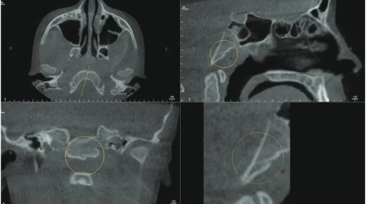
In the sagittal view the intracranial part of the clivus show- ed discontinuity, the radiographic presentation of this os- seous defect presentation can be described as a well-corti- cated, hypodense channel extending in the superior-inferi- or direction along the intracranial aspect of the clivus(Fig.
2). On initial inspection it appeared to be a fracture of the clivus. However, further close inspection revealed no other signs of osteolytic destruction on the posterior aspect of the clivus(intracranial surface). Based on the volumetric appearance of the defect, a radiographic impression of
Fig. 1. Multiplanar reconstruction images demonstrate a defect on the basiocciput of the clivus consistent with canalis basilaris medianus in an 11-year-old female.
- 143 -
Ali Z. Syed et al
CBM was made. This transclival defect can be best appre- ciated on the sagittal view. This entity depicted is an exam- ple of the complete type of CBM.
discussion
The term CBM is used to describe transclival defect of the basicocciput of the clivus. CBM presents itself in var- ious configurations such as keyhole defect, transverse or longitudinally fissures in the clivus.12 Radiological fea- tures of CBM can be described as a well-defined, corti- cated, osseous transclival defect located in the basiocciput of the clivus. In the axial view it is located in close ap- proximation to the anterior rim of the foramen magnum.10 CBM’s reported prevalence in the literature is is 2%-3%
for adults and 4%-5% in children.9,10
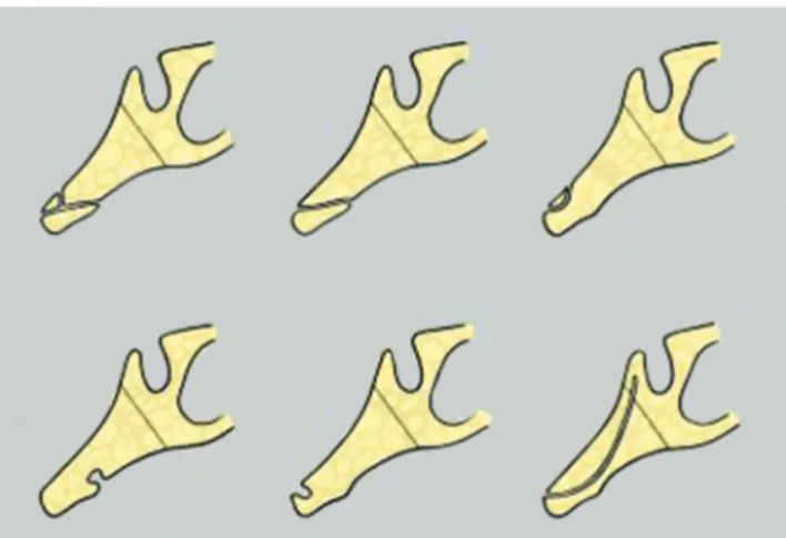
Six varieties of CBM(Fig. 3) were described in the lit- erature: three complete varieties(superior, inferior, and bi- furcated) and three incomplete varieties(a thin, long chan- nel; and either a superior recess; or an inferior recess).9,10 The complete type of CBM is characterized by the pre- sence of a channel or tract like hypodense region extend- ing either anterio-posteriorly or superior-inferior direction occasionally, connecting the intracranial and pharyngeal portions of the clivus. Incomplete type will traverse par- tially within the clivus and they fail to communicate with pharyngeal portion or intracranial of the portion of the clivus.
CBM in general is considered an anatomical variant without any clinical significance.8,9 However, few studies indicate that they could serve as potential pathways for the progression of the disease such as meningitis.13 Hemphill et al.11 and Martinez et al.12 reported a transclival bony defect that was associated with a meningocele and caused
recurrent meningitis.
Jacquemin et al.9 presented a case of the incomplete inferior type of CBM. The osseous defect was located at the level of pharyngeal fossa. Their patient presented with atypical bacterial meningitis. However, authors argued that a mere coexistence of CBM and meningitis may not have caused detrimental effects such as optic neuropa- thy and the episode of sterile meningitis as noted in their case.9
Lohman et al.13 reported a CBM in association with a Tornwaldt cyst for the first time in the literature. Their case demonstrated the potential overlap of these entities.
Literature is providing conflicting reports about CBM’s association with other pathological entities such as men- ingitis, Tornwaldt’s cyst and its detrimental effects as not- ed in the above description. Further future studies are rec- ommended to find its association with other pathological entities.
We reported two cases of CBM. First case described was incomplete type and second case was complete type of CBM, both cases were assessed using CBCT. In both the cases, the occurrence of CBM was not associated with meningitis or other problems. Our experience, as well as the literature as a whole, emphasizes the need for thorough analysis of skulls regardless of whether defects are detect- ed or not when diagnostic tools such as CBCT, CT, and MRI are readily available.2,5 We recommend that all data should be interpreted by a board-certified oral and maxil- lofacial radiologist to prevent misdiagnoses and to avoid potential complications.7
Fig. 2. A sagittal image demonstrates canalis basilaris medianus of
the complete type on the superior aspect of the clivus. Fig. 3. A schematic diagram depicting the different types of cana- lis basilaris medianus(CBM). The top row illustrates the complete forms of CBM and the bottom row illustrates incomplete forms of CBM. Top row: bifurcating type (left) CBM inferior type(middle), superior type(left), and. Bottom row: inferior recess(left), superior recess(middle), and channel(right).(Adapted from Currrano)
Evaluation of canalis basilaris medianus using cone-beam computed tomography
- 144 -
Acknowledgements
We thank Ms. Sarah G. Jawhari, senior dental student from CWRU School of Dental Medicine for helping with the illustrating the diagram depicting six types of canalis basilaris medianus.
references
1. Horner K. Cone-beam computed tomography: time for an evi- dence-based approach. Prim Dent J 2013; 2: 22-31.
2. Syed AZ, Sin C, Rios R, Mupparapu M. Incidental occurrence of an unusually large mastoid foramen on cone-beam comput- ed tomography and review of the literature. Imaging Sci Dent.
2016; 46: 39-45.
3. Haney E, Gansky SA, Lee JS, Johnson E, Maki K, Miller AJ, et al. Comparative analysis of traditional radiographs and cone- beam computed tomography volumetric images in the diagno- sis and treatment planning of maxillary impacted canines. Am J Orthod Dentofacial Orthop 2010; 137: 590-7.
4. Liang X, Jacobs R, Hassan B, Li L, Pauwels R, Corpas L, et al.
A comparative evaluation of cone beam computed tomogra- phy(CBCT) and multi-slice CT(MSCT) Part I. On subjective image quality. Eur J Radiol 2010; 75: 265-9.
5. Syed AZ, Mendes RA, Alnakhli TM, Pinto A. Thyroid papil- lary microcarcinoma: an incidental finding in a patient with
coronoid hyperplasia. BMJ Case Rep 2015; 2015. pii: bcr 2015212628
6. Newaz ZA, Barghan S, Katkar RA, Bennett JA, Nair MK.
Incidental findings of skull-base abnormalities in cone-beam computed tomography scans with consultation by maxillofa- cial radiologists. Am J Orthod Dentofacial Orthop 2015; 147:
127-31.
7. Zinman EJ, White SC, Tetradis S. Legal considerations in the use of cone beam computer tomography imaging. J Calif Dent Assoc 2010; 38: 49-56.
8. Cha JY, Mah J, Sinclair P. Incidental findings in the maxillofa- cial area with 3-dimensional cone-beam imaging. Am J Orthod Dentofacial Orthop 2007; 132: 7-14.
9. Jacquemin C, Bosley TM, al Saleh M, Mullaney P. Canalis basilaris medianus: MRI. Neuroradiology 2000; 42: 121-3.
10. Currarino G. Canalis basilaris medianus and related defects of the basiocciput. AJNR Am J Neuroradiol 1988; 9: 208-11.
11. Hemphill M, Freeman JM, Martinez CR, Nager GT Long DM, Crumrine P. A new, treatable source of recurrent meningitis:
basioccipital meningocele. Pediatrics 1982; 70: 941-3.
12. Martinez CR, Hemphill JM, Hodges FJ 3rd, Gayler BW, Nager GT, Long DM, et al. Basioccipital meningocele. AJNR Am J Neuroradiol 1981; 2: 100-2.
13. Lohman BD, Sarikaya B, McKinney AM, Hadi M. Not the typical Tornwaldt’s cyst this time? A nasopharyngeal cyst as- sociated with canalis basilaris medianus. Br J Radiol 2011;
84: e169-71.