https://doi.org/10.5624/isd.2019.49.4.317
Steatocystoma multiplex is a rare benign subcutaneous disease characterized by multiple dermal cyst-like lesions derived from the pilosebaceous glands. Its pathogenesis remains unclear, but it is predominantly referred to as a hamartomatous malformation of the pilosebaceous duct junction.1 Steatocystoma multiplex is most commonly di- agnosed in adolescence or early adulthood;2 however, it has been reported as early as birth3 and in patients as old as 78 years old.4 The condition appears to lack any sex or race predilection.5 Although usually asymptomatic, it can cause patients social stress related to their appearance if the lesions are numerous or large, especially on the face and neck. Some cases are sporadic; however, an autoso- mal dominant inherited type associated with mutations of the gene coding for keratin 17(KRT17) has also been de- scribed.6
Steatocystoma multiplex was first mentioned in a case report published by Jamieson in 1873 involving numerous cutaneous cysts scattered throughout the body.7 This con- dition has mainly been reported on the trunk and the prox- imal extremities, though it has been described as occurring on the oral mucosa8 as well. Few previous studies have reported lesions of the head and neck area concurrent with other characteristics resembling ectodermal dysplasia, es- pecially with regard to their radiographic imaging features.
This report aimed to describe a case of steatocystoma multiplex on the cervical area concurrent with several dental anomalies, with a focus on its clinical, radiological, and histopathological characteristics, as well as the possi- bility that this condition was inherited.
Case Report
A 32-year-old man was referred to Seoul National Uni- versity Dental Hospital for further evaluation and treat- ment of bilateral nodular lesions of the neck that had been present for several months. He had visited a local plastic
Steatocystoma multiplex: A case report of a rare entity
Nan-Young Shin 1, Ju Hee Kang 2, Jo-Eun Kim 2, Khantaly Symkhampa 1,3, Kyung-Hoe Huh 1, Won-Jin Yi 1, Min-Suk Heo 1,*, Sam-Sun Lee 1, Soon-Chul Choi 1
1Department of Oral and Maxillofacial Radiology and Dental Research Institute, School of Dentistry, Seoul National University, Seoul, Korea
2Department of Oral and Maxillofacial Radiology, Seoul National University Dental Hospital, Seoul, Korea
3Division of Oral and Maxillofacial Radiology, Department of Basic Science, Faculty of Dentistry, University of Health Sciences, Vientiane, Laos
AbStRACt
Steatocystoma multiplex is an uncommon benign skin disease, which typically manifests as numerous intradermal cysts that can be scattered anywhere on the body. Although usually asymptomatic, it can be significantly disfiguring.
One type of steatocystoma multiplex is known to be associated with the autosomal dominant inheritance of a mutation in the gene coding for keratin 17(KRT17). In such cases, it is often concurrent with other developmental abnormalities of the ectoderm-derived tissues, such as the nails, hair, and teeth. To the best of our knowledge, few cases have been reported of steatocystoma multiplex of the oral and maxillofacial region. This report describes a case of steatocystoma multiplex of both sides of the neck and multiple dental anomalies, with a focus on its clinical, radiological, and histopathological characteristics, as well as the possibility that the patient exhibited the familial type of this condition.(Imaging Sci Dent 2019; 49: 317-21)
Key woRdS: Steatocystoma Multiplex; Computed Tomography; Keratin-17
Copyright ⓒ 2019 by Korean Academy of Oral and Maxillofacial Radiology
This is an Open Access article distributed under the terms of the Creative Commons Attribution Non-Commercial License(http://creativecommons.org/licenses/by-nc/3.0) which permits unrestricted non-commercial use, distribution, and reproduction in any medium, provided the original work is properly cited.
Imaging Science in Dentistry·pISSN 2233-7822 eISSN 2233-7830 Received September 1, 2019; Revised September 17, 2019; Accepted September 23, 2019
*Correspondence to: Prof. Min-Suk Heo
Department of Oral and Maxillofacial Radiology and Dental Research Institute, School of Dentistry, Seoul National University, 101 Daehak-ro, Jongno-gu, Seoul 03080, Korea
Tel) 82-2-2072-3016, E-mail) hmslsh@snu.ac.kr
surgery clinic with a chief complaint of a large lesion and pain in the right cervical area. Documentation from the clinic stated that he had only agreed to a drainage proce- dure and had refused surgery to remove the lesions. Ac- cording to the patient, the pain and swelling had subsided as a result of the aspiration performed during his visit to the clinic. Additionally, while the patient had an operative history of cardiac septal defect repair performed when he was 10 months old, he gave no family history associated with skin lesions like those that he exhibited.
Panoramic radiography showed abnormal dentition.
First, the deciduous second molars were in a state of pro- longed retention and submergence on both sides of the maxilla and the right side of the mandible. Second, mul- tiple permanent teeth were missing, including the maxil- lary second premolars on both sides and 2 mandibular in- cisors on both sides and 1 mandibular molar on the right side. Finally, microdontia of 2 maxillary lateral incisors (peg lateral incisors) and 2 mandibular premolars was ob- served(Fig. 1).
To accurately evaluate the soft-tissue lesions, contrast- enhanced computed tomography(CECT) was performed.
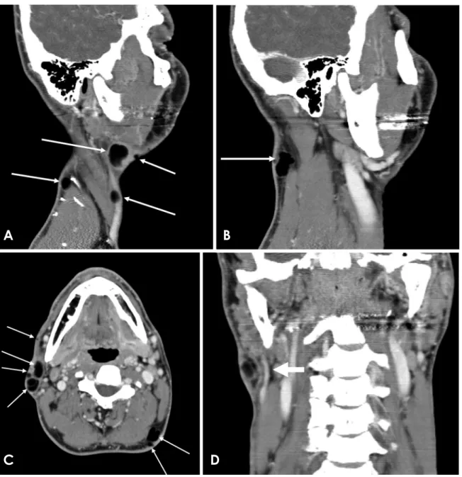
The images revealed multiple well-defined round to ovoid, smooth or scalloped, nodular lesions with diame- ters ranging from 2 to 20mm in the subcutaneous layer on both sides of the cervical region. The internal structure showed fat attenuation on the level of -110 to -100 Hounsfield units(HU) and some lobulation. Additionally, the large lesion of the right upper neck, where the patient experienced the pain, displayed peripheral enhancement with mild swelling, suggesting that it was accompanied by inflammation. The epidermis of that lesion was elevat- ed, and the adjacent platysma muscle and external jugular vein were displaced medially(Fig. 2). In other words, the
benign lesion exerted a pushing force on adjacent struc- tures. Based on these radiographic findings, the initial di- agnosis was lipomatosis.
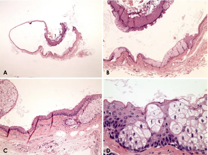
For microscopic examination, excisional biopsy was performed on a lesion(7×5×4mm) on the right side of the neck, and hematoxylin and eosin staining was used.
The histological finding was a subcutaneous cystic lesion containing sebum-like material. At the lowest magnifi- cation(×12.5), a cavity surrounded by a thin epithelial lining that formed part of the cyst wall was observed. The middle magnification settings(×40 and ×100) showed the specimen to have a wall lined by 3 to 5 layers of ke- ratinized stratified squamous epithelium lacking a granu- lar layer, and many sebaceous lobules were located very close to, as well as within, the cyst wall. In addition, a wavy luminal surface and sebum-like material or keratin debris were detected. The highest magnification(×400) showed numerous lobules of pilosebaceous glands on the cyst wall. The granular layer was completely absent(Fig.
3). Accordingly, the analysis resulted in a diagnosis of steatocystoma multiplex.
The patient revisited with bleeding of the above-men- tioned large lesion of the right neck 6 months after the biopsy. The aspiration contained serous fluid, blood, and sebum-like material. His discomfort was relieved with medication a few days later, and he required no further visits.
discussion
Steatocystoma multiplex consists of benign cystic le- sions derived from the sebaceous glands. The lesions are macroscopically smooth and uniform with a size of a few millimeters to centimeters along the long axis. Their col-
Fig. 1. A panoramic radiograph shows multiple examples of abnor- mal dentition, as follows: prolonged retention and submergence of the maxillary deciduous second molars on both sides and the mandibular deciduous second molar on the right side; absence of the maxillary second premolars on both sides, as well as 2 mandibular incisors on both sides and 1 mandibular molar on the right side; and microdontia of the maxillary lateral incisors on both sides and 2 mandibular premolars on both sides.
or is usually whitish or yellowish, which likely reflects sebum-like material in the cystic cavity. Therefore, small lesions can be clinically confused with acne conglobata or milia. Furthermore, it is difficult to differentiate ste- atocystoma multiplex from some dermal cystic lesions of different origin, such as dermoid/epidermoid cysts, mul- tiple lipomas, and xanthomatosis, on the basis of CECT
imaging findings, as these types of lesions are also char- acterized by low internal attenuation.9
Histopathologically, steatocystoma is defined as a der- mal cystic lesion containing sebum or sebum-like mate- rial and keratin debris, characterized by lobules originat- ing from sebaceous glands, and lacking a granular layer.
These features are critical to distinguish steatocystoma
A B
C D
Fig. 2. Contrast-enhanced computed tomographic imaging features of steatocystoma multiplex of the bilateral cervical subcutaneous area.
A. A sagittal image shows multiple well-defined round to ovoid, smooth-surfaced lesions of the right side(arrows). The internal density (-110 to -100 Hounsfield units) corresponds to fat attenuation. In the periphery of the largest lesion shown in the image, mild swelling with minimal enhancement is visible. B. A sagittal image reveals a scalloped lesion of the left side in the posterior cervical area(arrow).
C. An axial image reveals lateral cervical lesions of the right side and posterior cervical lesions of the left side(arrows). The second lesion from the front on the right side shows peripheral enhancement with mild swelling, which is consistent with the largest lesion shown in A.
The right rearmost lesion exhibits internal lobulation. D. A coronal image reveals that the epidermis is elevated, and the adjacent platysma muscle and external jugular vein are displaced medially by the lesion on the right side(thick arrow).
from eruptive vellus hair cysts, which have a similar ap- pearance, age of onset, and distribution pattern. An erup- tive vellus hair cyst is formed by several layers of squa- mous cells, with vellus hairs as well as keratinous materi- al in the cavity.10,11
As mentioned, the familial form of steatocystoma mul- tiplex has been described as a phenotypic variant of an au- tosomal dominant inherited disease associated with muta- tions of KRT17. Mutations of these genes have also been identified in patients with pachyonychia congenita type 2 (PC-2).12 Pachyonychia congenita(PC) has 2 main types, PC-1(the Jadassohn-Lewandowsky form) and PC-2(the Jackson-Lawler form). Common clinical features of PC-2 include hypertrophic nail dystrophy, natal teeth, and hair
abnormalities.13 Individuals with PC-2 may also develop dermal cysts, including steatocystoma multiplex.14
In PC-2 patients, various dental anomalies - for in- stance, early primary tooth loss,13 natal or neonatal teeth,15 Hutchinson-like teeth,16 and friable adult teeth17 - are also commonly recognized. The congenital absence of secondary dentition with persistent partial primary dentition, which was exhibited by our patient, was first described by Gass et al.18 They reported the identification via DNA analysis of a missense mutation in KRT17 in the described patient and his sister. Gass et al. showed that the amino acid substitution was located in the 2B domain, not in the 1A domain, which is the typical site of the mutations involved in steatocystoma multiplex and PC-
A B
C D
Fig. 3. Histological findings of the excisional biopsy specimen. A. Overall view of the biopsy specimen showing a cavity surrounded by a folded cyst wall in the subcutaneous tissue. The cyst is partially collapsed(H&E stain; original magnification, ×12.5). B. The intramural se- baceous lobules and crenulated surface of the cyst wall are indicative of steatocystoma multiplex. The cavity contains small amounts of kera- tinous material(H&E stain; original magnification, ×40). C. The wall is composed of 3 to 5 layers of squamous epithelium without a granular layer, and the luminal surface is wavy. Sebaceous lobules are located both very close to and within the cyst wall(H&E stain; original magnifi- cation, ×100). D. The cyst wall consists of several layers of cuboidal squamous epithelial cells with abundant sebaceous lobules. The granular layer is completely absent. A wavy eosinophilic homogeneous cuticle is also evident(H&E stain; original magnification, ×400).
2. That report can be said to extend the range of dental anomalies recognized in cases of PC-2.
Taken together, the features of our case were sufficient to suggest ectodermal dysplasia. Among them, associa- tion with KRT17 mutations or PC-2 have been established for steatocystoma multiplex and the dental anomalies described above, while no such association has been es- tablished for cardiac septal defect. Unfortunately, in our case, the patient’s family history was not obtained, and a genetic analysis was not conducted. Therefore, we cannot be certain that this case reflects a genetic condition; how- ever, it certainly represents steatocystoma multiplex with dental anomalies.
Steatocystoma multiplex is a benign disease that typi- cally does not cause any symptoms or discomfort. There- fore, it can be overlooked or excised simply for aesthetic reasons. It is likely that the pain that spurred the patient described herein to seek treatment stemmed from a sec- ondary infection. However, since the possibility of a KRT17 mutation cannot be excluded in cases presenting with multiple cysts consistent with steatocystoma multi- plex, patients should be carefully examined with regard to family history and the presence of other ectodermal ab- normalities, and a histological assessment should be per- formed as needed.
In conclusion, steatocystoma multiplex is an uncom- mon benign skin disease that is particularly rare in the head and neck region. Due to the high potential of associ- ation with a KRT17 mutation, a biopsy is recommended.
CECT is useful for defining the locations, shapes, internal structures, and effects on surrounding structures of these cyst-like lesions.
Conflicts of Interest: None
References
1. Plewig G, Wolff HH, Braun-Falco O. Steatocystoma multiplex:
anatomic reevaluation, electron microscopy, and autoradiogra- phy. Arch Dermatol Res 1982; 272: 363-80.
2. Sonnenblick EB, Buchness MR, Austin JH. CT demonstration of steatocystoma multiplex. J Comput Assist Tomogr 1986; 10:
357-9.
3. Park YM, Cho SH, Kang H. Congenital linear steatocystoma
multiplex of the nose. Pediatr Dermatol 2000; 17: 136-8.
4. Marzano AV, Tavecchio S, Balice Y, Polloni I, Veraldi S. Acral subcutaneous steatocystoma multiplex: a distinct subtype of the disease? Australas J Dermatol 2012; 53: 198-201.
5. Feinstein A, Friedman J, Schewach-Millet M. Pachyonychia congenita. J Am Acad Dermatol 1988; 19: 705-11.
6. Covello SP, Smith FJ, Sillevis Smitt JH, Paller AS, Munro CS, Jonkman MF, et al. Keratin 17 mutations cause either steato- cystoma multiplex or pachyonychia congenita type 2. Br J Der- matol 1998; 139: 475-80.
7. Jamieson WA. Case of numerous cutaneous cysts scattered over the body. Edinb Med J 1873; 19: 223-5.
8. Olsen DB, Mostofi RS, Lagrotteria LB. Steatocystoma simplex in the oral cavity: a previously undescribed condition. Oral Surg Oral Med Oral Pathol 1988; 66: 605-7.
9. Kim SJ, Park HJ, Oh ST, Lee JY, Cho BK. A case of steato- cystoma multiplex limited to the scalp. Ann Dermatol 2009; 21:
106-9.
10. Ohtake N, Kubota Y, Takayama O, Shimada S, Tamaki K. Re- lationship between steatocystoma multiplex and eruptive vellus hair cysts. J Am Acad Dermatol 1992; 26: 876-8.
11. Papakonstantinou E, Franke I, Gollnick H. Facial Steatocysto- ma multiplex combined with eruptive vellus hair cysts: a hy- brid? J Eur Acad Dermatol Venereol 2015; 29: 2051-3.
12. Xiao SX, Feng YG, Ren XR, Tan SS, Li L, Wang JM, et al. A novel mutation in the second half of the keratin 17 1A domain in a large pedigree with delayed-onset pachyonychia congenita type 2. J Invest Dermatol 2004; 122: 892-5.
13. Smith FJ, Corden LD, Rugg EL, Ratnavel R, Leigh IM, Moss C, et al. Missense mutations in keratin 17 cause either pachyony- chia congenita type 2 or a phenotype resembling steatocystoma multiplex. J Invest Dermatol 1997; 108: 220-3.
14. Moon SE, Lee YS, Youn JI. Eruptive vellus hair cyst and ste- atocystoma multiplex in a patient with pachyonychia congenita.
J Am Acad Dermatol 1994; 30: 275-6.
15. Leachman SA, Kaspar RL, Fleckman P, Florell SR, Smith FJ, McLean WH, et al. Clinical and pathological features of pachy- onychia congenita. J Investig Dermatol Symp Proc 2005; 10:
3-17.
16. Oh SW, Kim MY, Lee JS, Kim SC. Keratin 17 mutation in pachyonychia congenita type 2 patient with early onset steato- cystoma multiplex and Hutchinson-like tooth deformity. J Der- matol 2006; 33: 161-4.
17. Zamiri M, McLean WH, Hodgins MB, Munro CS. Pachyony- chia congenita type 2: abnormal dentition extending into adult- hood. Br J Dermatol 2008; 159: 500-1.
18. Gass JK, Wilson NJ, Smith FJ, Lane EB, Mclean WH, Rytina E, et al. Steatocystoma multiplex, oligodontia and partial persistent primary dentition associated with a novel keratin 17 mutation.
Br J Dermatol 2009; 161: 1396-8.