266
Open Access
The Correlation Between Left Ventricular Failure and
Right Ventricular Systolic Dysfunction Occurring in Thyrotoxicosis
Ji Yeon Hong, MD, Dae-Gyun Park, MD, Jong Jin Yoo, MD, Seung Min Lee, MD, Min-Kwan Kim, MD, Sung Eun Kim, MD, Jun-Hee Lee, MD, Kyoo-Rok Han, MD and Dong-Jin Oh, MD
Department of Internal Medicine, Hallym University College of Medicine, Seoul, Korea ABSTRACT
Background and Objectives: Heart failure rarely occurs in patients with thyrotoxicosis (6%), with half of the cases having left ventricular dysfunction (LVD). Although a few studies reported isolated right heart failure in thyro- toxicosis, there has been no evaluation of relationship between LVD and right ventricular dysfunction (RVD). Sub- jects and Methods: We enrolled 12 patients (mean age: 51±11 years, 9 females) diagnosed as having thyrotoxi- cosis with heart failure and LVD {left ventricular ejection fraction (LVEF) <40%}, and divided them into two groups {Group I with RVD defined as tricuspid annular plane excursion (TAPSE) less than 15 mm and Group II without RVD}. Clinical features, laboratory variables, and echocardiographic parameters were compared between two groups. Results: RVD was found in 6 (50%) patients. On admission, there were no significant differences be- tween the two groups in clinical features, laboratory variables, or echocardiographic parameters including atrial fibrillation {6 vs. 5, not significant (NS)}, heart rate (149±38 vs. 148±32/min, NS), LVEF (36.7±9.5 vs. 35.1±
6.3%, NS), or the tricuspid regurgitation peak pressure gradient (TRPPG, 30.9±2.0 vs. 36.3±9.3 mmHg, NS).
After antithyroid treatment, all achieved an euthyroid state and both ventricular functions were recovered. All data, including the recovery time of LVEF and the change of heart rate between two groups, displayed no significant differences. Conclusion: In half of patients, RVD was combined with thyrotoxicosis-associated LVD. There were no differences in clinical factors or hemodynamic parameters between patients with and without RVD. This sug- gests that RVD is not secondary to thyrotoxicosis-associated LVD. (Korean Circ J 2010;40:266-271)
KEY WORDS: Thyrotoxicosis; Right ventricular dysfunction; Left ventricular dysfunction.
Introduction
Thyrotoxicosis can be manifested by many cardiovas- cular symptoms including palpitation, exercise intoler- ance, dysrhythmia, systolic hypertension, cardiomegaly, peripheral edema, and angina.1) Over 90% of patients experience tachycardia, and 10% experience atrial fibrill- ation or atrial flutter. In addition, chronic or severe th-
yrotoxicosis can aggravate sinus tachycardia or atrial fi- brillation and cause heart failure accompanied by left ventricular systolic dysfunction.1-3) Heart failures by this mechanism have been explained as tachycardia-induced cardiomyopathy in previous studies.4)
Recently, it has been reported that thyrotoxicosis is presented first as right ventricular failure accompany- ing pulmonary artery hypertension and tricuspid regur- gitation.5) However, most studies on right ventricular dysfunction (RVD) related to thyrotoxicosis mention only isolated right ventricular failure,6-11) and there are almost no studies on about wheather both left ventricular dys- function (LVD) and right ventricular failure occur si- multaneously, or there is correlation between the two.
Therefore, to the purpose of this study was to identify factors that affect right ventricular systolic dysfunction occurring in thyrotoxicosis, and particularly examine whether RVD is related to LVD by tachycardia-induc- ed cardiomyopathy occurring in thyrotoxicosis patients.
Received: August 12, 2009
Revision Received: September 17, 2009 Accepted: September 30, 2009
Correspondence: Dae-Gyun Park, MD,Department of Internal Medicine, Hallym University College of Medicine, 445 Gil-dong, Gangdong-gu, Seoul 134-701, Korea
Tel: 82-2-2224-2379, Fax: 82-2-2225-2725 E-mail: dgpark@hallym.or.kr
This is an Open Access article distributed under the terms of the Creative Commons Attribution Non-Commercial License (http://creativecommons.
org/licenses/by-nc/3.0) which permits unrestricted non-commercial use, distribution, and reproduction in any medium, provided the original work is properly cited.
cc
Subjects and Methods
Subjects
This research is a retrospective study on 12 patients diagnosed with thyrotoxicosis and LVD {left ventricle ejection fraction (LVEF) <40%}. Among the patients (male:female=3:9, mean age=51±11) that were ad- mitted to Kangdong Sacred Heart Hospital of Hallym University College of Medicine for heart failure be- tween September 2005 to October 2007, 9 were diag- nosed with thyrotoxicosis for the first time and 3 were diagnosed in previous years, but not treated. In addit- ion, coronary artery disease, hypertension, valvular heart disease, drug use, and infection were ruled out as causes of LVD.
Methods
The 12 patients diagnosed with thyrotoxicosis and LVD were divided into a group (group 1) with RVD and a group (group 2) without RVD by using tricuspid an- nular plane excursion (TAPSE). The TAPSE estimated RV systolic function by measuring the level of systolic excursion of the lateral tricuspid valve annulus towards the apex in the four chamber view. If the measured TA- PSE was 15 mm or shorter, a patient was was defined as having RVD (group 1), and if it was 15 mm or longer, the patient was defined as non-RVD (group 2).
The sex, age, and clinical conditions, including the presence of underlying diseases and heart rates, echo- cardiographic findings, blood tests including troponin, B-type natriuretic peptide (BNP), and thyroid function tests at the time of admission were compared between 2 groups. In addition, when the two groups achieved an euthyroid state after antityhroid therapy, the clinical con- dition, echocardiographic parameters and blood treats were compared between two groups.
Left atrial dimension, diastolic left ventricular dimen- sion, LVEF, diastolic right ventricular dimension, maxi- mum pressure difference of tricuspid regurgitation, and postcaval dimension were measured with the use of tr- ansthoracic echocardiography. As for left atrial dimen- sion, maximal anteroposterior length was measured by M-mode echocardiography at the aortic level of the pa- rasternal short axis view. Diastolic left ventricular di- mensions were measured immediately before the QRS of an electrocardiogram with M-mode echocardiogra- phy at the parasternal short axis view. The right ven- tricular anteroposterior dimension was measured at the end of systole when right ventricular volume reached its maximum in the parasternal long axis view. While the inferior vena cava (IVC) dimension can be measured at a subcostal window, the widest length at exhalation was selected. Tricuspid regurgitation (TR) was classified into mild, moderate, or severe by measuring the degree of re- gurgitation. Mild TR is defined as when regurgitation
past the tricuspid valve reaches 1/3 entire distance of ri- ght atrium but not 1/2; moderate TR as when the regur- gitation reaches 1/2 the distance but not 2/3; and se- vere TR as when regurgitation reaches beyond 2/3 the distance, nearly to the rear wall of the right atrium.
Statistical analysis
For statistical analysis, Statistical Package for the So- cial Sciences (SPSS) 12.0 (SPSS Inc., Chicago, IL, USA) software for Windows was used. All data are presented in mean±standard deviation, and for discontinuous variables between the two groups, the chi square test and the Mann-Whitney test were used. In order to determine changes in clinical status, blood tests and echocardio- graphic indicators at the time of admission and after treatment of thyrotoxicosis, the Wilcoxon signed rank test was performed. Values were considered significant if the p was less than 0.05.
Results
Clinical characteristics (Table 1)
The mean age of all patients was 51±11, and there were more female (n=9, 75%) patients than male. Three patients had diabetes, hypertension or cerebrovascular disease; 3 patients smoked; and 2 patients had a history of alcohol use. Eleven patients showed atrial fibrillation with tachycardia, and the remaining patient showed si- nus tachycardia with a heart rate of 150/min. The cause of thyrotoxicosis in 11 patients was Graves’ disease, and 1 patient had suspected thyroid follicular adenomatosis.
Six patients showed RVD (50%), and clinical characte- ristics between the two groups were not significantly dif- ferent. Two patients underwent coronary angiography to rule out ischemic heart disease while hospitalized, and both belonged to RVD group. Neither had any lesions in the coronary artery, but one patient showed coronary vas- ospasm and the another showed myocardial bridging findings.
Blood test
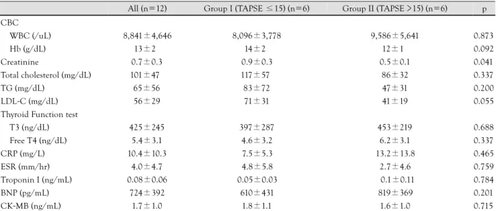
There was no significant difference found between the two groups in any blood tests obtained at admission, including free thyroxine (free T4), triiothyronine (T3), BNP, troponin, creatine kinase-MB (CK-MB) and C- reactive protein (CRP). Group 1 showed higher low den- sity lipoprotein-cholesterol level compared with group 2, but the difference was not statistically significant (p=
0.055). Serum creatinine values were significantly higher in group 1 (p<0.05), but mean value was within normal range (Table 2).
There was no significant difference between the two groups in tests for anti-thyroglobulin autoantibody, anti- thyroid peroxidase antibody (TPOAb) and thyroid stimu- lating hormone receptor antibody (TSHRAb) (Table 3).
Echocardiography (Table 4)
There were no statistically significant differences be- tween the two groups in the results of echocardiography on the left atrial dimension, diastolic left ventricular dimension, LVEF, diastolic right ventricular dimension, the maximum pressure gradient of TR, or the IVC dimen- sion. In the RVD group, the diastolic right ventricular dimension tended to be greater than normal group, but
the difference was not statistically significant (p=0.078).
Results after antithyroid drug treatment
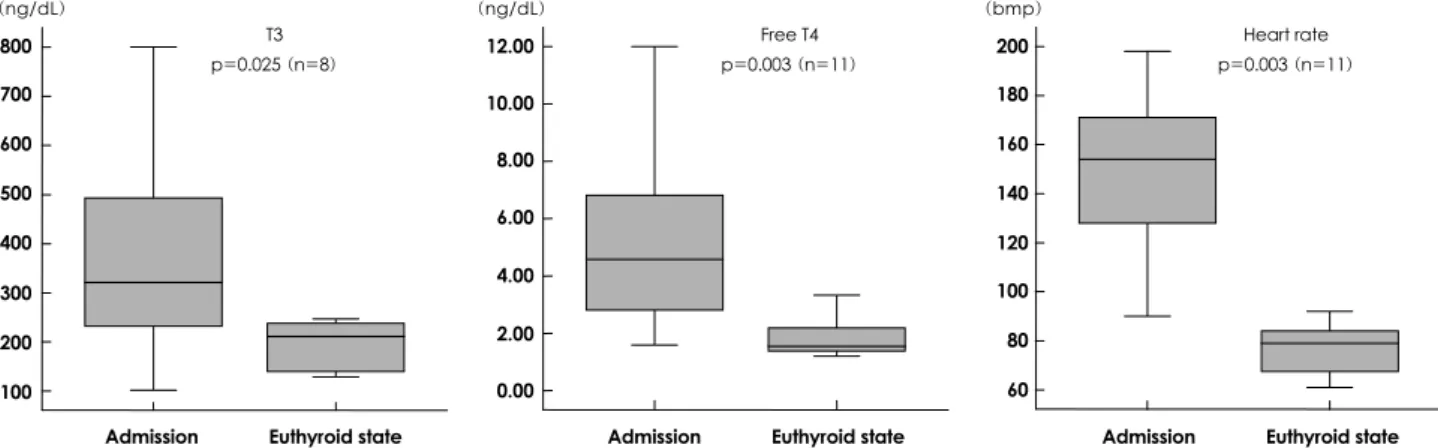
All patients took propylthiouracil (PTU) as antithy- roid drug agent, and the free T4 and T3 values of all patients reached normal levels in an average of 85.8±
70.5 days, this was a statistically significant change (Fig.
1). The 11 patients that showed atrial fibrillation with
Table 1. Characteristics of all patients on admission
All (n=12) Group I (TAPSE ≤15) (n=6) Group II (TAPSE >15) (n=6) p
Age (years) 51±11 50±6 52±3 0.936
Female 09 (75) 4 (67) 9 (84) 0.523
Hypertension 01 (8) 0 (0) 1 (16) 0.317
Diabetes mellitus 01 (8) 1 (16) 0 (0) 0.317
Cerebral vascular disease 01 (8) 1 (16) 0 (0) 0.317
Smoking history 03 (25) 2 (25) 1 (16) 0.902
Alcohol history 02 (16.3) 1 (16.3) 1 (16) 0.523
Etiology of hyperthyroidism
Graves’ disease 11 (92) 6 (100) 5 (82) 0.317
Others 01 (8) 0 (0) 1 (16) 0.317
SBP (mmHg) 123±26 122±19 148±32 0.747
DBP (mmHg) 78±19 78±14 77±24 0.511
AF on admission 11 (92) 6 (100) 5 (82) 0.317
HR at admission (bpm) 149±33 149±38 148±32 0.784
Values are in n (%) or mean±standard deviation. TAPSE: tricuspid annular plane excursion, SBP: systolic blood pressure, DBP: diastolic blood pressure, AF: atrial fibrillation, HR: heart rate
Table 2. Comparison of laboratory data between the 2 groups on admission
All (n=12) Group I (TAPSE ≤15) (n=6) Group II (TAPSE >15) (n=6) p CBC
WBC (/uL) 8,841±4,646 8,096±3,778 9,586±5,641 0.873
Hb (g/dL) 13±2 14±2 12±1 0.092
Creatinine 0.7±0.3 0.9±0.3 0.5±0.1 0.041
Total cholesterol (mg/dL) 101±47 117±57 86±32 0.337
TG (mg/dL) 65±56 83±72 47±31 0.200
LDL-C (mg/dL) 56±29 71±31 41±19 0.055
Thyroid Function test
T3 (ng/dL) 425±245 397±287 453±219 0.688
Free T4 (ng/dL) 5.4±3.1 4.6±3.2 6.2±3.1 0.337
CRP (mg/L) 10.4±10.3 7.5±5.3 13.2±13.8 0.465
ESR (mm/hr) 4.0±4.7 4.8±5.8 2.7±4.6 0.759
Troponin I (ng/mL) 0.08±0.06 0.05±0.03 0.1±0.11 0.784
BNP (pg/mL) 724±392 610±431 819±369 0.201
CK-MB (ng/mL) 1.7±1.0 1.8±1.1 1.6±1.0 0.715
Values are in n (%) or mean±standard deviation. TAPSE: tricuspid annular plane excursion, WBC: white blood cell, Hb: hemoglobin, TG: tri- glyceride, LDL-C: low density lipoprotein-cholesterol, T3: triiodothyronine, Free T4: free thyroxine, CRP: C-reactive protein, ESR: erythrocyte sedimentation rate, BNP: B-type natriuretic peptide, CK-MB: creatine kinase-MB, CBC: complete blood count
Table 3. Comparison of anti-thyroid antibodies between the 2 groups on admission
Total (%) Group I (TAPSE ≤15) (%) Group II (TAPSE >15) (%) p
TSHRAb (%) 45.7±21.7 37.2±20.2 60.0±20.6 0.1
Anti TPO (+) 6 (60) 2 (40) 4 (80) 0.524
Anti TG (+) 2 (28.7) 0 (0) 2 (50) 0.429
Values are in n (%) or mean±standard deviation. TAPSE: tricuspid annular plane excursion, TSHRAb: thyroid stimulating hormone receptor antibody, Anti TPO: anti peroxidase antibody, Anti TG: anti thyroglobulin antibody
tachycardia showed a significant decrease of heart rate from 149.3/min to 75.1/min in average, and two pa- tients were converted to normal sinus rhythm (Fig. 2).
LVEF and TAPSE were improved over 40% and 15 mm respectively in an average of 61.1±66.5 days, also a st- atistically significantly increase (Fig. 2). TR was signifi- cantly improved as thyroid function returned to nor- mal (Table 6). However no significant differences were found in blood pressure, BNP, diastolic left ventricular dimension, right ventricular dimension, the maximal pressure gradient of TR, or the IVC after treatment.
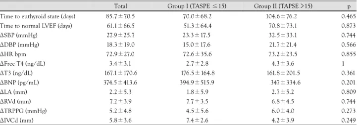
When the two groups were compared after restora-
tion to normal thyroid function, clinical parameters in- cluding blood pressure and heart rate did not show a significant difference, nor did echocardiographic para- meters, the thyroid function test (free T4, T3), BNP, left atrial dimension, LVEF, diastolic right ventricular dimension, the maximal pressure gradient of TR, or the IVC dimension (Table 5).
Discussion
Thyroid hormone (particularly T3) increases heart rate and left ventricular contractility, decreases systemic vas-
Table 4. Comparison of echocardiographic parameters between the 2 groups on admission
All (n=12) Group I (TAPSE ≤15) (n=6) Group II (TAPSE >15) (n=6) p
LA (mm) 43.7±6.4 44.3±3.6 43.2±8.7 0.748
LVDd (mm) 51.8±5.8 51.5±5.2 52.2±6.9 0.936
LVEF (%) 35.9±7.7 36.7±9.5 35.1±6.3 0.630
RVd (mm) 31.5±8.9 35.8±8.3 27.2±7.9 0.078
TRPPG (mmHg) 33.6±7.0 30.9±2.0 36.3±9.3 0.197
IVCd (mm) 20.4±4.9 19.6±2.2 21.3±4.6 0.296
Values are in n (%) or mean±standard deviation. TAPSE: tricuspid annular plane excursion, LA: left atrium, LVDd: left ventricular end diastolic diameter, LVEF: left ventricular ejection fraction, RVd: right ventricular diameter, TRPPG: tricuspid regurgitation peak pressure gra- dient, IVCd: inferior vena cava diameter
T3 p=0.025 (n=8) 800
700 600 500 400 300 200 100 (ng/dL)
Admission Euthyroid state 12.00 10.00 8.00 6.00 4.00 2.00 0.00 (ng/dL)
Free T4 p=0.003 (n=11)
Admission Euthyroid state 200 180 160 140 120 100 80 60 (bmp)
Admission Euthyroid state Heart rate p=0.003 (n=11)
Fig. 1. Comparisons of T3, free T4, and heart rate between admission and conversion to the euthyroid state. T3: triiodothyronine, free T4:
free thyroxine.
Fig. 2. Comparison of TAPSE and LVEF between admission and conversion to the euthyroid state. TAPSE: tricuspid annular plane excurs- ion, LVEF: left ventricular ejection fraction.
TAPSE p=0.003 (n=12) 35
30 25 20 15 10 5 (mm)
Admission Euthyroid state
LVEF p=0.002 (n=12) 60.0
50.0 40.0 30.0 20.0 (%)
Admission Euthyroid state
cular resistance, increases sodium re-absorption and ery- thropoietin secretion, and raises stroke volume up to 300%.1) However, during thyrotoxicosis, atrial fibrilla- tion or sinus tachycardia can occur, which may be caused by a calcium circulation distrubance to heart cells and structural changes in the heart.1-3) In case that atrial fi- brillation or sinus tachycardia lasts for an extended pe- riod due to uncontrolled thyrotoxicosis, 6% of the pa- tients experience heart failure, and among these cases half are accompanied by LVD; this is termed tachycar- dia induced cardiomyopathy.3)4)
Recently, a few studies have reported that thyrotoxi- cosis is manifested first by right heart failure with pul- monary artery hypertension and tricuspid regurgitation, but only isolated right ventricular failure was mention- ed,7-12) and no study has been reported in which both RVD and LVD occur simultaneously. The purpose of this study is to determine the causative factors that trigger RVD in thyrotoxicosis and to examine the relationship between RVD and LVD in tachycardia induced cardio- myopathy.
Among the patients that experienced LVD by thyro- toxicosis, half (50%) had RVD. All 12 patients showed improvement in clinical features, blood tests, and echo- cardiography after thyrotoxicosis treatment. On admis- sion, the RVD group (group 1) and the non-RVD group
(group 2) did not show any significant differences in blood tests or clinical features including heart rate and BNP. Echocardiographic parameters also did not show any significant differences between the two groups.
Even in the euthyroid state, there was no significant difference between the two groups in the results of all tests, including changes in heart rate and the change of LVEF. That suggests that RVD occuring in thyrotoxico- sis patients experiencing LVD has a different mecha- nism, compared with tachycardia induced cardiomyo- pathy known as mechanism of right heart failure in th- yrotoxicosis.
If RVD in thyrotoxicosis is not caused by a secon- dary change in tachycardia-induced cardiomyopathy or secon-dary pulmonary artery hypertension, the right ventricle itself may be considered as the cause of RVD.
Research indicating that the direct application of thy- roid hormone to myocardial cells causes toxic effects has been explained by myocardial stunning.13)14) In addition, Kiss et al.15) found that the function of calcium ions within myocardial cells is deteriorated in thyrotoxicosis patients, and demonstrated that thyroid hormone af- fects transcription within myocardial cells and inhibits the synthesis of required proteins, triggering heart fail- ure.16) In practice, thyroid hormone is known to affect many ion channels in the myocardial cell membrane, as well as gene expression within the nucleus of the myo- cardial cell.2) However, as thyroid hormone affects all myocardial cells and is not specific to the right ventricle, this data is not sufficient to explain the cause of RVD dysfunction. We hypothesize that when exposed to thy- roid hormone, sensitivity of thyroid hormone receptor (TR) isomers TRα1, TRα2, and TRβ1 within myo- cardial cell changes; this mayalter the effects of thyroid hormone and explain the mechanism of RVD.1)2)
Table 5. Comparison of clinical features, laboratory data, 2-dimensional echocardiographic parameters between the two groups in the eu- thyroid state
Total Group I (TASPE ≤15) Group II (TAPSE >15) p
Time to euthyroid state (days) 85.7±70.5 70.0±68.2 104.6±76.2 0.465
Time to normal LVEF (days) 61.1±66.5 51.3±64.4 70.8±73.1 0.873
ΔSBP (mmHg) 27.9±25.7 23.3±17.5 32.5±33.1 0.744
ΔDBP (mmHg) 18.3±19.0 15.0±17.6 21.7±21.4 0.566
HR
Δ bpm 72.9±27.0 72.6±35.6 73.2±23.5 0.855
Free T4 (ng/dL)
Δ 3.4±3.1 2.7±2.8 4.3±3.6 1
T3 (ng/dL)
Δ 167.1±170.6 176.5±164.8 161.8±201.5 0.361
BNP (pg/mL)
Δ 374.5±413.6 394.9±515.9 347±334.6 0.201
LA (mm)
Δ 2.2±5.3 1.8±5.9 2.7±5.2 0.809
RVd (mm)
Δ 7.2±3.9 7.7±3.5 6.8±4.5 0.744
TRPPG (mmHg)
Δ 5.2±4.8 4.5±5.6 6.0±4.0 0.273
IVCd (mm)
Δ 5.8±3.6 7.4±2.6 4.2±3.9 0.249
Values are mean±standard deviation. Δ: change from admission to the euthyroid state, LVEF: left ventricular ejection fraction, SBP: systolic blood pressure, DBP: diastolic blood pressure, HR: heart rate, free T4: free thyroxine, T3: triiodothyronine, BNP: B-type Natriuretic Peptide, LA: left atrium, RVd: right ventricular diameter, TRPPG: tricuspid regurgitation peak pressure gradient, IVCd: inferior vena cava diameter, TAPSE: tricuspid annular plane excursion
Table 6. Comparison of tricuspid regurgitation between admiss- ion and conversion to the euthyroid state (p=0.02)
TR severity Admission (n=12) Euthyroid state (n=11)
Trace 0 1
Mild 7 9
Moderate 3 1
Severe 2 0
TR: tricuspid regurgitation
The limitation of this research is that we only studied 12 patients, so it is hard to draw statistically significant conclusions. However, only few case reports have been presented on thyrotoxicosis and right heart failure, and even in the largest study, performed by Cohen and Sch- attner7) only 8 patients were examined. In addition, since this is a retrospective study, cardiac catheterization for the accurate measurement of pulmonary artery pressure was not performed.
In conclusion, we demonstrate that RVD occurring in thyrotoxicosis is not related to secondary hemodynamic change by left ventricular systolic dysfunction. We hypo- thesize there may be a direct toxic effect of thyroid hor- mone on right ventricle, but further study is needed to validate this idea. However, we believe it is reasonable given that thyroid hormone affects gene expression in the nuclear of myocardial cells and works on various ion channels existing in the myocardial cell membrane. A sensitivity difference to thyroid hormone within cells could affect right heart failure. I think it is needed fur- ther study about molecular biology.
REFERENCES
1) Dahl P, Danzi S, Kein I. Thyrotoxic cardiac disease. Curr Heart Fail Rep 2008;5:170-6.
2) Klein I, Ojamaa K. Thyroid hormone and the cardiovascular sys- tem. N Engl J Med 2001;344:501-9.
3) Umana E, Solares CA, Alpert MA. Tachycardia-induced cardio- myopathy. Am J Med 2003;114:51-5.
4) Siu CW, Yeung CY, Lau CP, Kung A, Tse HF. Incidence, clinical characteristics and outcome of congestive heart failure as the ini- tial presentation in patients with primary hyperthyroidism. Heart 2007;93:483-7.
5) Di Giovambattista R. Hyperthyroidism as a reversible cause of
right ventricular overload and congestive heart failure. Cardio- vasc Ultrasound 2008;6:29.
6) Lozano HF, Sharma CN. Reversible pulmonary hypertension, tri- cuspid regurgitation and right-sided heart failure associated with hyperthyroidism: case report and review of the literature. Cardiol Rev 2004;12:299-305.
7) Cohen J, Schattner A. Right heart failure and hyperthyroidism: a neglected presentation. Am J Med 2003;115:76-7.
8) Park JH, Shong MH, Lee JH, Choi SW, Jeoung JO, Seong IW. Re- versible severe tricuspid regurgitation with right heart failure as- sociated with thyrotoxicosis. Thyroid 2006;16:813-4.
9) Lozano HF, Sharma CN. Reversible pulmonary hypertension, tri- cuspid regurgitation and right-sided heart failure associated hy- perthyroidism: case report and review of the literature. Cardiol Rev 2004;12:299-305.
10) Nakchbandi I, Wirth J, Inzucchi S. Pulmonary hypertension ca- used by Graves’ thyrotoxicosis: normal pulmonary hemodynamics restored by (131)I treatment. Chest 1999;116:1483-5.
11) Syrius V, Plastiras SC, Paterakis T, Moyssakis I, Vlachoyianno- poulos P. Severe reversible right heart failure in a patient with hyperthyroidism. Int J Clin Pract 2008;62:334-6.
12) Kang B, Cho DK, Byun KH, Eun LY, Cho YH. Isolated pulmonary arterial hypertension-Janus’ faces of hyperthyroidism. Korean Circ J 2009;39:168-70.
13) Pereira N, Parisi A, Dec GW, Choo J, Hajjar R, Gordon PC. Myo- cardial stunning in hyperthyroidism. Clin Cardiol 2000;23:298- 300.
14) Kwak JJ, Choi YJ, Kwon KH, Park SH. A case of myocardial stunning in hyperthyroidism. Korean Circ J 2004;34:516-9.
15) Kiss E, Jakab G, Kranias EG, Edes I. Thyroid hormone-induced alterations in phospholamban protein expression: regulatory eff- ects on sarcoplasmic reticulum Ca2+ transport and myocardial re- laxation. Circ Res 1994;75:245-51.
16) Garcia-Gonzalez MJ, Dominguez-Rodriguez A, Garcia CR. Acute right ventricular dysfunction after cardioversion or hyperthyroid cardiomyopathy in an unrecognized thyrotoxicosis patient? Am J Emerg Med 2007;25:723-4.